This website uses cookies to ensure you get the best experience on our website.


- Table of Contents
Plan MOSPD1 IHC-P around observed cytoplasmic staining (HPA tissue IHC) and annotated ER/Golgi membranes (UniProt). Compare high staining in Purkinje cells with undetected staining in adipocytes (HPA tissue IHC), and validate the catalog antibody in human sections because its reported IHC test used mouse tissue (datasheet).

Expected localisation, validated protocols, controls and antibodies — the at-a-glance facts below, then the full design guide.
| Expected localisation | Cytoplasmic staining (HPA tissue IHC); ER/Golgi membranes (UniProt) | |
| Staining pattern | High cytoplasmic staining in Purkinje cells (HPA tissue IHC) | |
| Antigen retrieval | Citrate pH 6.0 HIER, 95–98 °C, 20 min (rule: cytoplasmic / membrane antigen) | |
| Positive control | Cerebellum+4 more · see all | |
| Negative control | Adipose tissue+1 more · see all |
| Fixation | Keep paraffin-section fixation consistent (standard IHC practice; not target-specific); Matched tissue-IHC evidence does not establish the fixation claim. Validate the specimen-specific method before use. (selected-SKU IHC image A15298) | |
| Caveat | Human IHC is untested for the catalog antibody (datasheet) | |
| Regulation | No expression regulator specified (UniProt) | |
| Isoform / epitope | 3 isoforms; epitope coverage and membrane side unknown (UniProt) |
The catalog antibody’s IHC-P protocol is accompanied by one published MOSPD1 IHC protocol for breast tumor tissue (PMC11453086).
| Sample | Tissue sections; selected-image fixative not specified (standard IHC workflow) |
| Fixation | Image fixative and duration unreported (datasheet A15298); verify before use. |
| Sectioning | 4–5 µm sections on charged slides (standard) |
| Deparaffinisation | Xylene, graded ethanol series to water (standard) |
| Antigen retrieval | Heat-induced epitope retrieval in citrate buffer, pH 6.0, 20 min at 95–98 °C (standard rule: cytoplasmic / membrane antigen) |
| Peroxidase block | 3% H2O2, 10 min, room temperature (standard) |
| Blocking | 10% normal serum of the secondary host, 30 min, room temperature (standard) |
| Primary antibody | Rabbit anti-MOSPD1, 1:100–1:500 starting range (standard) |
| Primary incubation | Overnight at 4 °C (standard) |
| Detection | HRP-polymer secondary, DAB chromogen 5–10 min (standard) |
| Counterstain | Hematoxylin, blue, dehydrate and mount (standard) |
| Expected result | MOSPD1-positive staining in purkinje cells of cerebellum (HPA tissue IHC: High). HPA tissue profile: General cytoplasmic expression. No signal in the no-primary control. |
In paraffin IHC, expect mainly cytoplasmic MOSPD1 staining, strongest in cerebellar Purkinje cells and moderate in several other listed cell types (HPA tissue IHC: general cytoplasmic expression; Purkinje cells High). MOSPD1 is annotated at endoplasmic reticulum and Golgi membranes, with two transmembrane segments (UniProt Q9UJG1). Treat the tissue pattern as a guide: HPA rates its IHC evidence Approved, with medium agreement between staining and RNA data and external verification pending (HPA tissue IHC).
| Purkinje cell cytoplasm stains strongly; surrounding tissue is less prominent. | This matches the clearest listed positive cell type (HPA tissue IHC: cerebellar Purkinje cells High). Assess signal within identified cells, since HPA reports a general cytoplasmic IHC pattern rather than a distinctive structure visible in every paraffin section (HPA tissue IHC). |
| Glandular, basal, glial or squamous epithelial cells show moderate cytoplasmic staining. | Moderate signal fits the listed cells in adrenal gland, appendix, breast, bronchus, caudate, cerebral cortex and cervix (HPA tissue IHC: Medium). Compare like cell types within a section; the tissue name alone does not specify that every cell should stain (HPA tissue IHC). |
| A putative IHC positive is confined to nuclei, with no convincing cytoplasmic signal. | Recheck compartment assignment and controls: nuclear-only chromogen differs from the general cytoplasmic tissue IHC profile (HPA tissue IHC). Do not automatically dismiss nuclear signal across methods, because approved ICC-IF images place MOSPD1 in the nucleoplasm and vesicles (HPA subcellular ICC-IF). |
| Strong colour appears in an unexpected cell type, or spreads evenly beyond cell boundaries. | Check for cross-reactivity, endogenous detection activity or background before scoring it as MOSPD1 (general IHC practice). HPA reports adipocytes and ovarian stroma cells as Not detected, while several other cells have Low signal; those observations are useful comparisons, not absolute specificity tests (HPA tissue IHC). |
| A known positive compartment has no detectable chromogen. | Absent Purkinje cell staining conflicts with the strongest listed IHC observation (HPA tissue IHC: Purkinje cells High). First evaluate staining controls and tissue preservation (general IHC practice); HPA's Approved rating has medium RNA agreement and awaits external verification, so a single negative section is not proof of absent protein (HPA tissue IHC). |
| Compartment evidence | UniProt places MOSPD1 at endoplasmic reticulum and Golgi membranes and records transmembrane segments at residues 159–179 and 191–211 (UniProt Q9UJG1). HPA tissue IHC describes general cytoplasmic expression; it does not resolve those organelles in a routine chromogenic section (HPA tissue IHC; general IHC practice). |
| Cell type and signal range | HPA lists Purkinje cells as High, seven specified cell populations as Medium, and other populations as Low or Not detected (HPA tissue IHC). Score the cell population examined rather than assigning one intensity to an entire tissue (general IHC practice). |
| Antibody evidence | The HPA antibody HPA031158 is Approved for IHC and ICC, while the tissue IHC summary reports medium staining-to-RNA consistency and pending external verification (HPA antibodies; HPA tissue IHC). An Approved result supports pattern comparison but does not independently establish every signal's specificity (HPA tissue IHC). |
| Isoforms and epitope | UniProt lists three MOSPD1 isoforms and an MSP domain at residues 16–143 (UniProt Q9UJG1). The supplied evidence gives no antibody epitope, so it cannot establish which isoforms the IHC antibody detects or predict an isoform-specific staining pattern. |
| Processing and modification | The supplied UniProt record lists a 1–213 chain, no signal peptide or propeptide, and no annotated glycosylation or modified residues (UniProt Q9UJG1). These annotations provide no basis to invoke cleavage, shedding or a modification-dependent IHC pattern; they also do not establish fixation sensitivity. |
| IF/ICC question: should its pattern match IHC exactly? | No exact match is established: HPA reports approved nucleoplasm and vesicle localisation in ICC-IF images from A-431, U-251MG and U2OS, while tissue IHC is generally cytoplasmic (HPA subcellular ICC-IF; HPA tissue IHC). Interpret each application with its own controls and readout (general IHC/IF practice). |
| Situation | Likely cause | Next action |
|---|---|---|
| Purkinje cells are unstained. | The expected strong reference pattern is missing (HPA tissue IHC: Purkinje cells High); an IHC workflow or sample problem is possible (general IHC practice). | Verify tissue identity, positive and negative controls, primary antibody use and detection reagents; review antigen retrieval against the IHC-validated antibody's instructions (general IHC practice). No MOSPD1-specific retrieval or fixation response is supplied. |
| Every cell shows similar weak colour. | Diffuse colour can reflect nonspecific background or detection activity (general IHC practice), and obscures HPA's cell-specific intensity differences (HPA tissue IHC). | Inspect the no-primary control, blocking, washing and chromogen development; adjust these general IHC steps before comparing cell populations (general IHC practice). |
| Colour appears mainly outside cells or along section edges. | The distribution does not resemble HPA's general cytoplasmic IHC profile (HPA tissue IHC); staining artefact is possible (general IHC practice). | Inspect section quality and control slides, then repeat with consistent washing and detection conditions if needed (general IHC practice). |
| Adipocytes or ovarian stroma cells stain strongly. | That contrasts with Not detected observations for those specific cell populations (HPA tissue IHC); cross-reactivity or endogenous activity is possible (general IHC practice). | Confirm cell identity and examine no-primary and detection controls. Compare the same run with a listed positive population before assigning the colour to MOSPD1 (HPA tissue IHC; general IHC practice). |
| Chromogenic signal seems exclusively nuclear. | Nuclear-only staining differs from HPA's general cytoplasmic tissue IHC profile, although nucleoplasm is an approved ICC-IF location (HPA tissue IHC; HPA subcellular ICC-IF). | Check morphology, counterstain and control slides, then score IHC and IF/ICC within their respective evidence sets; do not transfer an ICC-IF localisation call directly to paraffin IHC (general IHC/IF practice). |
| A listed moderate-positive population stains inconsistently. | HPA reports Medium staining in specified cell types and only medium agreement between IHC staining and RNA expression (HPA tissue IHC). Section-to-section variation or workflow variation may also affect an IHC comparison (general IHC practice). | Compare identified cells in matched sections and review staining controls across the run. Record the observed intensity rather than forcing a Medium score; seek independent confirmation if the conclusion depends on a disputed pattern (HPA tissue IHC; general IHC practice). |


Comprehensive Human Protein Atlas IHC scoring per tissue (reliability: Approved — Medium consistency between antibody staining and RNA expression data. Pending external verification.). Rows are taken directly from the HPA tissue chart — click any row's HPA link to view the source.
| Tissue | Cell type | Level | Evidence | Source |
|---|---|---|---|---|
| Cerebellum | Purkinje cells | High | Protein (IHC) | HPA → |
| Adrenal gland | Glandular cells | Medium | Protein (IHC) | HPA → |
| Appendix | Glandular cells | Medium | Protein (IHC) | HPA → |
| Breast | Glandular cells | Medium | Protein (IHC) | HPA → |
| Bronchus | Basal cells | Medium | Protein (IHC) | HPA → |
Troubleshoot MOSPD1 staining in paraffin sections by checking retrieval, compartment, controls, and scoring before interpreting signal.
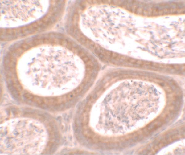
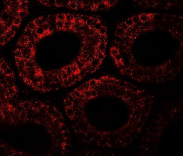
A15298 has mouse testis IHC and IF images (catalog image captions). Human, mouse, and rat reactivity is listed, but IHC and IF validation is reported only in mouse samples (catalog applications and validation notes).
A15298 will render with an IHC image of mouse testis stained at 2.5 μg/ml (A15298 IHC image caption). Its IF image shows mouse testis stained at 20 μg/ml (A15298 IF image caption).
Which to pick: Choose A15298 for tissue IHC: IHC-P is listed and mouse testis IHC is shown, but the image caption does not report the section processing or fixative (A15298 applications; A15298 IHC image caption). A15298 also has mouse testis IF data; ICC validation is unreported (A15298 IF image caption; catalog validation notes). For human or rat samples, A15298 lists reactivity in those species, but its IHC and IF validation is reported only in mouse samples (catalog reactivity and validation notes).