This website uses cookies to ensure you get the best experience on our website.


- Table of Contents
Plan chromogenic IHC for MPI with an IHC-validated antibody (datasheet: M00175-2). Use cytoplasmic staining as the expected tissue pattern and lung macrophages as a high-staining reference (HPA tissue IHC).

Expected localisation, validated protocols, controls and antibodies — the at-a-glance facts below, then the full design guide.
| Expected localisation | Cytoplasmic staining (HPA tissue IHC) | |
| Staining pattern | Cytoplasmic staining; high in lung macrophages (HPA tissue IHC) | |
| Antigen retrieval | EDTA pH 8.0 HIER, heat-mediated (datasheet M00175-2) | |
| Positive control | Adrenal gland+4 more · see all | |
| Negative control | Adipose tissue+3 more · see all |
| Fixation | Matched tissue-IHC evidence does not establish the fixation claim. Validate the specimen-specific method before use. (selected-SKU IHC image M00175-2) | |
| Caveat | Lung macrophages may dominate the signal (HPA tissue IHC) | |
| Regulation | No specific inducer reported (UniProt) | |
| Isoform / epitope | Two isoforms; epitope differences unspecified (UniProt) |
The catalog antibody’s IHC-P protocol is accompanied by one published MPI immunohistochemistry protocol in a prostate cancer tissue microarray (PMC9491030).
| Sample | Paraffin-embedded human lung cancer tissue; fixative not specified (datasheet M00175-2) |
| Fixation | Image fixative and duration unreported (datasheet M00175-2); verify before use. |
| Sectioning | 4–5 µm sections on charged slides (standard) |
| Deparaffinisation | Xylene, graded ethanol series to water (standard) |
| Antigen retrieval | Heat retrieval: EDTA pH 8.0 (datasheet M00175-2); 20 min, 95–100 °C (standard) |
| Peroxidase block | 3% H2O2, 10 min, room temperature (standard) |
| Blocking | 10% goat serum (datasheet M00175-2) |
| Primary antibody | Mouse monoclonal (clone 5G5) anti-MPI, 2μg/ml (datasheet M00175-2) |
| Primary incubation | Overnight at 4 °C (datasheet M00175-2) |
| Detection | Streptavidin-biotin complex (SABC), DAB chromogen (datasheet M00175-2) |
| Counterstain | Hematoxylin, blue, dehydrate and mount (standard) |
| Expected result | MPI-positive staining in glandular cells of adrenal gland (HPA tissue IHC: High). HPA tissue profile: General cytoplasmic expression. No signal in the no-primary control. |
MPI is expected mainly in the cytoplasm of stained cells (UniProt P34949: cytoplasm; HPA: general cytoplasmic IHC expression). In paraffin IHC, strong examples include lung macrophages, pancreatic exocrine glandular cells and testicular Leydig cells (HPA: High in each). The HPA tissue profile is Approved, with medium consistency between antibody staining and RNA expression; MPI has no transmembrane segment (HPA: tissue reliability; UniProt P34949 topology).
| Cytoplasmic staining in lung macrophages or pancreatic exocrine cells, with cell outlines and nuclei readable. | This fits the expected compartment and documented High cell-specific staining (UniProt P34949: cytoplasm; HPA: High in these cells). Compare cells within the section; a strong field alone does not establish which cells carry the signal (standard IHC interpretation). |
| Staining is chiefly nuclear, or forms a dominant sharply outlined membrane pattern. | Treat this as a compartment mismatch for IHC: HPA describes general cytoplasmic tissue staining, and UniProt places MPI in the cytoplasm (HPA: tissue profile; UniProt P34949: subcellular location). HPA ICC-IF also reports an additional plasma membrane location, so a minor membrane component alone is not proof of artefact (HPA: subcellular). |
| Strong colour appears mainly in adipocytes, smooth muscle cells or soft-tissue fibroblasts. | These cell types are listed as Not detected by HPA tissue IHC (HPA: adipose tissue, smooth muscle and soft tissue). Recheck cell identity and controls before calling MPI positive; unexpected colour may reflect cross-reactivity or detection background (standard IHC interpretation). |
| Colour spreads evenly across stroma, empty spaces and cell types instead of resolving cytoplasm. | A uniform haze does not match the cell-resolved cytoplasmic profile (HPA: general cytoplasmic expression). Assess reagent-only controls and background before scoring cells; nonspecific antibody binding or endogenous detection activity can produce diffuse chromogenic signal (standard IHC practice). |
| No cytoplasmic signal is visible in a well-preserved, expected-positive cell population. | Check a documented High population such as lung macrophages or pancreatic exocrine cells first (HPA: tissue IHC). An absent result may reflect assay failure or a sample-specific difference; HPA calls its tissue profile Approved with medium RNA-staining consistency, so one negative section cannot settle biological absence (HPA: reliability description; standard IHC interpretation). |
| Cell population and tissue | Expect intensity to vary by cell type: adrenal glandular cells, lung macrophages, pancreatic exocrine cells, salivary glandular cells and Leydig cells are High, while adipocytes are Not detected in HPA IHC (HPA: tissue IHC). Score identified cells rather than a whole section as uniformly positive (standard IHC interpretation). |
| Compartment and topology | MPI is annotated as cytoplasmic and has no transmembrane segment (UniProt P34949: location and topology). The expected IHC reading is predominantly intracellular cytoplasmic staining (HPA: tissue profile); membrane-like staining needs context because HPA ICC-IF reports an additional plasma membrane location (HPA: subcellular). |
| Evidence and antibody validation | The tissue profile is Approved but has medium consistency with RNA data, and HPA007200 is listed as IHC Approved (HPA: tissue reliability and antibody record). These designations support comparison with the reference pattern; they do not make every unexpected cell or compartment a confirmed biological finding (standard IHC interpretation). |
| Isoforms and processing | UniProt lists 2 isoforms and annotates the mature chain as residues 2–423, with no signal peptide or propeptide (UniProt P34949: isoforms and processing). No antibody epitope or isoform-specific staining claim is supplied; interpret the HPA images as a reference pattern, without assigning a signal to either isoform. |
| IF/ICC: what localisation should be expected? | Mainly cytosol, with an additional plasma membrane location in HPA ICC-IF (HPA: subcellular, both locations Approved). This helps interpret an IF image but does not establish that a membrane-dominant chromogenic tissue pattern is expected; the HPA tissue IHC profile is generally cytoplasmic (HPA: tissue IHC). |
| Situation | Likely cause | Next action |
|---|---|---|
| Known-positive cells show no colour. | The expected cell population may be absent from the section, or the IHC run may have failed (standard IHC practice); lung macrophages and pancreatic exocrine cells are documented High examples (HPA: tissue IHC). | Confirm the relevant cells are present, then review a same-run positive control and the detection steps (standard IHC practice). Record a technical failure separately from a cell-level negative result. |
| Signal is faint in a selected tissue. | The chosen cells may normally stain weakly: HPA lists small-intestinal glandular cells and rectal glandular cells as Low (HPA: tissue IHC). | Compare with a documented High cell population and score cell type and intensity together (HPA: tissue IHC; standard IHC interpretation). Do not use a low-staining population as the sole sensitivity check. |
| Nuclei dominate the stain. | That compartment conflicts with cytoplasmic MPI annotation and HPA's general cytoplasmic tissue pattern (UniProt P34949: location; HPA: tissue IHC); the cause is unresolved from the supplied records. | Review the counterstain and reagent-only control, then verify whether the chromogen truly occupies nuclei (standard IHC practice). Score the nuclear pattern as unexpected until resolved. |
| Brown colour covers many cell types and extracellular areas. | Diffuse colour may come from nonspecific binding or endogenous chromogenic detection activity rather than cell-localized MPI (standard IHC practice; HPA: general cytoplasmic tissue profile). | Inspect negative controls and the detection system; if peroxidase detection is used, check its endogenous-activity control (standard IHC practice). Reassess only discrete cytoplasmic signal. |
| A cell type listed as Not detected appears strongly positive. | Possible explanations include mistaken cell identification, cross-reactivity or background; HPA lists adipocytes, parathyroid glandular cells, smooth muscle cells and soft-tissue fibroblasts as Not detected (HPA: tissue IHC; standard IHC interpretation). | Confirm morphology and compare a same-run documented positive population and negative controls (standard IHC practice). Report the discrepancy without treating the HPA category as proof that every specimen must be negative. |
| A positive cell shows some edge signal. | HPA ICC-IF reports an additional plasma membrane location, while tissue IHC is generally cytoplasmic (HPA: subcellular and tissue IHC). The records do not establish whether this section's edge signal is specific. | Judge the edge signal alongside cytoplasmic staining, cell identity and controls (standard IHC interpretation). Keep a membrane-dominant result separate from the expected IHC pattern. |


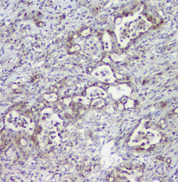
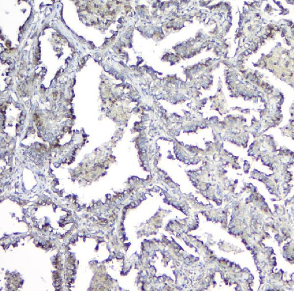
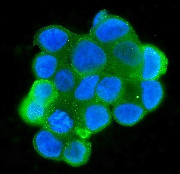


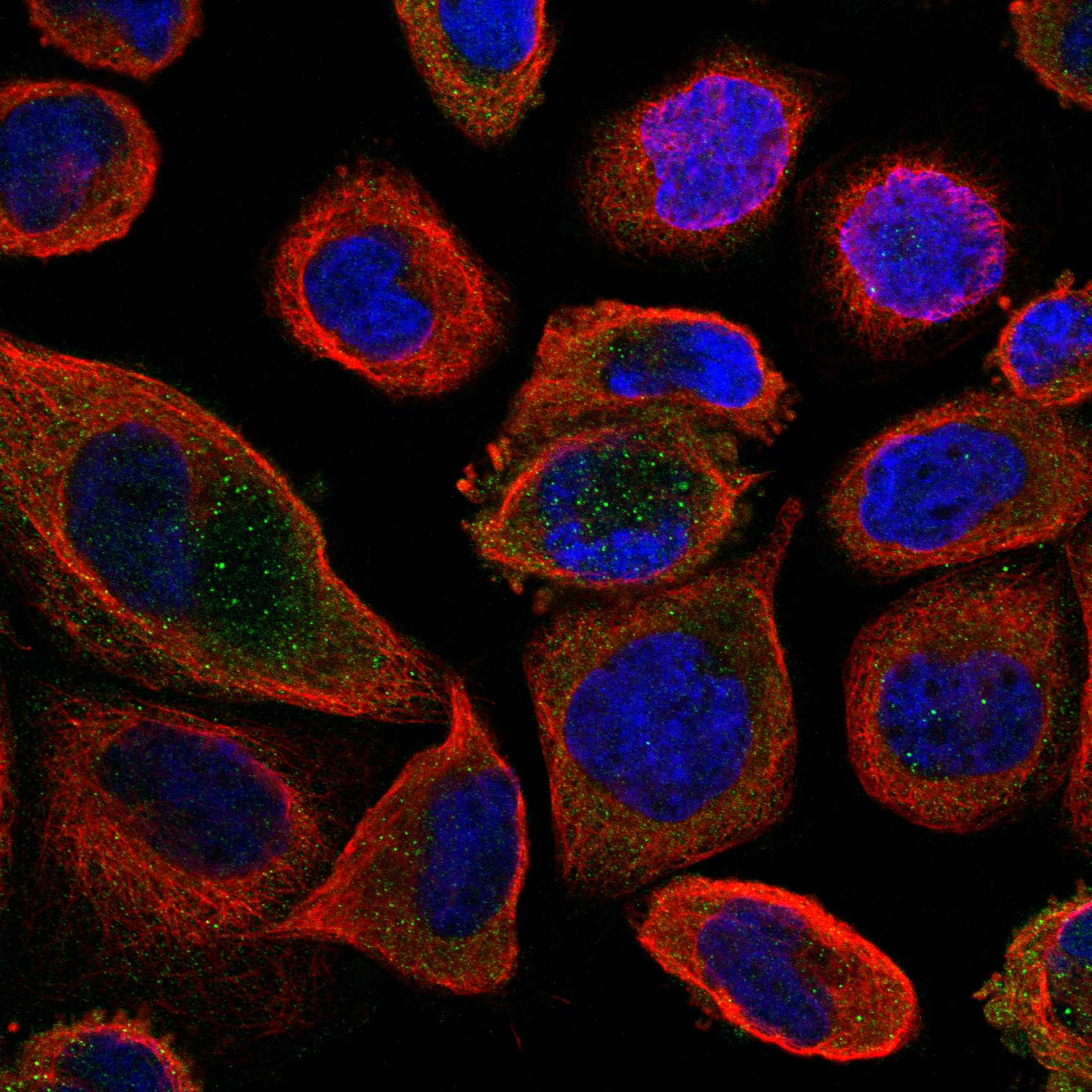

Comprehensive Human Protein Atlas IHC scoring per tissue (reliability: Approved — Medium consistency between antibody staining and RNA expression data.). Rows are taken directly from the HPA tissue chart — click any row's HPA link to view the source.
| Tissue | Cell type | Level | Evidence | Source |
|---|---|---|---|---|
| Adrenal gland | Glandular cells | High | Protein (IHC) | HPA → |
| Lung | Macrophages | High | Protein (IHC) | HPA → |
| Pancreas | Exocrine glandular cells | High | Protein (IHC) | HPA → |
| Salivary gland | Glandular cells | High | Protein (IHC) | HPA → |
| Testis | Leydig cells | High | Protein (IHC) | HPA → |
| Tissue | Cell type | Level | Evidence | Source |
|---|---|---|---|---|
| Adipose tissue | Adipocytes | Not detected | Protein (IHC) | HPA → |
| Parathyroid gland | Glandular cells | Not detected | Protein (IHC) | HPA → |
| Smooth muscle | Smooth muscle cells | Not detected | Protein (IHC) | HPA → |
| Soft tissue | Fibroblasts | Not detected | Protein (IHC) | HPA → |
Troubleshoot MPI staining in paraffin sections by checking retrieval, cytoplasmic localisation, cell identity and assay controls before interpreting signal strength.
Anti-MPI antibodies have IHC images from paraffin human tumor sections and rat brain (IHC image captions), plus IF/ICC images from human MCF-7 and T-47D cells (IF image captions).
M00175-2 shows IHC in paraffin human lung, breast, renal clear cell carcinoma and ovarian serous adenocarcinoma sections, plus IF/ICC in T-47D cells (M00175-2 image captions). A00175 shows IHC in paraffin human intestinal cancer, lung cancer and rat brain sections (A00175 image captions); M00175 shows IF/ICC in MCF-7 cells (M00175 image caption).
Which to pick: For paraffin-section IHC, choose M00175-2, a mouse monoclonal with EDTA retrieval at pH 8 in its human lung cancer image (M00175-2 catalog entry and IHC caption), or A00175, a rabbit antibody with citrate retrieval at pH 6 in its human intestinal cancer image (A00175 catalog entry and IHC caption); fixation is unreported in both captions. For IF/ICC, M00175 has an MCF-7 image (M00175 IF caption), while M00175-2 has a T-47D image (M00175-2 IF caption). For work across species, A00175 lists human, mouse and rat reactivity (A00175 catalog entry), with IHC images in human and rat tissue (A00175 IHC captions); the supplied images do not establish mouse tissue IHC performance.