MPP7 / MAGUK p55 subfamily member 7 · IHC design guide

Design Immunohistochemistry for MPP7

Plan chromogenic MPP7 IHC in paraffin sections using the cytoplasmic and membranous tissue pattern as a reference (HPA tissue IHC). Start the human-reactive IHC-P catalog antibody at 1:25 (datasheet), and interpret staining with the reported off-target binding in mind (HPA tissue IHC).

Evidence assembled Oct 2026 · For research use; verify linked source records and product datasheet before use
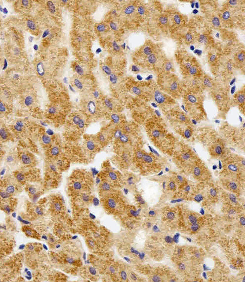
Immunohistochemistry protocol sheet for MPP7 (IHC for MPP7): expected localisation Cytoplasmic and membranous tissue staining (HPA tissue IHC), antibody A07143, validated IHC image, and IHC protocol steps
Printable MPP7 IHC protocol sheet — expected localisation Cytoplasmic and membranous tissue staining (HPA tissue IHC), antibody A07143, controls and protocol steps. Open the full MPP7 IHC guide →

MPP7 Immunohistochemistry Experimental Design Guide

Expected localisation, validated protocols, controls and antibodies — the at-a-glance facts below, then the full design guide.

Must know before staining
Expected localisation Cytoplasmic and membranous tissue staining (HPA tissue IHC)
Staining pattern Epithelial cells show cytoplasmic and membranous staining (HPA tissue IHC)
Antigen retrieval Citrate pH 6.0 HIER, 95–98 °C, 20 min (rule: cytoplasmic / membrane antigen)
Positive control ⓘ Bronchus+4 more · see all
Negative control ⓘ Adipose tissue+4 more · see all
Important caveats
Reasons your staining may differ from the expected pattern.
Fixation Keep formalin fixation consistent across sections. (standard IHC practice; not target-specific)
Caveat Presumed off-target staining complicates interpretation (HPA tissue IHC)
Regulation Expression regulation is unreported (UniProt)
Isoform / epitope 2 isoforms; check epitope coverage (UniProt)
Section 1

Recommended MPP7 IHC & IF Protocols

The catalog antibody’s IHC-P protocol is paired with one published protocol using an esophageal squamous cell carcinoma tissue microarray (PMC9501138).

Recommended immunohistochemistry (IHC-P) protocol parameters
SampleParaffin-embedded H. kidney tissue; fixative not specified (datasheet A07143)
FixationImage fixative and duration unreported (datasheet A07143); verify before use.
Sectioning4–5 µm sections on charged slides (standard)
DeparaffinisationXylene, graded ethanol series to water (standard)
Antigen retrievalHeat-induced epitope retrieval in citrate buffer, pH 6.0, 20 min at 95–98 °C (standard rule: cytoplasmic / membrane antigen)
Peroxidase block3% H2O2, 10 min, room temperature (standard)
Blocking10% normal serum of the secondary host, 30 min, room temperature (standard)
Primary antibodyRabbit anti-MPP7, 1:25 (datasheet A07143)
Primary incubationOvernight at 4 °C (standard)
DetectionHRP-polymer secondary, DAB chromogen 5–10 min (standard)
CounterstainHematoxylin, blue, dehydrate and mount (standard)
Expected resultMPP7-positive staining in respiratory epithelial cells of bronchus (HPA tissue IHC: High). HPA tissue profile: General cytoplasmic and membranous expression. No signal in the no-primary control.
💡Decision noteStart with citrate pH 6.0 HIER at 95–98 °C for 20 min (page antigen-retrieval rule); the paper reports antigen repair without conditions (PMC9501138).
Section 2

What Is the Expected MPP7 Staining Pattern?

MPP7 is a junction-associated, cytoplasmic and membranous protein; epidermal cells show predominantly lateral membrane localisation (UniProt Q5T2T1). In tissue IHC, expect epithelial staining, including high signal in bronchial respiratory epithelium and vaginal squamous epithelium (HPA tissue IHC). MPP7 has no transmembrane segment (UniProt Q5T2T1 topology). HPA rates its tissue IHC profile Approved, while reporting low agreement with RNA data and presumed off-target staining that was disregarded (HPA tissue IHC).

What am I looking at on my slide?
Membranous staining outlines contacts between epithelial cells, with some cytoplasmic signal.This fits MPP7 at cell junctions and the cell cortex (UniProt Q5T2T1) and HPA’s general membranous and cytoplasmic tissue profile (HPA tissue IHC). Judge the pattern within the identified cell population; uniform staining of every cell is not required by these sources.
Strong nuclear staining dominates the slide, with little junctional staining.Reassess specificity: junctions are the supported main IF location, whereas nucleoplasmic localisation is uncertain (HPA subcellular ICC-IF). UniProt places MPP7 at junctions, the lateral membrane and in cytoplasm (UniProt Q5T2T1). A nuclear signal alone is therefore insufficient to call the expected IHC pattern.
Adipocytes or bone-marrow hematopoietic cells stain as strongly as the intended positive epithelium.Those cell populations were not detected in HPA tissue IHC (HPA tissue IHC). Consider cross-reactivity or endogenous chromogenic activity before interpreting their signal as MPP7; HPA reports presumed off-target staining that it disregarded (HPA tissue IHC).
Chromogen forms a diffuse haze across cells and extracellular areas.The haze cannot be assigned confidently to the reported epithelial compartments (HPA tissue IHC; UniProt Q5T2T1). In a chromogenic assay, assess background with appropriate negative controls and inspect blocking, washes and detection chemistry (general IHC practice).
No epithelial signal appears in a bronchus control section.HPA reports high staining in bronchial respiratory epithelial cells (HPA tissue IHC), so an absent control signal calls the run into question. Check section integrity, retrieval and detection before treating a negative experimental section as evidence that MPP7 is absent (general IHC practice).
💡Expected MPP7 appearanceCall a convincing positive result when epithelial cell contacts show membranous staining, with possible cytoplasmic signal and high staining in HPA-positive cell populations; dominant nuclear staining or strong signal in HPA-negative cells needs scrutiny (UniProt Q5T2T1; HPA tissue IHC; HPA subcellular ICC-IF).
How each factor affects the staining
Compartment and topologyMPP7 is reported at tight and adherens junctions, the lateral membrane and cell cortex (UniProt Q5T2T1); it has no transmembrane segment (UniProt Q5T2T1 topology). Interpret a cell-border signal by its location, without assuming that MPP7 spans the membrane.
Choice of tissue and cell populationHPA reports high staining in bronchial respiratory epithelium, fallopian-tube glandular cells, placental trophoblasts, late spermatids, thyroid glandular cells and vaginal squamous epithelium (HPA tissue IHC). It reports low esophageal squamous epithelial staining despite tissue-enhanced esophageal RNA (HPA tissue IHC).
Antibody and evidence limitsHPA037598 is Approved for IHC; HPA037597 is Supported for ICC-IF, with no IHC status listed for that antibody (HPA antibodies). HPA also reports low staining-to-RNA agreement and disregarded presumed off-target staining (HPA tissue IHC).
Isoforms and epitope uncertaintyUniProt lists 2 MPP7 isoforms (UniProt Q5T2T1). The supplied sources do not map the catalog antibody’s epitope or show isoform-specific IHC staining; do not assign a tissue pattern to one isoform from these data.
Why is my staining missing, weak or wrong?
SituationLikely causeNext action
The intended positive epithelium is negative.A failed run is possible when an HPA high population, such as bronchial respiratory epithelium, has no signal (HPA tissue IHC).Check that the expected cells are present, then review the antibody dilution, retrieval conditions and chromogenic detection with a positive control (general IHC practice). No target-specific retrieval setting is supplied.
Signal is mainly nuclear.Nucleoplasmic localisation is uncertain in ICC-IF; cell junctions are its supported main location (HPA subcellular ICC-IF).Compare nuclear staining with cell-border staining in an HPA-positive epithelial population and check negative controls before scoring it as MPP7 (HPA tissue IHC; general IHC practice).
Unexpected cells stain strongly.Cross-reactivity or endogenous detection activity may account for staining in cell types reported as not detected, such as adipocytes (HPA tissue IHC; general IHC practice).Identify the stained cells on the counterstained section; use appropriate negative controls and, for peroxidase detection, check endogenous peroxidase blocking (general IHC practice).
The whole section has diffuse chromogen.Diffuse background obscures the reported membranous and cytoplasmic pattern (HPA tissue IHC); inadequate blocking, washing or detection control can contribute (general IHC practice).Compare with a negative control, inspect chromogen development and detection reagents, and optimize blocking and washes for the assay (general IHC practice).
Esophagus stains weakly despite its RNA designation.HPA calls esophageal RNA tissue-enhanced but reports low staining in esophageal squamous epithelial cells (HPA tissue IHC).Use the observed IHC level when judging the slide; compare with an HPA high epithelial population, such as bronchial respiratory epithelium (HPA tissue IHC).
IF/ICC: should a nuclear signal count as MPP7?HPA supports cell-junction localisation in ICC-IF and labels additional nucleoplasmic localisation uncertain (HPA subcellular ICC-IF).Interpret IF/ICC against junctional staining and its own controls; do not infer an IHC protocol or a definitive nuclear IHC pattern from the ICC-IF images (HPA subcellular ICC-IF; general IF practice).

Sample controls for MPP7 IHC & IF

🧪Run bronchus first: respiratory epithelial cells should stain strongly (HPA: High in respiratory epithelial cells). Use adipose tissue as a negative comparator (HPA: Not detected in adipocytes); on the bronchus slide, use adjacent non-epithelial cells as background comparators and check that they lack the epithelial junctional staining pattern (UniProt Q5T2T1 localization).
Positive control tissue: Bronchus (Respiratory epithelial cells, HPA High)
Negative control tissue: Adipose tissue (HPA Not detected)
ICC-IF cell lines (HPA subcellular resource): HPA ICC-IF images show MPP7 in CACO-2, RT-4, U2OS, with annotated localisation: Cell Junctions (supported) (HPA subcellular).
Technical controls: Include a no-primary, secondary-only slide and an isotype IgG control matched to the primary antibody’s host species and clonality; use MPP7-knockout material as a biological negative where available (standard IHC practice). For chromogenic bronchus IHC, quench endogenous peroxidase and inspect inflammatory cells for residual background (HPA: High in bronchial respiratory epithelial cells; standard IHC practice).
⚠️Feasibility: A target-specific fixation window and the effect of fixation are unreported, and the selected A07143 paraffin-section caption does not state a fixative (A07143 tissue-IHC caption). Retrieval dependence is also unreported; optimize antigen retrieval for the paraffin sections and assess junctional localization when scoring (UniProt Q5T2T1 localization; standard IHC practice). ICC-IF images support cell-junction localization, but the supplied evidence does not establish that frozen sections or IF are easier; bronchial inflammatory cells may add peroxidase background in chromogenic IHC (HPA subcellular: Cell Junctions supported; standard IHC practice).

HPA tissue IHC evidence for MPP7

Comprehensive Human Protein Atlas IHC scoring per tissue (reliability: Approved — Low consistency between antibody staining and RNA expression data. Presumed off target binding observed and disregarded.). Rows are taken directly from the HPA tissue chart — click any row's HPA link to view the source.

Positive expression · recommended positive controls

TissueCell typeLevelEvidenceSource
Bronchus Respiratory epithelial cells High Protein (IHC) HPA →
Fallopian tube Glandular cells High Protein (IHC) HPA →
Placenta Trophoblastic cells High Protein (IHC) HPA →
Testis Elongated or late spermatids High Protein (IHC) HPA →
Thyroid gland Glandular cells High Protein (IHC) HPA →

Undetected expression · recommended negative controls

TissueCell typeLevelEvidenceSource
Adipose tissue Adipocytes Not detected Protein (IHC) HPA →
Bone marrow Hematopoietic cells Not detected Protein (IHC) HPA →
Caudate Glial cells Not detected Protein (IHC) HPA →
Cerebellum Cells in granular layer Not detected Protein (IHC) HPA →
Cerebral cortex Endothelial cells Not detected Protein (IHC) HPA →
Section 3

Advanced MPP7 IHC Tips

Troubleshoot MPP7 staining by evaluating retrieval, compartment, cellular context and controls together; tissue staining alone may be misleading (HPA: presumed off-target binding).

How should I retrieve MPP7 antigen in paraffin sections?
Start with citrate buffer at pH 6.0, heated to 95–98 °C for 20 min (page antigen-retrieval protocol). Let sections cool in buffer, then compare staining in a positive epithelial control and a no-primary control processed alongside the specimen (standard IHC practice; HPA: high staining in bronchial respiratory epithelial cells). If junctional staining remains weak, vary heating time cautiously before testing another retrieval buffer, because excessive heating can damage morphology and increase diffuse staining (standard IHC practice). Score whether signal outlines cell contacts rather than merely becoming darker throughout the cytoplasm (UniProt Q5T2T1: cell-junction and lateral-membrane localisation; HPA: general cytoplasmic and membranous tissue profile).
How can I assess whether fixation caused weak MPP7 staining?
Target-specific fixation sensitivity is unknown: the A07143 kidney caption describes a paraffin section but does not state its fixative (A07143 tissue-IHC caption). Record the specimen's fixative, fixation duration and processing history, then compare sections with similar handling before changing antibody concentration (standard IHC practice). Run the same retrieval and staining batch on a documented positive control, such as bronchial respiratory epithelium, while checking tissue preservation and cell-contact morphology (HPA: high staining in bronchial respiratory epithelial cells; standard IHC practice). If staining differs across processing batches, repeat with matched fixation conditions; neither tissue expression patterns nor protein topology establishes a fixation effect for MPP7 (standard IHC interpretation).
Where should convincing MPP7 staining appear in tissue?
Look for signal concentrated at epithelial cell contacts, especially along lateral borders, with possible cortical or cytoplasmic staining (UniProt Q5T2T1: lateral membrane, junction and cortex localisation; HPA: general cytoplasmic and membranous tissue profile). In epidermal cells, lateral-membrane enrichment is specifically reported (UniProt Q5T2T1: epidermal localisation). Compare sharply outlined contacts with diffuse staining on the same slide, and inspect a no-primary control before treating broad color development as specific (standard IHC practice). Nucleoplasmic signal warrants particular caution in chromogenic sections because its reported additional localisation is uncertain and comes from subcellular imaging (HPA subcellular: nucleoplasm uncertain; standard IHC interpretation).
Could isoforms or epitope accessibility explain inconsistent MPP7 IHC?
MPP7 has 2 annotated isoforms and contains paired L27, PDZ, SH3 and guanylate kinase-like domains (UniProt Q5T2T1: isoforms and domains). The A07143 caption identifies an N-terminal antibody, but gives no residue-level epitope or isoform coverage, so do not assign a negative section to one isoform (A07143 tissue-IHC caption). Compare retrieval conditions and independently documented antibodies whose epitopes are known, using matched sections and identical scoring rules (standard IHC practice). A reported phosphoserine at residue 409 establishes a modification site, not a demonstrated cause of altered IHC recognition by this antibody (UniProt Q5T2T1: modified residues; standard IHC interpretation).
How can IF help assess an ambiguous MPP7 IHC pattern?
Use IF on matched material to examine whether MPP7 signal follows cell contacts, and multiplex with an epithelial marker to identify the expected cells (UniProt Q5T2T1: junctional localisation; standard IF practice). Select a red or far-red fluorophore after checking tissue autofluorescence and single-label controls, particularly when green background obscures borders (standard IF practice). Because MPP7 has no transmembrane segment and is associated with the cell cortex, test gentle permeabilisation for access to intracellular epitopes; the exact N-terminal antibody epitope is unspecified (UniProt Q5T2T1: topology and cortex; A07143 tissue-IHC caption). Compare IF findings with IHC cautiously: junctional localisation is supported in subcellular imaging, whereas reported nucleoplasmic localisation is uncertain (HPA subcellular).
What should I change when MPP7 DAB staining is diffuse?
First inspect the no-primary control, tissue edges and areas of poor preservation, then verify that endogenous peroxidase was blocked before DAB development (standard chromogenic IHC practice). The A07143 paraffin-kidney example used primary antibody at 1:100, a peroxidase-conjugated secondary at 1:400 and DAB, but it does not report a fixative (A07143 tissue-IHC caption). If background persists, titrate the primary around the demonstrated dilution, shorten development and improve blocking or washes while retaining a positive control (standard IHC practice). Treat widespread staining without a credible cell-contact pattern cautiously because presumed off-target binding was reported in tissue IHC (HPA: reliability description; UniProt Q5T2T1: junctional localisation).
How should I quantify MPP7 staining across sections? ⚠ ANSWER MARKED FOR VERIFICATION
Define the cell population and compartment before scoring, then record the percentage of positive cells and staining intensity as an H-score (standard IHC practice; UniProt Q5T2T1: junctional and cytoplasmic localisation). Score junction-associated and diffuse cytoplasmic signal separately, using the same threshold, exposure-independent brightfield settings and region-selection rules across slides (standard IHC practice). Normalise positive cells to all evaluable cells of the specified population, or report positive junction length per tissue area in mm² when borders can be segmented reliably (standard image-analysis practice). Exclude damaged edges and necrotic regions, and report controls because presumed off-target staining limits interpretation of absolute signal (standard IHC practice; HPA: reliability description).
How do I distinguish genuine MPP7 signal from artefact?
Favor reproducible cell-contact staining in morphologically intact cells over staining confined to section edges or necrotic areas (UniProt Q5T2T1: junctional localisation; standard IHC interpretation). Compare the cell type with documented tissue patterns: respiratory epithelial cells in bronchus show high staining, while adipocytes in adipose tissue are reported as not detected (HPA tissue IHC). Check no-primary and peroxidase-block controls when DAB appears in unexpected compartments, and do not treat isolated nuclear staining as confirmation because nucleoplasmic localisation is uncertain (standard IHC practice; HPA subcellular). Require a consistent pattern across controls or an independent reagent before making a strong expression claim, given reported presumed off-target binding and low RNA–staining consistency (HPA: reliability description).
Boster reagents

Best MPP7 / MAGUK p55 subfamily member 7 IHC Antibodies

A07143 has IHC images from paraffin-embedded H. kidney, H. liver and M. kidney sections (A07143 image captions). Its listed reactivity is human (catalog: reactivity).

Real IHC data Immunohistochemical analysis of paraffin-embedded H. kidney section using MPP7 Antibody (N-term) (Cat#A07143). A07143 was diluted at 1:100 dilution. A peroxidase-conjugated goat anti-rabbit IgG at 1:400 dilution was used as the secondary antibody, followed by DAB staining.
Anti-MPP7 Antibody (N-term)
Cat # A07143

A07143 is listed for human IHC-P (catalog: applications and reactivity). Its IHC captions show paraffin-embedded H. kidney, H. liver and M. kidney sections (A07143 image captions).

Which to pick: For tissue IHC, choose A07143, a rabbit polyclonal listed for human IHC-P (catalog: host, clonality, applications and reactivity); its own captions document paraffin sections, with the fixative unreported (A07143 image captions). No listed SKU has IF/ICC validation or IF images (catalog: applications and IF image alts). For cross-species work, the A07143 M. kidney image is figure evidence, while the catalog lists human reactivity only (A07143 IHC caption; catalog: reactivity).

Each figure is that product's own IHC / IF validation image from its datasheet.

References

  1. UniProt Consortium. UniProt entry Q5T2T1 (MPP7_HUMAN, MAGUK p55 subfamily member 7).
  2. Human Protein Atlas. MPP7 tissue IHC expression (reliability: Approved).
  3. Human Protein Atlas. MPP7 subcellular location (ICC-IF): Mainly localized to the cell junctions. In addition localized to the nucleoplasm..
  4. Human Protein Atlas. MPP7 antibody validation summary (2 antibodies).
  5. MPP7 as a Novel Biomarker of Esophageal Cancer: MPP7 Knockdown Inhibits Esophageal Cancer Cell Migration and Invasion. Life (Basel, Switzerland) 2022 — PMC9501138.
  6. MPP7 inhibits tumor metastasis through promoting snail degradation in clear cell renal cell carcinoma. Cellular & molecular biology letters 2026 — PMC12781724.
  7. MPP7 is a potential prognostic marker and is associated with cancer metabolism and immune infiltration in clear cell renal cell carcinoma: a bioinformatics analysis based on the TCGA database. Translational andrology and urology 2023 — PMC10170268.
  8. PubMed PMID:14702039 — UniProt-cited evidence.
  9. PubMed PMID:15164054 — UniProt-cited evidence.
  10. PubMed PMID:15489334 — UniProt-cited evidence.