This website uses cookies to ensure you get the best experience on our website.


- Table of Contents
Plan cell type scoring around MRE11’s ubiquitous nuclear tissue staining (HPA tissue IHC). This paraffin IHC guide pairs that pattern with the catalog antibody’s 2–5 μg/ml range (datasheet A32234).

Expected localisation, validated protocols, controls and antibodies — the at-a-glance facts below, then the full design guide.
| Expected localisation | Ubiquitous nuclear tissue staining (HPA tissue IHC) | |
| Staining pattern | Widespread nuclear staining; high in adipocytes (HPA tissue IHC) | |
| Antigen retrieval | EDTA pH 8.0 HIER, heat-mediated (datasheet A32234) | |
| Positive control | Adipose tissue+4 more · see all | |
| Negative control | None in HPA (detected in all 45 tissues); use no-primary + isotype controls |
| Fixation | Keep fixation consistent across paraffin specimens (selected-SKU IHC image A32234) | |
| Caveat | Staining has medium concordance with RNA data (HPA tissue IHC) | |
| Regulation | DNA damage recruits MRE11 to break sites (UniProt) | |
| Isoform / epitope | 3 isoforms; epitope coverage is unspecified (UniProt) |
The catalog antibody uses EDTA pH 8.0 heat retrieval (datasheet A32234). Four published MRE11 IHC methods provide the tissue protocols below (PMC4057395; PMC5223425; PMC13544048; PMC13186548).
| Sample | Paraffin-embedded human bladder epithelial carcinoma tissue; fixative not specified (datasheet A32234) |
| Fixation | Image fixative and duration unreported (datasheet A32234); verify before use. |
| Sectioning | 4–5 µm sections on charged slides (standard) |
| Deparaffinisation | Xylene, graded ethanol series to water (standard) |
| Antigen retrieval | Heat retrieval: EDTA pH 8.0 (datasheet A32234); 20 min, 95–100 °C (standard) |
| Peroxidase block | 3% H2O2, 10 min, room temperature (standard) |
| Blocking | 10% goat serum (datasheet A32234) |
| Primary antibody | Rabbit anti-MRE11, 2-5 μg/ml (datasheet A32234) |
| Primary incubation | Overnight at 4 °C (datasheet A32234) |
| Detection | Streptavidin-biotin complex (SABC), DAB chromogen (datasheet A32234) |
| Counterstain | Hematoxylin, blue, dehydrate and mount (standard) |
| Expected result | MRE11-positive staining in adipocytes of adipose tissue (HPA tissue IHC: High). HPA tissue profile: Ubiquitous nuclear expression. No signal in the no-primary control. |
MRE11 should appear predominantly in nuclei across many cell types (HPA: ubiquitous nuclear expression; UniProt P49959: nucleus). HPA reports high staining in adipocytes, adrenal and appendix glandular cells, bone marrow hematopoietic cells, bronchial respiratory epithelial cells, caudate glial cells, and cerebellar granular-layer cells (HPA: tissue IHC). Its lack of a transmembrane segment supports an intracellular pattern (UniProt P49959: topology). HPA rates the tissue pattern Supported, with medium consistency against RNA data (HPA: tissue IHC).
| Predominantly nuclear chromogenic staining in the listed positive cell types. | This agrees with the reported tissue pattern (HPA: ubiquitous nuclear expression and listed High cell types) and nuclear location (UniProt P49959). Judge intensity against the matched control and local background; HPA's High category does not specify a universal chromogen intensity or scoring cutoff (HPA: tissue IHC). |
| Distinct nuclear staining with some variation among cells or regions. | Nuclear localization remains the main interpretation criterion (HPA: tissue IHC; UniProt P49959: nucleus). MRE11 also localizes to DNA double-strand breaks and telomeres (UniProt P49959: subcellular location), but routine chromogenic IHC cannot identify those sites from nuclear color alone (standard IHC practice). |
| Predominantly cytoplasmic, membranous, or extracellular staining with little nuclear signal. | Treat this as a suspect compartment pattern or staining artefact (HPA: ubiquitous nuclear expression; UniProt P49959: nucleus, no transmembrane segment). Review the antibody control and detection background before assigning biological meaning; these sources do not establish an expected membrane or secreted MRE11 pattern (UniProt P49959: topology and processing). |
| Strong staining restricted to an unexpected cell population, especially without staining in listed positive cells. | Consider cross-reactivity or endogenous detection activity, and inspect cell identity and compartment with the counterstain (standard IHC practice). Because HPA describes ubiquitous nuclear expression, an unlisted cell type is not automatically negative (HPA: tissue IHC). A nuclear signal in that cell type needs control-based assessment, not rejection by tissue name alone (HPA: tissue IHC). |
| Diffuse color across nuclei, cytoplasm, and surrounding tissue, or no nuclear signal in a listed positive tissue. | Diffuse color weakens confidence in compartment scoring; check background with a no-primary control (standard IHC practice). An absent signal in a listed High cell type warrants review of tissue integrity, detection and antibody conditions (HPA: tissue IHC; standard IHC practice). HPA's Supported rating does not guarantee every section will stain (HPA: reliability summary). |
| Compartment and target topology | Nuclear signal is the primary localization check (HPA: ubiquitous nuclear expression; UniProt P49959: nucleus). MRE11 has no annotated transmembrane segment or signal peptide, so a dominant cell-surface or secreted pattern lacks support from this record (UniProt P49959: topology and processing). |
| Tissue distribution and evidence strength | HPA lists multiple High cell populations and low RNA tissue specificity, supporting a broad choice of positive tissue (HPA: tissue IHC). Its tissue reliability is Supported with medium staining–RNA consistency; interpret discrepant sections with controls rather than treating RNA as a cell-level IHC readout (HPA: reliability summary; standard IHC practice). |
| Antibody validation scope | Two listed rabbit polyclonal antibodies, HPA002691 and CAB004081, each have Supported IHC status (HPA: antibody validation). This supports use of their reported patterns as a reference; it does not validate an unlisted antibody, establish an optimal dilution, or prove that every chromogenic signal is specific (HPA: antibody validation; standard IHC practice). |
| Isoforms and epitope coverage | Three isoforms are listed (UniProt P49959: isoforms 1–3). Epitope positions and isoform recognition are absent from the supplied antibody data (HPA: antibody summaries); therefore, staining differences cannot be assigned to a specific isoform. Check the chosen antibody's epitope documentation before making an isoform-specific interpretation (standard IHC practice). |
| Retrieval and detection workflow | Antigen retrieval and endogenous enzyme blocking are general considerations for chromogenic paraffin IHC (standard IHC practice). The supplied HPA and UniProt records give no MRE11-specific retrieval condition, fixation sensitivity, or endogenous detection profile (HPA: supplied tissue and antibody data; UniProt P49959: supplied record). Optimize these steps with appropriate controls. |
| IF/ICC comparison | For the separate IF/ICC guide, HPA reports enhanced nucleoplasmic localization and Supported ICC status for both listed antibodies (HPA: subcellular and antibody data). That observation can inform a localization check, but this IHC section supplies no IF/ICC protocol or fluorescence intensity threshold (HPA: supplied subcellular data). |
| Situation | Likely cause | Next action |
|---|---|---|
| No nuclear stain in a listed High cell type. | The result conflicts with the HPA tissue reference, although a failed section alone cannot identify the failed step (HPA: tissue IHC; standard IHC practice). | Confirm cell identity and tissue preservation, then check a concurrent positive control, antibody application, retrieval and detection steps (standard IHC practice). Do not infer MRE11-specific fixation sensitivity from this result (HPA: supplied tissue data). |
| Only cytoplasmic or membranous color is visible. | The dominant compartment disagrees with nuclear localization and the absence of a transmembrane segment (HPA: tissue IHC; UniProt P49959: topology). | Compare with a no-primary control, inspect counterstained nuclei, and review detection background and antibody specificity (standard IHC practice). Record the pattern as unresolved if nuclear enrichment is still absent. |
| Color appears across the whole section or obscures cell boundaries. | Diffuse background can arise from the staining workflow and prevents reliable compartment assessment (standard IHC practice). | Check the no-primary control and review blocking, washing, detection reagent exposure and chromogen development (standard IHC practice). Reassess nuclear localization only where cells and local background remain distinguishable. |
| Unexpected cells stain while listed positive cells do not. | Cross-reactivity or endogenous detection activity is possible, but HPA's ubiquitous nuclear profile means cell identity alone cannot establish a false positive (HPA: tissue IHC; standard IHC practice). | Verify cell identity with morphology and counterstain; compare compartments in both populations and run a no-primary control (standard IHC practice). Seek independent antibody or orthogonal evidence before assigning the unexpected signal to MRE11 (standard IHC practice). |
| Two antibodies give different distributions. | The supplied data list separate antibodies with Supported IHC status, without epitopes or isoform coverage (HPA: antibody validation; UniProt P49959: three isoforms). | Compare matched sections, controls and nuclear localization, then check each antibody's epitope information (standard IHC practice). Do not attribute the difference to a particular isoform without evidence of recognition. |
| An IF/ICC image seems more punctate than the IHC slide. | HPA describes nucleoplasmic IF localization, while UniProt reports recruitment to DNA double-strand breaks; neither source sets a required chromogenic puncta pattern (HPA: subcellular; UniProt P49959: localization). | Score the paraffin IHC slide for nuclear enrichment and interpretable background (HPA: tissue IHC; standard IHC practice). Use the separate IF/ICC guide for fluorescence-specific controls and interpretation. |


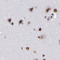

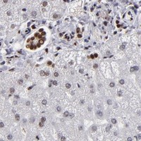


Comprehensive Human Protein Atlas IHC scoring per tissue (reliability: Supported — Medium consistency between antibody staining and RNA expression data.). Rows are taken directly from the HPA tissue chart — click any row's HPA link to view the source.
| Tissue | Cell type | Level | Evidence | Source |
|---|---|---|---|---|
| Adipose tissue | Adipocytes | High | Protein (IHC) | HPA → |
| Adrenal gland | Glandular cells | High | Protein (IHC) | HPA → |
| Appendix | Glandular cells | High | Protein (IHC) | HPA → |
| Bone marrow | Hematopoietic cells | High | Protein (IHC) | HPA → |
| Breast | Adipocytes | High | Protein (IHC) | HPA → |
| Tissue | Cell type | Level | Evidence | Source |
|---|---|---|---|---|
| None in HPA: MRE11 is detected in all 45 scored tissues. Use a no-primary (secondary-only) and an isotype control instead. | ||||
These questions focus on interpreting MRE11 nuclear staining in paraffin sections while keeping antibody specific observations separate from general IHC practice.
Both catalog antibodies have human paraffin-section IHC images (catalog IHC captions); A32234 also has IF/ICC data in T-47D cells (A32234 IF caption). Human, Mouse and Rat reactivity is listed for both (catalog reactivity).
A32234 will render with an IHC image from human bladder epithelial carcinoma tissue (A32234 IHC caption); its application list includes IF and ICC, with an IF image from T-47D cells (A32234 applications; A32234 IF caption). A32234-1 will render with an IHC image from human laryngeal squamous cell carcinoma tissue (A32234-1 IHC caption); its application list includes IHC (A32234-1 applications).
Which to pick: For paraffin-section IHC, either SKU has a human tissue image using EDTA retrieval at pH 8.0 and 2 μg/ml primary antibody; fixation is unreported in both captions (A32234 IHC caption; A32234-1 IHC caption). Choose A32234 for IF/ICC because those applications and a T-47D IF image are listed for that SKU (A32234 applications; A32234 IF caption). For cross-species tissue IHC, A32234-1 explicitly lists Human, Mouse and Rat for paraffin-section IHC, although its supplied IHC images show human tissue (A32234-1 dilution data; A32234-1 IHC captions).