This website uses cookies to ensure you get the best experience on our website.


- Table of Contents
Plan chromogenic MRPL13 IHC in paraffin sections using the granular cytoplasmic tissue pattern as a reference (HPA tissue IHC). Compare high-staining kidney tubule cells with adipocytes, where signal was not detected (HPA tissue IHC).

Expected localisation, validated protocols, controls and antibodies — the at-a-glance facts below, then the full design guide.
| Expected localisation | Granular cytoplasm (HPA tissue IHC); mitochondrion (UniProt) | |
| Staining pattern | Granular cytoplasmic staining in most tissues (HPA tissue IHC) | |
| Antigen retrieval | Citrate pH 6.0 HIER, 95–98 °C, 20 min (rule: cytoplasmic / membrane antigen) | |
| Positive control | Appendix+4 more · see all | |
| Negative control | Adipose tissue+1 more · see all |
| Fixation | Keep fixation consistent across samples (standard IHC practice; not target-specific) | |
| Caveat | Some cell types have no detected signal (HPA tissue IHC) | |
| Regulation | No specific regulation annotated (UniProt) | |
| Isoform / epitope | No isoforms annotated; mature chain spans residues 2–178 (UniProt) |
Compare the catalog antibody’s IHC-P protocol (datasheet) with the published MRPL13 kidney-section protocol (PMC13411704).
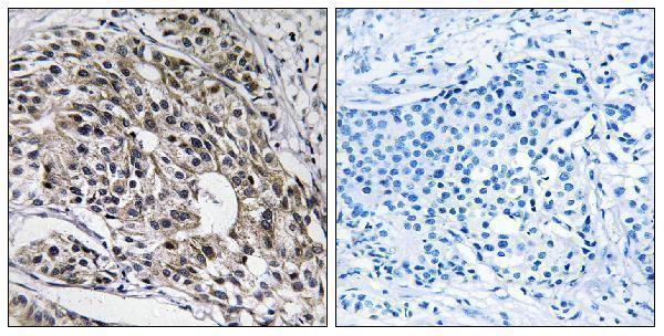
| Sample | Paraffin-embedded human breast carcinoma tissue; fixative not specified (datasheet A13508-1) |
| Fixation | Image fixative and duration unreported (datasheet A13508-1); verify before use. |
| Sectioning | 4–5 µm sections on charged slides (standard) |
| Deparaffinisation | Xylene, graded ethanol series to water (standard) |
| Antigen retrieval | Heat-induced epitope retrieval in citrate buffer, pH 6.0, 20 min at 95–98 °C (standard rule: cytoplasmic / membrane antigen) |
| Peroxidase block | 3% H2O2, 10 min, room temperature (standard) |
| Blocking | 10% normal serum of the secondary host, 30 min, room temperature (standard) |
| Primary antibody | Rabbit anti-MRPL13, 1:100-1:300 (datasheet A13508-1) |
| Primary incubation | Overnight at 4 °C (standard) |
| Detection | HRP-polymer secondary, DAB chromogen 5–10 min (standard) |
| Counterstain | Hematoxylin, blue, dehydrate and mount (standard) |
| Expected result | MRPL13-positive staining in glandular cells of appendix (HPA tissue IHC: High). HPA tissue profile: Cytoplasmic expression with a granular pattern in most tissues. No signal in the no-primary control. |
MRPL13 is a mitochondrial protein without a transmembrane segment (UniProt Q9BYD1). In paraffin-section IHC, expect granular cytoplasmic staining in many tissues, particularly the epithelial and tubular cell populations that HPA scores high (HPA tissue IHC). HPA rates the tissue staining Approved, with medium consistency between antibody staining and RNA expression (HPA tissue IHC).
| Granular cytoplasmic staining in bronchial respiratory epithelium, kidney tubules, or pancreatic exocrine glands. | This matches the reported cytoplasmic granular pattern and high staining in those cells (HPA tissue IHC). Assess the cellular pattern as well as intensity: MRPL13 is mitochondrial, but chromogenic IHC alone does not prove that each granule is a mitochondrion (UniProt Q9BYD1; HPA tissue IHC). |
| Strong staining appears predominantly in nuclei or outlines the plasma membrane. | That compartment conflicts with the reported mitochondrial location and granular cytoplasmic IHC profile (UniProt Q9BYD1; HPA tissue IHC). Treat it as suspect; review morphology, detection controls, and staining conditions before interpreting it as MRPL13. |
| Strong staining appears in adipocytes or ovarian stromal cells. | HPA reports MRPL13 as not detected in those populations (HPA tissue IHC). Consider cross-reactivity or endogenous detection activity, particularly if a control also stains. A single discrepant field cannot establish that the cells express MRPL13. |
| Brown reaction product is diffuse across cells and surrounding tissue, with little granular structure. | This does not resemble HPA's granular cytoplasmic profile (HPA tissue IHC). Diffuse signal may arise from background in a chromogenic IHC workflow (general IHC practice). Check the control and staining distribution before scoring cells as positive. |
| No convincing signal appears in a section containing kidney tubules or colon glandular cells. | Those cell populations are scored high by HPA, so an entirely blank result warrants a technical check (HPA tissue IHC). Examine tissue preservation, the positive control, retrieval and detection steps; absence of signal alone does not establish biological loss of MRPL13. |
| Subcellular location and topology | MRPL13 is mitochondrial and has no annotated transmembrane segment (UniProt Q9BYD1). Interpret a granular cytoplasmic pattern in that context; the record does not define an IHC epitope or establish which retrieval condition works best. |
| Cell population and tissue choice | HPA scores respiratory epithelium, several glandular populations, kidney tubules, and cholangiocytes high, while adipocytes and ovarian stromal cells are not detected (HPA tissue IHC). Compare like cell populations when evaluating signal. |
| Breadth and strength of expression | HPA describes granular cytoplasmic staining in most tissues and low tissue specificity by RNA (HPA tissue IHC). Do not require uniformly strong staining across a section: HPA also scores skeletal myocytes, smooth muscle cells, and several other populations low. |
| Evidence behind the staining call | The HPA tissue profile is Approved with medium staining-to-RNA consistency; the listed HPA060899 antibody is Approved for IHC, with no Enhanced designation supplied (HPA tissue IHC; HPA antibodies). Use that level of support when interpreting unexpected staining. |
| IF/ICC evidence boundary | HPA supplies no main ICC-IF location, no cell-line images, and no ICC validation status for the listed antibody (HPA subcellular; HPA antibodies). The IHC pattern can guide an IF expectation, but it is not an observed ICC-IF result. |
| Situation | Likely cause | Next action |
|---|---|---|
| A known-positive epithelial or tubular area is blank. | The outcome conflicts with HPA high staining in those cells; the provided sources do not identify a target-specific fixation or retrieval failure (HPA tissue IHC). | Check section morphology and a working positive control, then review the catalog antibody's IHC-P directions, retrieval, dilution, and detection steps (general IHC practice). |
| Granular staining is obscured by diffuse brown background. | Background can obscure cell-level scoring in chromogenic IHC (general IHC practice); the diffuse pattern differs from HPA's granular cytoplasmic profile (HPA tissue IHC). | Inspect a detection control and review blocking, washes, primary-antibody dilution, and chromogen development (general IHC practice). Score only distinguishable cellular signal. |
| Nuclei or cell borders stain more strongly than cytoplasm. | The distribution conflicts with mitochondrial localisation and the reported granular cytoplasmic IHC pattern (UniProt Q9BYD1; HPA tissue IHC). | Check counterstain and tissue morphology, compare the detection control, and reassess whether the apparent signal belongs to the target cells (general IHC practice). |
| Adipocytes or ovarian stromal cells appear strongly positive. | HPA reports those populations as not detected; cross-reactivity or endogenous detection activity is possible, but the images alone do not identify a cause (HPA tissue IHC). | Compare with a detection control and with a high-staining cell population on an appropriate section; review whether staining follows cell boundaries and the expected granular pattern (HPA tissue IHC; general IHC practice). |
| Weak cells are called negative because adjacent cells stain strongly. | HPA reports both high and low staining among different cell populations, and describes low tissue specificity by RNA (HPA tissue IHC). | Score the named cell population separately and record weak granular cytoplasmic signal when discernible; avoid applying a strong-cell threshold to every tissue compartment (HPA tissue IHC). |
| What should an IF/ICC result look like? | UniProt places MRPL13 in mitochondria, but HPA provides no ICC-IF images, main ICC-IF location, or ICC validation status for HPA060899 (UniProt Q9BYD1; HPA subcellular; HPA antibodies). | Use mitochondrial localisation as a provisional expectation and assess IF controls on the separate IF/ICC guide page; do not treat the HPA IHC profile as IF validation (UniProt Q9BYD1; HPA tissue IHC; general IF practice). |


Comprehensive Human Protein Atlas IHC scoring per tissue (reliability: Approved — Medium consistency between antibody staining and RNA expression data.). Rows are taken directly from the HPA tissue chart — click any row's HPA link to view the source.
| Tissue | Cell type | Level | Evidence | Source |
|---|---|---|---|---|
| Appendix | Glandular cells | High | Protein (IHC) | HPA → |
| Bronchus | Respiratory epithelial cells | High | Protein (IHC) | HPA → |
| Colon | Glandular cells | High | Protein (IHC) | HPA → |
| Duodenum | Glandular cells | High | Protein (IHC) | HPA → |
| Gallbladder | Glandular cells | High | Protein (IHC) | HPA → |
Troubleshoot MRPL13 chromogenic IHC in paraffin sections by assessing retrieval, granular cytoplasmic staining, controls, and cell-specific scoring (A13508-1 caption; HPA tissue IHC).
A13508-1 has a paraffin-section human IHC image (image caption), lists IF as an application, and lists human, mouse and rat reactivity (catalog: A13508-1); no IF image is supplied (catalog: IF images).
The sole figure-backed card is A13508-1: its image shows IHC on paraffin-embedded human breast carcinoma tissue with synthesized-peptide blocking (A13508-1 image caption). A13508-1 also lists IF as an application and human, mouse and rat reactivity, but supplies no IF image (catalog: A13508-1 applications/reactivity/IF images).
Which to pick: For tissue IHC, choose A13508-1 if a paraffin-section example is useful; its caption shows human breast carcinoma tissue, but does not report the fixative (A13508-1 image caption). For IF, A13508-1 is the IF-listed rabbit polyclonal option; ICC testing and an IF image are unreported (catalog: A13508-1 applications/host/IF images). For mouse or rat work, A13508-1 lists both species; A13508-3 is a human-reactive rabbit polyclonal IHC option with no supplied IHC image (catalog: A13508-1 reactivity; A13508-3 applications/reactivity/host/IHC images).