This website uses cookies to ensure you get the best experience on our website.


- Table of Contents
Plan chromogenic IHC for MRPS17 using its observed cytoplasmic tissue pattern (HPA tissue IHC) and mitochondrial location (UniProt). Compare medium staining in adrenal glandular cells with adipocytes reported as not detected, and assess antibody specificity (HPA tissue IHC).

Expected localisation, validated protocols, controls and antibodies — the at-a-glance facts below, then the full design guide.
| Expected localisation | Cytoplasmic in tissue sections (HPA tissue IHC) | |
| Staining pattern | General cytoplasmic staining across cell types (HPA tissue IHC) | |
| Antigen retrieval | Tris-EDTA pH 9.0 HIER, heat-mediated (datasheet A14408) | |
| Positive control | Adrenal gland+4 more · see all | |
| Negative control | Adipose tissue+4 more · see all |
| Fixation | Keep fixation consistent across sections (standard IHC practice; not target-specific) | |
| Caveat | Antibody may recognize proteins from multiple genes (HPA tissue IHC) | |
| Regulation | No expression regulation annotated (UniProt) | |
| Isoform / epitope | No isoforms; check epitope against mature chain 21–130 (UniProt) |
Compare the catalog antibody's IHC-P protocol (datasheet A14408) with published MRPS17 protocols for gastric cancer (PMC8247386) and lung adenocarcinoma (PMC13525032).
| Sample | Paraffin-embedded human liver cancer tissue; fixative not specified (datasheet A14408) |
| Fixation | Image fixative and duration unreported (datasheet A14408); verify before use. |
| Sectioning | 4–5 µm sections on charged slides (standard) |
| Deparaffinisation | Xylene, graded ethanol series to water (standard) |
| Antigen retrieval | Heat retrieval: Tris-EDTA pH 9.0 (datasheet A14408); 20 min, 95–100 °C (standard) |
| Peroxidase block | 3% H2O2, 10 min, room temperature (standard) |
| Blocking | 10% normal serum of the secondary host, 30 min, room temperature (standard) |
| Primary antibody | Rabbit anti-MRPS17, 1:100 - 1:300 (datasheet A14408) |
| Primary incubation | Overnight at 4 °C (standard) |
| Detection | HRP-polymer secondary, DAB chromogen 5–10 min (standard) |
| Counterstain | Hematoxylin, blue, dehydrate and mount (standard) |
| Expected result | MRPS17-positive staining in glandular cells of adrenal gland (HPA tissue IHC: Medium). HPA tissue profile: General cytoplasmic expression. No signal in the no-primary control. |
MRPS17 is a mitochondrial small ribosomal subunit protein with no transmembrane segment (UniProt Q9Y2R5). In paraffin section IHC, expect cytoplasmic staining rather than a resolved mitochondrial outline: HPA describes general cytoplasmic expression and medium staining in selected glandular, epithelial, hematopoietic and neuronal cells (HPA tissue IHC). HPA rates its tissue antibody Approved, while cautioning that it targets protein from more than one gene and awaits external verification (HPA tissue IHC).
| Cytoplasmic staining in adrenal gland or duodenal glandular cells, bronchial respiratory epithelium, or cerebellar Purkinje cells. | These are examples of medium IHC staining in the specified cells (HPA tissue IHC). A cytoplasmic pattern agrees with HPA and is compatible with mitochondrial MRPS17 (HPA tissue IHC; UniProt Q9Y2R5); it does not establish target specificity on its own. |
| Predominantly nuclear staining, or staining confined to a cell surface outline. | Neither pattern matches HPA's general cytoplasmic profile or UniProt's mitochondrial assignment and absence of a transmembrane segment (HPA tissue IHC; UniProt Q9Y2R5). Treat it as questionable staining and compare with controls before scoring. |
| Strong signal in adipocytes, or follicle cells of the ovary, despite little signal in an expected positive cell population. | HPA reports MRPS17 as not detected in those cell populations (HPA tissue IHC). Consider cross-reactivity or endogenous detection activity; the Approved tissue antibody has a multi-gene targeting caution, so this pattern cannot be assigned confidently to MRPS17 (HPA tissue IHC). |
| Diffuse color across cells and extracellular spaces, with little distinction between expected positive and negative areas. | This is a poor basis for cellular scoring (general IHC practice). Check background in a no-primary control and reassess blocking, antibody concentration and detection conditions (general IHC practice); HPA specifically disregarded observed plasma staining (HPA tissue IHC). |
| No cytoplasmic signal in an adequately preserved section containing HPA medium-staining cells. | Absence in those cells conflicts with the reported IHC pattern (HPA tissue IHC). First check tissue identity, section quality and positive-control performance, then optimize the general IHC workflow; a single blank section does not establish biological absence (general IHC practice). |
| Compartment and resolution | UniProt places MRPS17 in mitochondria, while HPA reports general cytoplasmic tissue IHC staining (UniProt Q9Y2R5; HPA tissue IHC). Score the observed cytoplasmic distribution without requiring individual mitochondria to be visible in a chromogenic section. |
| Processing and membrane topology | UniProt lists a 130-residue precursor, a mature chain spanning residues 21–130, and no transmembrane segment (UniProt Q9Y2R5). These annotations support an intracellular interpretation; they do not identify the antibody epitope or predict an antigen-retrieval condition. |
| Tissue choice and antibody confidence | HPA reports low tissue RNA specificity, medium staining in selected cells, and not-detected results in others (HPA tissue IHC). Its Approved tissue result carries pending external verification and a multi-gene targeting warning; use positive and comparison tissues to judge the pattern, without treating either as proof of specificity (HPA tissue IHC). |
| What does IF/ICC establish here? | HPA provides no main ICC-IF location or cell-line images for MRPS17, and lists no ICC validation for HPA047159 (HPA subcellular; HPA antibodies). UniProt's mitochondrial assignment is a localization expectation, not an IF image result (UniProt Q9Y2R5). |
| Situation | Likely cause | Next action |
|---|---|---|
| Expected positive cells are blank. | The section or detection run may have failed; HPA reports medium staining in the specified positive cells (HPA tissue IHC). | Confirm cell identity and morphology, then check a working positive control and the detection reagents before adjusting general IHC conditions (general IHC practice). |
| Signal is mainly nuclear or forms a crisp cell surface outline. | That distribution disagrees with the HPA cytoplasmic profile and UniProt mitochondrial location (HPA tissue IHC; UniProt Q9Y2R5). | Review the no-primary control and cellular localization, and seek an independent specificity check before calling the signal MRPS17 (general IHC practice; HPA tissue IHC antibody caution). |
| Adipocytes or ovarian follicle cells stain prominently. | HPA lists these populations as not detected; non-specific binding or endogenous detection activity may be contributing (HPA tissue IHC; general IHC practice). | Compare with an HPA medium-staining cell population and no-primary control; investigate the detection system if the unexpected signal persists (HPA tissue IHC; general IHC practice). |
| Color is widespread, including plasma or spaces between cells. | Background can obscure cellular staining (general IHC practice); HPA observed plasma staining and disregarded it (HPA tissue IHC). | Do not score plasma as MRPS17; inspect no-primary background, then review blocking, washing and antibody concentration using general IHC controls (HPA tissue IHC; general IHC practice). |
| Two sections show different signal intensity. | Differences in cell composition or staining-run conditions can change an IHC comparison (general IHC practice); HPA levels are assigned to particular cell populations (HPA tissue IHC). | Compare the same identified cell type under matched staining and detection conditions, with controls in each run (general IHC practice). |
| A cytoplasmic positive result is being treated as definitive target validation. | HPA calls the tissue result Approved but flags multi-gene targeting and pending external verification (HPA tissue IHC). | Report the pattern as consistent with MRPS17 and use an independent specificity check for a target-specific conclusion (HPA tissue IHC antibody caution; general IHC practice). |


Comprehensive Human Protein Atlas IHC scoring per tissue (reliability: Approved — Caution, targets protein from more than one gene. Pending external verification. Staining in plasma observed and disregarded.). Rows are taken directly from the HPA tissue chart — click any row's HPA link to view the source.
| Tissue | Cell type | Level | Evidence | Source |
|---|---|---|---|---|
| Adrenal gland | Glandular cells | Medium | Protein (IHC) | HPA → |
| Bone marrow | Hematopoietic cells | Medium | Protein (IHC) | HPA → |
| Bronchus | Respiratory epithelial cells | Medium | Protein (IHC) | HPA → |
| Cerebellum | Purkinje cells | Medium | Protein (IHC) | HPA → |
| Cerebral cortex | Neuronal cells | Medium | Protein (IHC) | HPA → |
| Tissue | Cell type | Level | Evidence | Source |
|---|---|---|---|---|
| Adipose tissue | Adipocytes | Not detected | Protein (IHC) | HPA → |
| Caudate | Glial cells | Not detected | Protein (IHC) | HPA → |
| Epididymis | Glandular cells | Not detected | Protein (IHC) | HPA → |
| Ovary | Follicle cells | Not detected | Protein (IHC) | HPA → |
| Parathyroid gland | Glandular cells | Not detected | Protein (IHC) | HPA → |
Troubleshoot MRPS17 staining in paraffin sections by checking retrieval, compartment, controls, and scoring before interpreting signal intensity.
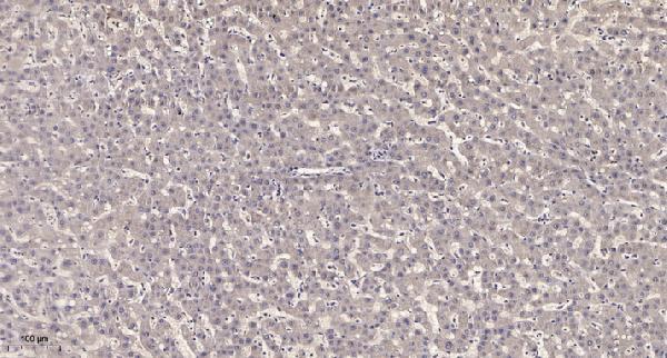
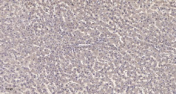
The catalog antibodies cover human and mouse IHC and IF applications (catalog: A14408, A14408S17); their figures show paraffin-section IHC of human liver cancer (catalog image captions). No IF figure is supplied (catalog).
A14408 will render with a human liver cancer paraffin-section IHC figure; its listed applications include IHC, IF and ICC, with human and mouse reactivity (catalog: A14408). A14408S17 will render with its own human liver cancer paraffin-section IHC figure; its listed applications include IHC and IF, with human and mouse reactivity (catalog: A14408S17).
Which to pick: For tissue IHC, either A14408 or A14408S17 has its own paraffin-section IHC example using primary antibody at 1:200 overnight at 4°C and Tris-EDTA retrieval at pH 9.0; the fixative is unreported in both captions (catalog image captions: A14408, A14408S17). For IF/ICC, choose A14408 because both applications are listed, while A14408S17 lists IF but not ICC; neither has an IF figure in the payload (catalog: A14408, A14408S17). Both are listed as polyclonal rabbit antibodies reactive with human and mouse, so either is a cross-species candidate (catalog: A14408, A14408S17).