This website uses cookies to ensure you get the best experience on our website.


- Table of Contents
Plan MRPS5 paraffin-section IHC around cytoplasmic tissue staining (HPA tissue IHC) and mitochondrial localization (UniProt). Start with the catalog antibody’s IHC dilution range of 1:100–1:300 (datasheet), using kidney tubule cells as a high-staining reference and adipocytes as an undetected reference (HPA tissue IHC).

Expected localisation, validated protocols, controls and antibodies — the at-a-glance facts below, then the full design guide.
| Expected localisation | Cytoplasmic tissue staining (HPA tissue IHC); mitochondrial protein (UniProt) | |
| Staining pattern | Widespread cytoplasmic staining across cell types (HPA tissue IHC) | |
| Antigen retrieval | Citrate pH 6.0 HIER, 95–98 °C, 20 min (rule: cytoplasmic / membrane antigen) | |
| Positive control | Breast+4 more · see all | |
| Negative control | Adipose tissue+4 more · see all |
| Fixation | Keep fixation consistent across sections (standard IHC practice; not target-specific) | |
| Caveat | Staining has low consistency with RNA expression (HPA tissue IHC) | |
| Regulation | No specific regulator annotated (UniProt) | |
| Isoform / epitope | 2 isoforms; epitope differences unreported (UniProt) |
Compare the catalog antibody’s IHC-P protocol with a published human clear cell renal cell carcinoma protocol (PMC11614452: immunohistochemical staining).
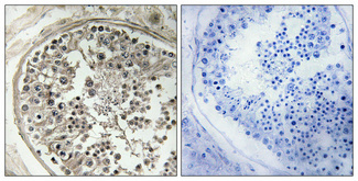
| Sample | Paraffin-embedded Human testis tissue; fixative not specified (datasheet A14183S5) |
| Fixation | Image fixative and duration unreported (datasheet A14183S5); verify before use. |
| Sectioning | 4–5 µm sections on charged slides (standard) |
| Deparaffinisation | Xylene, graded ethanol series to water (standard) |
| Antigen retrieval | Heat-induced epitope retrieval in citrate buffer, pH 6.0, 20 min at 95–98 °C (standard rule: cytoplasmic / membrane antigen) |
| Peroxidase block | 3% H2O2, 10 min, room temperature (standard) |
| Blocking | 10% normal serum of the secondary host, 30 min, room temperature (standard) |
| Primary antibody | Rabbit anti-MRPS5, 1:100-1:300 (datasheet A14183S5) |
| Primary incubation | Overnight at 4 °C (standard) |
| Detection | HRP-polymer secondary, DAB chromogen 5–10 min (standard) |
| Counterstain | Hematoxylin, blue, dehydrate and mount (standard) |
| Expected result | MRPS5-positive staining in myoepithelial cells of breast (HPA tissue IHC: High). HPA tissue profile: Ubiquitous cytoplasmic expression. No signal in the no-primary control. |
MRPS5 is a mitochondrial small ribosomal subunit protein with no transmembrane segment (UniProt P82675). In paraffin tissue sections, expect predominantly cytoplasmic staining, including strong staining in kidney tubular cells and duodenal glandular cells (HPA: tissue IHC). HPA rates its IHC antibody Approved, but reports low consistency with RNA expression and pending external verification (HPA: reliability).
| Cytoplasmic staining is strongest in kidney tubular cells or duodenal glandular cells (HPA: High in both). | This fits the reported tissue pattern (HPA: tissue IHC). A punctate cytoplasmic appearance would be compatible with mitochondrial localization, but chromogenic IHC alone cannot confirm organelle identity (UniProt P82675; HPA: mitochondria supported in ICC-IF). |
| A strong, predominantly nuclear or membrane-rim signal replaces cytoplasmic staining (HPA: ubiquitous cytoplasmic profile). | That compartment is unexpected for MRPS5 (UniProt P82675: mitochondrion; no transmembrane segment). Check morphology and detection controls before scoring it as specific; aberrant staining can reflect an IHC artefact (general IHC practice). |
| Strong staining appears in adipocytes or liver cholangiocytes (HPA: Not detected in those cells). | Treat the result as discordant with HPA's cell-level observations, rather than proof that the whole tissue is negative. Cross-reactivity or endogenous detection activity is possible; compare a no-primary control (HPA: tissue IHC; general IHC practice). |
| Colour spreads across tissue and blank areas without a discernible cell pattern (general IHC practice). | Diffuse background prevents a reliable MRPS5 call. Review blocking, washes and detection controls before interpreting intensity (general IHC practice); HPA reports cell-associated cytoplasmic staining (HPA: tissue IHC). |
| No signal appears in kidney tubular cells despite intact tissue morphology (HPA: High in kidney tubular cells). | A failed run is plausible, but a negative result alone cannot identify its cause. Check the antibody and detection steps, then review retrieval conditions as a general IHC troubleshooting step (HPA: tissue IHC; general IHC practice). |
| Which cells provide useful comparisons? (HPA: tissue IHC) | Kidney tubular and small-intestinal glandular cells are High; adipocytes are Not detected (HPA: tissue IHC). Score the named cells, since HPA levels do not describe every cell in a section. |
| How firm is the tissue pattern? (HPA: reliability) | The IHC antibody is Approved, with low consistency between staining and RNA expression and external verification pending (HPA: reliability); use discordant staining as a reason to investigate. |
| Do isoforms or processing change the expected slide pattern? (UniProt P82675) | Two isoforms are listed, and the annotated chain spans residues 1–430 (UniProt P82675). Epitope coverage by isoform is unspecified, so no isoform-specific IHC pattern can be predicted. |
| Does protein topology suggest a surface or shed signal? (UniProt P82675) | MRPS5 has no annotated transmembrane segment, signal peptide or glycosylation sites (UniProt P82675); these annotations offer no basis for expecting membrane-rim staining or extracellular deposition. |
| IF/ICC: where should signal localize? (HPA: subcellular ICC-IF) | Mainly mitochondria; additional midbody and primary-cilium locations are uncertain (HPA: subcellular ICC-IF). This is an IF localization answer, not a paraffin IHC protocol or proof of those structures in tissue. |
| Situation | Likely cause | Next action |
|---|---|---|
| Kidney tubular cells are unstained (HPA: High in kidney tubular cells). | Primary antibody, retrieval or detection may have failed (general IHC practice); HPA provides no MRPS5-specific retrieval requirement. | Review the validated IHC conditions and detection controls, then adjust retrieval only as a general IHC optimization (general IHC practice). |
| Nuclei dominate while cytoplasm is faint (HPA: ubiquitous cytoplasmic profile). | The pattern conflicts with mitochondrial localization (UniProt P82675); nonspecific signal or interpretation error is possible (general IHC practice). | Recheck cell boundaries and the no-primary control; score MRPS5 only where a credible cytoplasmic pattern remains (general IHC practice; HPA: tissue IHC). |
| Adipocytes show strong stain (HPA: Not detected in adipocytes). | Cross-reactivity or endogenous detection activity may contribute (general IHC practice); HPA's negative call is cell-specific (HPA: tissue IHC). | Compare no-primary and detection controls, and confirm that the scored cells are adipocytes (general IHC practice; HPA: tissue IHC). |
| Diffuse colour obscures glandular cells (general IHC practice). | Excess background can arise during antibody incubation or detection (general IHC practice); it cannot establish MRPS5 distribution. | Check blocking, antibody concentration and washes using the validated IHC workflow, then reassess cellular contrast (general IHC practice). |
| A negative tissue shows some positive cells (HPA: cell-level tissue IHC). | HPA's Not detected calls name particular cells, such as liver cholangiocytes, rather than every cell in that tissue (HPA: tissue IHC). | Identify the stained cell type before calling the result discordant; compare the matching HPA cell-level entry (HPA: tissue IHC). |
| The result changes with fixation or retrieval (general IHC practice). | The supplied record does not establish MRPS5-specific fixation sensitivity or epitope masking (UniProt P82675; HPA: tissue IHC). | Document processing conditions and compare matched controls; avoid assigning a target-specific mechanism from this change alone (general IHC practice). |


Comprehensive Human Protein Atlas IHC scoring per tissue (reliability: Approved — Low consistency between antibody staining and RNA expression data. Pending external verification.). Rows are taken directly from the HPA tissue chart — click any row's HPA link to view the source.
| Tissue | Cell type | Level | Evidence | Source |
|---|---|---|---|---|
| Breast | Myoepithelial cells | High | Protein (IHC) | HPA → |
| Duodenum | Glandular cells | High | Protein (IHC) | HPA → |
| Gallbladder | Glandular cells | High | Protein (IHC) | HPA → |
| Kidney | Cells in tubules | High | Protein (IHC) | HPA → |
| Small intestine | Glandular cells | High | Protein (IHC) | HPA → |
| Tissue | Cell type | Level | Evidence | Source |
|---|---|---|---|---|
| Adipose tissue | Adipocytes | Not detected | Protein (IHC) | HPA → |
| Adrenal gland | Glandular cells | Not detected | Protein (IHC) | HPA → |
| Caudate | Glial cells | Not detected | Protein (IHC) | HPA → |
| Cerebellum | Cells in granular layer | Not detected | Protein (IHC) | HPA → |
| Cervix | Glandular cells | Not detected | Protein (IHC) | HPA → |
Troubleshoot MRPS5 chromogenic IHC in paraffin sections by checking retrieval, processing, cytoplasmic localisation and cell-specific controls before interpreting staining intensity.
A14183S5 has real IHC data from human paraffin-embedded testis (catalog IHC caption). IF is listed for human and mouse, but no IF figure is supplied (catalog applications, reactivity and IF image alts).
A14183S5 will render with its own IHC figure showing human paraffin-embedded testis at 1:100 (catalog IHC caption). The catalog also lists IF and human and mouse reactivity, without an IF figure or mouse tissue image (catalog applications, reactivity and image alts).
Which to pick: For tissue IHC, choose A14183S5: its own image shows human paraffin-embedded testis, and the fixative is unreported (catalog IHC caption). For IF/ICC, A14183S5 lists IF at 1:50, but ICC validation and an IF figure are unreported (catalog applications, IF dilution and IF image alts). For cross-species work, A14183S5 lists human and mouse reactivity; it has a rabbit host and unreported clonality, while its IHC image documents human tissue only (catalog reactivity, host and clone; catalog IHC caption).