MS4A6A / Membrane-spanning 4-domains subfamily A member 6A · IHC design guide

Design Immunohistochemistry for MS4A6A

Use adrenal or breast glandular cells, which show high staining, as IHC reference points (HPA tissue IHC). Compare the reported nuclear pattern with membrane topology, and start the catalog antibody at 2.5 μg/mL (HPA tissue IHC; UniProt; datasheet).

Evidence assembled Oct 2026 · For research use; verify linked source records and product datasheet before use
Immunohistochemistry protocol sheet for MS4A6A (IHC for MS4A6A): expected localisation Nuclear staining in tissue (HPA tissue IHC); membrane protein (UniProt), antibody A09251, validated IHC image, and IHC protocol steps
Printable MS4A6A IHC protocol sheet — expected localisation Nuclear staining in tissue (HPA tissue IHC); membrane protein (UniProt), antibody A09251, controls and protocol steps. Open the full MS4A6A IHC guide →

MS4A6A Immunohistochemistry Experimental Design Guide

Expected localisation, validated protocols, controls and antibodies — the at-a-glance facts below, then the full design guide.

Must know before staining
Expected localisation Nuclear staining in tissue (HPA tissue IHC); membrane protein (UniProt)
Staining pattern General nuclear staining across tissue cells (HPA tissue IHC)
Antigen retrieval Citrate pH 6.0 HIER, 95–98 °C, 20 min (rule: cytoplasmic / membrane antigen)
Positive control ⓘ Adrenal gland+4 more · see all
Negative control ⓘ None in HPA (detected in all 45 tissues); use no-primary + isotype controls
Important caveats
Reasons your staining may differ from the expected pattern.
Fixation Keep fixation consistent across sections (standard IHC practice; not target-specific); Matched tissue-IHC evidence does not establish the fixation claim. Validate the specimen-specific method before use. (selected-SKU IHC image A09251)
Caveat Nuclear staining conflicts with membrane topology (HPA tissue IHC; UniProt)
Regulation Variable in some blood cell lines (UniProt)
Isoform / epitope 5 isoforms; check extracellular versus cytoplasmic epitope (UniProt)
Section 1

Recommended MS4A6A IHC & IF Protocols

The catalog antibody’s IHC-P protocol is paired with a published MS4A6A protocol for human tissue sections (PMC9290968).

Recommended immunohistochemistry (IHC-P) protocol parameters
SampleTissue sections; selected-image fixative not specified (standard IHC workflow)
FixationImage fixative and duration unreported (datasheet A09251); verify before use.
Sectioning4–5 µm sections on charged slides (standard)
DeparaffinisationXylene, graded ethanol series to water (standard)
Antigen retrievalHeat-induced epitope retrieval in citrate buffer, pH 6.0, 20 min at 95–98 °C (standard rule: cytoplasmic / membrane antigen)
Peroxidase block3% H2O2, 10 min, room temperature (standard)
Blocking10% normal serum of the secondary host, 30 min, room temperature (standard)
Primary antibodyRabbit anti-MS4A6A, 2.5 μg/mL (datasheet A09251)
Primary incubationOvernight at 4 °C (standard)
DetectionHRP-polymer secondary, DAB chromogen 5–10 min (standard)
CounterstainHematoxylin, blue, dehydrate and mount (standard)
Expected resultMS4A6A-positive staining in glandular cells of adrenal gland (HPA tissue IHC: High). HPA tissue profile: General nuclear expression. No signal in the no-primary control.
💡Decision noteStart with citrate pH 6.0 retrieval at 95–98 °C for 20 min for this membrane target (UniProt Q9H2W1 topology; page retrieval rule).
Section 2

What Is the Expected MS4A6A Staining Pattern?

In paraffin-section IHC, MS4A6A has a four-pass membrane topology (UniProt Q9H2W1 topology), yet HPA describes a general nuclear staining pattern (HPA tissue IHC). High staining is reported in glandular cells of adrenal gland, breast, cervix, endometrium and epididymis, among other listed cells (HPA tissue IHC: High). HPA rates its tissue staining Approved, pending external verification (HPA tissue IHC: reliability). Interpret compartment and cell identity together.

What am I looking at on my slide?
Strong nuclear chromogen in listed glandular cells.This matches HPA's reported general nuclear pattern and High glandular-cell staining (HPA tissue IHC). Because the protein is annotated as membrane-localized with four transmembrane segments (UniProt Q9H2W1), nuclear staining alone does not establish target-specific localization; seek independent confirmation.
Crisp cell-border or intracellular punctate staining in a listed High-staining cell population.A membrane-associated pattern is plausible from topology (UniProt Q9H2W1); vesicles are reported in ICC-IF (HPA subcellular: approved). Neither source establishes this exact appearance in paraffin tissue. Score cell identity, compartment and controls before calling it MS4A6A-specific.
Nuclear-only staining is treated as proof of membrane-localized MS4A6A.That interpretation conflicts with the membrane annotation (UniProt Q9H2W1) despite resembling HPA's tissue IHC profile (HPA tissue IHC: general nuclear expression). Consider nonspecific staining or antibody-dependent localization; the supplied evidence cannot resolve the discrepancy.
Strong signal in cells outside the listed High populations, especially with broad background.Unexpected distribution raises cross-reactivity or endogenous chromogenic activity as possibilities (general IHC practice). HPA reports Low staining in cardiomyocytes, not absence (HPA tissue IHC: Low); an unexpected result warrants controls rather than an automatic false-positive label.
No staining in a section containing a listed High-staining cell population.This differs from HPA's observed High staining in those cells (HPA tissue IHC: High), but the HPA antibody is only Approved and pending external verification (HPA tissue IHC: reliability). Check tissue preservation, retrieval, antibody incubation and detection controls (general IHC practice).
💡Expected MS4A6A appearanceA provisional positive call is staining in a listed High-staining cell population (HPA tissue IHC: High), with its compartment recorded: HPA reports a general nuclear pattern (HPA tissue IHC), whereas membrane localization is expected from topology (UniProt Q9H2W1); widespread cell-independent chromogen suggests background (general IHC practice).
How each factor affects the staining
Compartment evidenceUniProt annotates membrane localization and four transmembrane segments (UniProt Q9H2W1); HPA tissue IHC reports general nuclear expression (HPA tissue IHC). This disagreement limits compartment-based specificity claims.
Antibody validationThe tissue IHC antibody HPA011391 is Approved, with external verification pending; HPA076623 is Approved for ICC, with no IHC status supplied (HPA antibodies; HPA tissue IHC). Do not transfer one application's status to the other.
Tissue and cell selectionHPA reports High staining in several glandular populations, bronchial respiratory epithelium and selected brain cells, and Low staining in cardiomyocytes (HPA tissue IHC). These are observed levels, not independently established control performance.
Isoforms and epitopeFive isoforms are listed (UniProt Q9H2W1). The supplied record gives no antibody epitope, so it cannot establish which isoforms either HPA antibody detects or whether a particular retrieval condition exposes that epitope.
IF/ICC pattern?HPA reports approved vesicular ICC-IF localization but cautions that it is based on antibodies targeting proteins from multiple genes (HPA subcellular). Treat it as provisional context for IF/ICC, not a validated paraffin-IHC pattern.
Why is my staining missing, weak or wrong?
SituationLikely causeNext action
Only nuclei stain, with no convincing membrane-associated signal.The appearance matches HPA's tissue profile but conflicts with UniProt membrane topology (HPA tissue IHC; UniProt Q9H2W1). Specificity remains unresolved.Record the nuclear pattern without calling it definitive MS4A6A localization; compare an independent antibody or orthogonal evidence where available (general IHC practice).
Chromogen appears throughout the section or in many unrelated cell types.Nonspecific binding or endogenous detection activity may contribute (general IHC practice); HPA's reported High staining is assigned to specific cell populations (HPA tissue IHC).Inspect no-primary and detection-only controls, then optimize blocking, washing and detection conditions (general IHC practice).
A listed High-staining population gives little or no signal.The result differs from the HPA observation (HPA tissue IHC: High). Retrieval, incubation or detection may be inadequate, though no MS4A6A-specific fixation sensitivity is supplied.Check section quality and positive detection controls; optimize retrieval and primary-antibody conditions within a general IHC workflow (general IHC practice).
Cardiomyocytes show strong staining.HPA reports Low staining in cardiomyocytes (HPA tissue IHC: Low), so strong signal merits review; Low does not mean a validated negative control.Compare staining with background controls and verify cell identification before attributing the signal to MS4A6A (general IHC practice).
Nuclear and membrane-like signals vary between antibodies.HPA tissue IHC reports nuclear expression, UniProt annotates membrane localization, and the listed antibodies have different application statuses (HPA tissue IHC; UniProt Q9H2W1; HPA antibodies).Keep antibody and application results separate; compare matched sections and use independent specificity evidence before reconciling the patterns (general IHC practice).
ICC-IF vesicles are used to score an IHC section as positive.HPA's vesicular result comes from ICC-IF and carries a multiple-gene antibody caution (HPA subcellular); it does not validate vesicular staining in paraffin sections.Score the IHC section on its own cell distribution, compartment and controls, and report the ICC-IF observation separately (general IHC practice).

Sample controls for MS4A6A IHC & IF

🧪Run caudate first and score its neuronal cells for MS4A6A staining (HPA: High in caudate neuronal cells). HPA detects MS4A6A in all 45 scored tissues, so use no-primary and isotype controls for the negative reference; nearby cells without specific staining should show background-level signal, but should not be presumed biologically negative (HPA tissue data).
Positive control tissue: Adrenal gland (Glandular cells, HPA High)
Negative control tissue: None in HPA: MS4A6A is detected in all 45 scored tissues. Use a no-primary (secondary-only) and an isotype control instead.
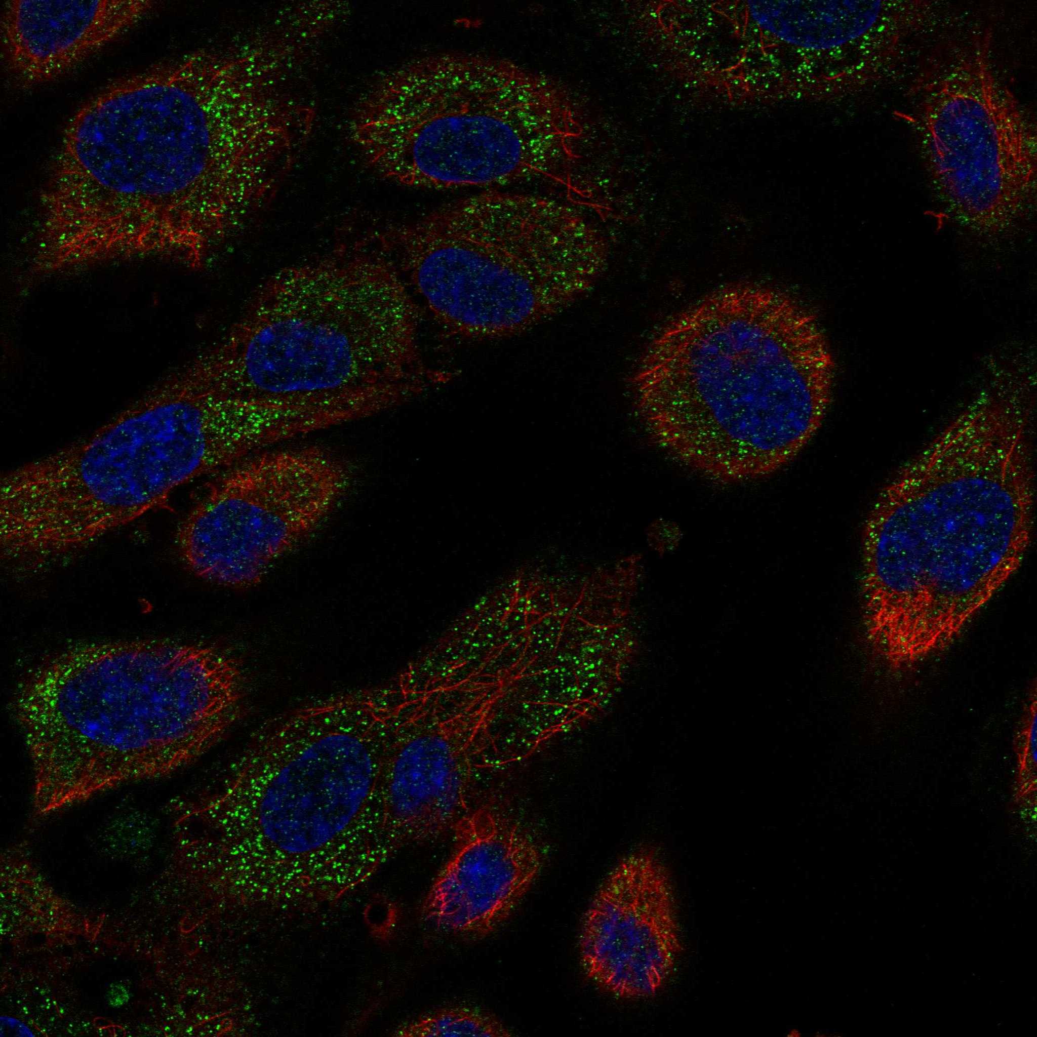
ICC-IF cell lines (HPA subcellular resource): HPA ICC-IF images show MS4A6A in HUVEC/TERT2, U2OS, with annotated localisation: Vesicles (approved) (HPA subcellular).
Technical controls: Include a no-primary, secondary-only section and an isotype control matched to the primary antibody’s host species and immunoglobulin class; assess specificity with knockout material or a cognate-peptide block if available (standard IHC practice). For chromogenic detection in caudate sections, quench endogenous peroxidase and compare any residual signal with the negative controls (standard IHC practice).
⚠️Feasibility: A target-specific fixation window and antigen-retrieval requirement are unreported in the supplied IHC evidence; the A09251 rat-brain IHC caption reports 2.5 µg/mL but does not state a fixative (A09251 caption). The evidence does not establish whether frozen sections or IF/ICC would be easier; MS4A6A is a membrane protein (UniProt Q9H2W1 topology), and HPA cautions that its vesicle ICC-IF assignment is based on antibodies targeting proteins from multiple genes (HPA subcellular). If adapting caudate staining to IF, distinguish specific signal from brain lipofuscin autofluorescence (standard IF practice).

HPA tissue IHC evidence for MS4A6A

Comprehensive Human Protein Atlas IHC scoring per tissue (reliability: Approved — Pending external verification.). Rows are taken directly from the HPA tissue chart — click any row's HPA link to view the source.

Positive expression · recommended positive controls

TissueCell typeLevelEvidenceSource
Adrenal gland Glandular cells High Protein (IHC) HPA →
Breast Glandular cells High Protein (IHC) HPA →
Bronchus Respiratory epithelial cells High Protein (IHC) HPA →
Caudate Neuronal cells High Protein (IHC) HPA →
Cerebellum Cells in molecular layer High Protein (IHC) HPA →

Undetected expression · recommended negative controls

TissueCell typeLevelEvidenceSource
None in HPA: MS4A6A is detected in all 45 scored tissues. Use a no-primary (secondary-only) and an isotype control instead.
Section 3

Advanced MS4A6A IHC Tips

Use compartment-aware controls when evaluating MS4A6A in chromogenic IHC; published localisation profiles differ (UniProt Q9H2W1: membrane; HPA tissue IHC: general nuclear expression).

How should I retrieve MS4A6A in paraffin sections when staining is weak?
Begin with citrate at pH 6.0, heated to 95–98 °C for 20 min (page retrieval rule: cytoplasmic / membrane antigen). Let sections cool in retrieval buffer, then compare the staining with a matched section processed under the same conditions without retrieval (standard IHC practice). If staining remains weak, test a different retrieval duration on adjacent sections while holding antibody concentration and detection conditions constant (standard IHC practice). MS4A6A has 4 predicted transmembrane segments and both cytoplasmic and extracellular regions, so the antibody’s epitope matters when interpreting a retrieval response (UniProt Q9H2W1 topology). Record tissue preservation and background alongside signal intensity; stronger colour alone does not establish specificity (standard IHC practice).
Can I change fixation to improve MS4A6A staining?
Target-specific fixation sensitivity is unknown from the supplied evidence; the selected A09251 rat brain caption does not state a fixative (A09251 tissue-IHC caption). For paraffin sections, first keep fixation and tissue processing consistent across cases, then vary one preparation step at a time (standard IHC practice). Compare sections from the same specimen when assessing whether a fixation change improves staining, because different specimens can vary in cell composition (standard IHC practice). Check morphology, expected cellular distribution, and matched negative controls before accepting increased colour as recovered antigen (standard IHC practice). Neither the HPA staining profile nor predicted MS4A6A topology establishes a fixation preference (HPA tissue IHC; UniProt Q9H2W1 topology).
Should MS4A6A staining be membranous, vesicular, or nuclear?
Score membranous, cytoplasmic or punctate, and nuclear chromogen separately rather than combining them into one positive category (standard IHC practice). A membrane assignment follows the predicted 4 transmembrane segments (UniProt Q9H2W1 topology), while HPA reports general nuclear expression in tissue IHC (HPA tissue IHC). HPA also reports approved vesicular localisation by ICC/IF, with a caution that those antibodies target proteins from multiple genes (HPA subcellular). These observations do not by themselves identify which compartment your IHC antibody detects. Compare the same compartment across matched sections and use an independent specificity control before treating unexpected nuclear or punctate staining as MS4A6A (standard IHC practice).
How can antibody epitope position affect MS4A6A IHC results?
MS4A6A has 5 reported isoforms, but the supplied record does not map their differences onto the antibody epitope (UniProt Q9H2W1 isoforms). The canonical topology places residues 1–46 and 207–248 inside the cell, with extracellular loops at 68–84 and 138–185 (UniProt Q9H2W1 topology). Check the catalog antibody’s stated immunogen or epitope before interpreting a negative section or comparing antibodies (standard IHC practice). When that information is unavailable, document the antibody identifier and retrieval conditions, and avoid assigning staining to a particular isoform (standard IHC practice). Compare antibodies against distinct documented regions only with matched sections and controls (standard IHC practice).
How should I cross-check an IHC pattern with multiplex IF?
Use IF on a matched specimen as a separate localisation check, and pair MS4A6A with a validated marker for the cell population being assessed (standard IF practice). For example, glandular cells are reported as high in adrenal gland tissue IHC, making a glandular-cell marker relevant there (HPA tissue IHC: adrenal gland). Choose spectrally separated fluorophores, favouring a far-red channel when shorter-wavelength tissue autofluorescence obscures signal, and include single-colour controls (standard IF practice). Set permeabilisation according to the documented epitope: cytoplasmic regions require access through the membrane, whereas extracellular loops may be accessible without it (UniProt Q9H2W1 topology; standard IF practice). Compare puncta cautiously because HPA’s vesicle assignment carries a multiple-gene antibody caution (HPA subcellular).
What controls help diagnose diffuse or nuclear DAB background?
Run a no-primary section through the same chromogenic detection steps to reveal background from secondary reagents or endogenous tissue activity (standard IHC practice). Include a peroxidase block before DAB development and compare development times across matched sections (standard chromogenic IHC practice). If diffuse colour persists, titrate the primary antibody and review blocking and wash conditions while preserving the same exposure and scoring thresholds (standard IHC practice). Nuclear staining needs careful interpretation because HPA reports general nuclear expression, whereas UniProt assigns MS4A6A to the membrane (HPA tissue IHC; UniProt Q9H2W1 subcellular). Distinguish discrete cellular staining from tissue pigment, edge staining, and precipitated chromogen using morphology and controls (standard IHC practice).
How should I quantify MS4A6A across paraffin sections? ⚠ ANSWER MARKED FOR VERIFICATION
Define the cell population and compartment before scoring, then apply the same rules to every section (standard IHC practice). An H-score combines the percentage of cells at each intensity level; record the underlying percentages so another reader can audit the result (standard IHC practice). If the question concerns cell abundance, report positive cells per mm² of viable tissue and normalise to the area or total eligible cells assessed (standard IHC practice). Keep nuclear, membranous, and punctate scores separate because the supplied localisation sources differ (HPA tissue IHC; HPA subcellular; UniProt Q9H2W1 subcellular). Exclude folds and damaged regions using prespecified criteria, with matched staining and image settings across comparisons (standard IHC practice).
When is an apparent MS4A6A-positive cell convincing?
Call a cell positive only when its chromogen exceeds matched negative-control background and its morphology permits a clear compartment assignment (standard IHC practice). Interpret membrane staining against MS4A6A’s predicted transmembrane structure, while recording nuclear staining separately because HPA reports a general nuclear pattern (UniProt Q9H2W1 topology; HPA tissue IHC). Check cell identity against the population expected in that specimen; HPA reports high staining in several glandular-cell populations but low staining in cardiomyocytes (HPA tissue IHC). Reject patterns confined to section edges or necrotic areas, and investigate colour retained in no-primary controls as possible endogenous enzyme activity (standard IHC practice). Confirm a surprising cell type or compartment with an independent specificity control before drawing a biological conclusion (standard IHC practice).
Boster reagents

Best MS4A6A / Membrane-spanning 4-domains subfamily A member 6A IHC Antibodies

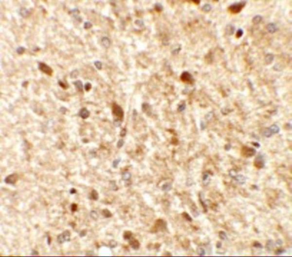
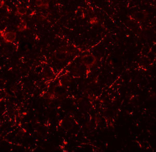
A09251 has IHC and IF images from rat brain tissue (IHC and IF image captions). Human and rat reactivity is listed, but the supplied IHC and IF images show rat tissue only (catalog reactivity; image captions).

Real IHC data Immunohistochemistry of MS4A6A in rat brain tissue with MS4A6A antibody at 2.5 μg/mL.
Anti-MS4A6A Antibody
Cat # A09251

A09251 is listed for IHC-P and IF (catalog applications). Its IHC and IF images show rat brain tissue at 2.5 μg/mL and 20 μg/mL, respectively (image captions).

Which to pick: Choose A09251 for paraffin-section IHC: IHC-P is listed, and its own IHC caption shows staining in rat brain tissue at 2.5 μg/mL; the fixative is unreported (catalog applications; IHC image caption). For IF, A09251 has a rat brain tissue image at 20 μg/mL; ICC validation is not reported (IF image caption; catalog applications). A09251 lists both human and rat reactivity, although the supplied IHC and IF images show rat tissue only; clonality is unreported (catalog reactivity; image captions; catalog clone field).

Each figure is that product's own IHC / IF validation image from its datasheet.

References

  1. UniProt Consortium. UniProt entry Q9H2W1 (M4A6A_HUMAN, Membrane-spanning 4-domains subfamily A member 6A).
  2. Human Protein Atlas. MS4A6A tissue IHC expression (reliability: Approved).
  3. Human Protein Atlas. MS4A6A subcellular location (ICC-IF): Localized to vesicles. Caution: Based on antibodies targeting proteins from multiple genes..
  4. Human Protein Atlas. MS4A6A antibody validation summary (2 antibodies).
  5. Identifying MS4A6A(+) macrophages as potential contributors to the pathogenesis of nonalcoholic fatty liver disease, periodontitis, and type 2 diabetes mellitus. Heliyon 2024 — PMC11033123.
  6. Single-cell immunoprofiling reveals a dysfunctional-like immune microenvironment and malignant phenotype in aging penile squamous cell carcinoma. Cell biology and toxicology 2026 — PMC13457360.
  7. MS4A6A is a new prognostic biomarker produced by macrophages in glioma patients. Frontiers in immunology 2022 — PMC9472524.
  8. Differential expression and regulation of MS4A family members in myeloid cells in physiological and pathological conditions. Journal of leukocyte biology 2022 — PMC9290968.
  9. PubMed PMID:11245982 — UniProt-cited evidence.
  10. PubMed PMID:11401424 — UniProt-cited evidence.
  11. PubMed PMID:11486273 — UniProt-cited evidence.