MTA3 / Metastasis-associated protein MTA3 · IHC design guide

Design Immunohistochemistry for MTA3

Plan chromogenic MTA3 IHC in paraffin sections using nuclear staining as the tissue benchmark (HPA tissue IHC). The guide covers cell-level interpretation and the catalog antibody’s 1:50–1:200 IHC dilution range (datasheet: 1:50–1:200).

Evidence assembled Oct 2026 · For research use; verify linked source records and product datasheet before use
Immunohistochemistry protocol sheet for MTA3 (IHC for MTA3): expected localisation Nuclear tissue staining (HPA tissue IHC); cytoplasm annotated (UniProt), antibody A05612, validated IHC image, and IHC protocol steps
Printable MTA3 IHC protocol sheet — expected localisation Nuclear tissue staining (HPA tissue IHC); cytoplasm annotated (UniProt), antibody A05612, controls and protocol steps. Open the full MTA3 IHC guide →

MTA3 Immunohistochemistry Experimental Design Guide

Expected localisation, validated protocols, controls and antibodies — the at-a-glance facts below, then the full design guide.

Must know before staining
Expected localisation Nuclear tissue staining (HPA tissue IHC); cytoplasm annotated (UniProt)
Staining pattern General nuclear staining across tissues (HPA tissue IHC)
Antigen retrieval Tris-EDTA pH 9.0 HIER, 95–98 °C, 20 min (rule: nuclear antigen)
Positive control ⓘ Adipose tissue+4 more · see all
Negative control ⓘ Heart muscle+1 more · see all
Important caveats
Reasons your staining may differ from the expected pattern.
Fixation Keep fixation consistent across sections (standard IHC practice; not target-specific)
Caveat Adipocytes and glia can stain strongly; check cell identity (HPA tissue IHC)
Regulation Lymphoid tissue–enhanced RNA (HPA tissue RNA)
Isoform / epitope 2 isoforms; epitope coverage unspecified (UniProt)
Section 1

Recommended MTA3 IHC & IF Protocols

Compare the catalog antibody’s IHC-P protocol with four published MTA3 IHC protocols (PMC5514899; PMC13351380; PMC3853379; PMC3643958).

Recommended immunohistochemistry (IHC-P) protocol parameters
SampleParaffin-embedded rat liver tissue; fixative not specified (datasheet A05612)
FixationImage fixative and duration unreported (datasheet A05612); verify before use.
Sectioning4–5 µm sections on charged slides (standard)
DeparaffinisationXylene, graded ethanol series to water (standard)
Antigen retrievalHeat-induced epitope retrieval in Tris-EDTA buffer, pH 9.0, 20 min at 95–98 °C (standard rule: nuclear antigen)
Peroxidase block3% H2O2, 10 min, room temperature (standard)
Blocking10% normal serum of the secondary host, 30 min, room temperature (standard)
Primary antibodyRabbit anti-MTA3, 1:50-1:200 (datasheet A05612)
Primary incubationOvernight at 4 °C (standard)
DetectionHRP-polymer secondary, DAB chromogen 5–10 min (standard)
CounterstainHematoxylin, blue, dehydrate and mount (standard)
Expected resultMTA3-positive staining in adipocytes of adipose tissue (HPA tissue IHC: High). HPA tissue profile: General nuclear expression. No signal in the no-primary control.
💡Decision noteStart with the page’s Tris-EDTA pH 9.0 retrieval at 95–98 °C for 20 min (page antigen retrieval). Published conditions can guide tissue-specific optimization.
Section 2

What Is the Expected MTA3 Staining Pattern?

MTA3 should stain mainly nuclei across several tissue cell types (HPA tissue IHC: general nuclear expression; Approved, with medium consistency against RNA data). Nucleoplasmic localization is supported by ICC-IF, while UniProt also lists cytoplasm (HPA subcellular; UniProt Q9BTC8). Its lack of a transmembrane segment supports an intracellular pattern (UniProt Q9BTC8 topology).

What am I looking at on my slide?
Clear nuclear staining in colon, duodenum, or appendix glandular cells.This matches the reported general nuclear pattern and High staining in those glandular cells (HPA tissue IHC). Compare stained nuclei with nearby unstained areas and the counterstain before scoring; intensity alone does not establish antibody specificity (general IHC practice).
Staining is predominantly cytoplasmic, with little nuclear signal.Recheck the result: HPA reports general nuclear tissue staining and supported nucleoplasmic ICC-IF localization, although UniProt also lists cytoplasm (HPA tissue IHC; HPA subcellular; UniProt Q9BTC8). Cytoplasmic signal alone is therefore insufficient to call MTA3 positive.
Strong staining appears in cardiomyocytes or smooth muscle cells.These cells were Not detected in the supplied tissue images (HPA tissue IHC). Check whether the signal follows tissue structures or reflects nonspecific antibody binding or endogenous detection activity (general IHC practice). The HPA observations are comparators, not an absolute absence claim.
Color covers nuclei, cytoplasm, and spaces between cells indiscriminately.A diffuse deposit cannot support nuclear scoring (general IHC practice). Assess a no-primary control for detection background, then inspect blocking, washes, and chromogen development (general IHC practice). HPA's reported pattern is general nuclear expression (HPA tissue IHC).
No nuclear signal appears in a section selected as a positive comparator.High staining is reported for glandular cells in colon, duodenum, and appendix, among other listed cells (HPA tissue IHC). First verify that the relevant cells are present; then check the staining run and antibody conditions (general IHC practice). A blank section alone does not establish absent MTA3.
💡Expected MTA3 appearanceCall a result positive when discrete nuclear staining is visible in a listed High cell population, such as colon glandular cells (HPA tissue IHC); diffuse deposit or strong staining in cardiomyocytes is suspect (general IHC practice; HPA tissue IHC: cardiomyocytes Not detected).
How each factor affects the staining
Compartment and functionMTA3 participates in the chromatin-remodeling NuRD complex, consistent with a nuclear readout (UniProt Q9BTC8 function; HPA tissue IHC: general nuclear expression). UniProt also lists cytoplasm, so assess any cytoplasmic signal against the dominant nuclear pattern (UniProt Q9BTC8).
Tissue and cell contextHPA reports High staining in adipocytes, glial cells, and several glandular cell populations, but Not detected in cardiomyocytes and smooth muscle cells (HPA tissue IHC). UniProt's statement of no expression in nonepithelial cells conflicts with some of those observations (UniProt Q9BTC8 tissue specificity); do not use epithelial identity alone to score slides.
Strength of the validation evidenceThe supplied tissue profile is Approved with medium consistency between antibody staining and RNA expression; HPA039433 is IHC Approved, not listed as IHC Enhanced (HPA tissue IHC; HPA antibodies). Treat an unexpected cell pattern as a finding to verify, rather than proof of a new distribution.
Isoforms and epitope coverageTwo MTA3 isoforms are listed, but the payload does not map the IHC antibody's epitope to either one (UniProt Q9BTC8 isoforms; HPA antibodies). Isoform-specific staining cannot be inferred from these sources; interpret the reported tissue pattern at the MTA3 level.
Processing and modificationUniProt lists one chain spanning residues 1–594, no signal peptide or propeptide, no glycosylation sites, and four example phosphorylation sites (UniProt Q9BTC8). These facts do not establish shedding, a stain intensity change, or fixation sensitivity for this antibody.
Why is my staining missing, weak or wrong?
SituationLikely causeNext action
Known-positive glandular cells are blank.The chosen section may lack the scored cells, or the IHC run may have failed (general IHC practice).Confirm glandular cells in a listed High tissue, then review retrieval, primary antibody conditions, and detection controls for the run (HPA tissue IHC; general IHC practice).
Nearly every compartment looks brown.Nonspecific binding, endogenous detection activity, or excessive chromogen development can obscure localization (general IHC practice).Compare a no-primary control, check the detection block and wash steps, and shorten development if the run controls support it (general IHC practice).
Cardiomyocytes or smooth muscle cells stain strongly.The result departs from the supplied Not detected observations; nonspecific binding or endogenous activity is possible (HPA tissue IHC; general IHC practice).Inspect the no-primary control and tissue morphology, then repeat with an independent specificity control before scoring those cells as MTA3 positive (general IHC practice).
Nuclear staining is weak beside a strong cytoplasmic haze.Background may hide the expected nuclear pattern; UniProt's cytoplasmic annotation does not validate a cytoplasmic-only IHC result (general IHC practice; UniProt Q9BTC8; HPA tissue IHC).Compare nuclear counterstain and control sections; reassess blocking, washes, and antibody concentration using the catalog IHC-P instructions if available (general IHC practice).
Adipocytes or glial cells stain despite an expectation of epithelial restriction.That expectation follows UniProt's tissue-specificity statement, while HPA reports High staining in these nonepithelial cells (UniProt Q9BTC8; HPA tissue IHC).Record the observed cell type and compartment, cite the disagreement, and seek an independent specificity check before drawing a biological conclusion (general IHC practice).
IF/ICC shows puncta in addition to nuclear fluorescence. Is that expected?HPA reports supported nucleoplasmic localization plus approved vesicle localization in ICC-IF (HPA subcellular).A nucleoplasmic signal with additional vesicular puncta can fit that report; diffuse fluorescence without a clear nuclear signal needs control review (HPA subcellular; general IF practice).

Sample controls for MTA3 IHC & IF

🧪Run adrenal gland first; its glandular cells should stain (HPA: High in adrenal gland glandular cells). Use heart muscle as the negative tissue (HPA: Not detected in cardiomyocytes); on the positive slide, cells established as target-negative should retain counterstain without specific nuclear signal (standard IHC control practice).
Positive control tissue: Adipose tissue (Adipocytes, HPA High)
Negative control tissue: Heart muscle (HPA Not detected)
ICC-IF cell lines (HPA subcellular resource): HPA ICC-IF images show MTA3 in A-431, U-251MG, U2OS, with annotated localisation: Nucleoplasm (supported) (HPA subcellular).
Technical controls: Include a no-primary, secondary-only control and an isotype control matched to the primary antibody’s host species, immunoglobulin class, and clonality; use validated MTA3 knockout material or a peptide block, if available, as a biological specificity control (standard IHC control practice). For chromogenic staining, quench endogenous peroxidase in the adrenal section (standard IHC practice).
⚠️Feasibility: A target-specific fixation window or fixation effect is unreported in the supplied evidence, and the selected A05612 rat-liver paraffin-section caption does not state a fixative (A05612 tissue-IHC caption). That caption uses microwave retrieval in 10 mM PBS, pH 7.2, before staining at 1:100; it does not establish that retrieval is required for adrenal gland or that frozen sections or IF are easier (A05612 tissue-IHC caption). Check the adrenal section’s peroxidase control for endogenous chromogenic signal (standard IHC practice).

HPA tissue IHC evidence for MTA3

Comprehensive Human Protein Atlas IHC scoring per tissue (reliability: Approved — Medium consistency between antibody staining and RNA expression data.). Rows are taken directly from the HPA tissue chart — click any row's HPA link to view the source.

Positive expression · recommended positive controls

TissueCell typeLevelEvidenceSource
Adipose tissue Adipocytes High Protein (IHC) HPA →
Adrenal gland Glandular cells High Protein (IHC) HPA →
Appendix Glandular cells High Protein (IHC) HPA →
Breast Adipocytes High Protein (IHC) HPA →
Caudate Glial cells High Protein (IHC) HPA →

Undetected expression · recommended negative controls

TissueCell typeLevelEvidenceSource
Heart muscle Cardiomyocytes Not detected Protein (IHC) HPA →
Smooth muscle Smooth muscle cells Not detected Protein (IHC) HPA →
Section 3

Advanced MTA3 IHC Tips

These troubleshooting answers focus on chromogenic IHC in paraffin sections, with one IF/ICC entry for readers comparing the two applications.

How should I retrieve MTA3 when nuclear staining is weak?
Use Tris-EDTA pH 9.0 HIER at 95–98 °C for 20 min (page retrieval setting). Let sections cool in retrieval buffer before washing, and compare runs with the same heating and cooling conditions (standard IHC practice). If staining remains weak, test 10 mM PBS at pH 7.2 with microwave retrieval on a matched section; that method was used for paraffin-embedded rat liver with SKU A05612 (selected A05612 tissue-IHC caption). Keep antibody dilution and detection constant during the comparison so a change in nuclear signal can be attributed to retrieval (standard IHC practice). Score nuclei separately from diffuse background because MTA3 is mainly nucleoplasmic (HPA subcellular).
Could fixation explain absent MTA3 staining in my paraffin sections?
Target-specific fixation sensitivity is unknown: the selected A05612 tissue-IHC caption reports paraffin embedding but does not state the fixative (selected A05612 tissue-IHC caption). Record the fixative, fixation duration, tissue thickness and processing history for each section before comparing staining results (standard IHC practice). If a known positive control stains while the test section does not, compare matched material processed under documented conditions before assigning the difference to MTA3 expression (standard IHC practice). Use the same retrieval and detection settings across that comparison, including the page’s 20 min HIER step (page retrieval setting; standard IHC practice). HPA staining patterns do not establish how MTA3 responds to fixation (HPA tissue IHC: Approved, medium consistency).
Should I count cytoplasmic staining as MTA3 positive?
Prioritize nuclear staining in paraffin sections: MTA3 is mainly nucleoplasmic in the subcellular reference data (HPA subcellular: supported nucleoplasm). UniProt also lists cytoplasmic localisation, while HPA reports additional vesicular localisation, so record a reproducible extranuclear pattern separately (UniProt Q9BTC8 subcellular; HPA subcellular: approved vesicles). MTA3 has no transmembrane segment, making a crisp plasma-membrane rim a reason to investigate staining specificity (UniProt Q9BTC8 topology). Compare nuclei and cytoplasm within the same cells at a consistent exposure or chromogen endpoint, and inspect a primary-antibody-omission control for nonspecific signal (standard IHC practice). Do not combine nuclear and cytoplasmic scores when assessing the expected staining pattern (HPA subcellular: supported nucleoplasm).
How can I assess whether the antibody misses an MTA3 isoform or modified epitope?
MTA3 has 2 reported isoforms, but the supplied record does not map the catalog antibody’s epitope to either one (UniProt Q9BTC8 isoforms; supplied antibody evidence). Check the antibody’s documented immunogen or epitope before claiming isoform coverage, and retain that uncertainty in any IHC interpretation (standard antibody-validation practice). UniProt annotates phosphorylation at residues 428, 430, 455 and 519, without establishing an effect on this antibody’s binding (UniProt Q9BTC8 modified residues). Compare staining after the page’s pH 9.0 retrieval against a matched positive control while keeping detection conditions fixed (page retrieval setting; standard IHC practice). A negative section alone cannot distinguish absent protein from an inaccessible epitope (standard IHC practice).
How should I investigate MTA3 localisation by multiplex IF?
Pair MTA3 with a marker identifying the cell population under study, and assess colocalisation at the single-cell level (standard multiplex IF practice). Expect predominantly nucleoplasmic signal, while documenting reproducible vesicular or cytoplasmic signal separately (HPA subcellular: supported nucleoplasm, approved vesicles; UniProt Q9BTC8 subcellular). Choose spectrally separated fluorophores after measuring tissue autofluorescence; favor a channel with lower background for a weak MTA3 signal (standard IF practice). Permeabilise after fixation to give antibody access to the intracellular nuclear epitope, then compare a mild and a stronger condition without assuming this antibody’s IF sensitivity (UniProt Q9BTC8 topology; standard IF practice). Include single-stain and no-primary controls when judging overlap or background (standard multiplex IF practice).
What should I check when MTA3 IHC shows diffuse brown background?
Inspect a section processed without primary antibody to identify background from the chromogenic detection workflow (standard IHC practice). For a peroxidase-based method, check endogenous peroxidase blocking before DAB development and shorten chromogen exposure if control sections darken (standard chromogenic IHC practice). Recheck blocking, washes and antibody dilution on matched sections; the selected A05612 rat-liver image used 1:100, which is evidence for that image’s conditions (selected A05612 tissue-IHC caption; standard IHC practice). Judge improvement by the contrast between nuclei and adjacent unstained areas, since the reference localisation is mainly nucleoplasmic (HPA subcellular: supported nucleoplasm). Record pigment, tissue folds and edge staining separately so they are not scored as cellular MTA3 signal (standard IHC practice).
How should I score MTA3 staining across paraffin sections? ⚠ ANSWER MARKED FOR VERIFICATION
Define the analysis region and cell population before scoring, then report nuclear percentage positive and an intensity-based H-score where staining supports those measures (standard IHC quantification practice; HPA subcellular: supported nucleoplasm). An H-score weights the percentages of cells at intensities 0–3, giving a range of 0–300 (standard IHC quantification practice). For spatial comparisons, report positive nuclei per mm² alongside total evaluable nuclei or tissue area, and exclude folds and necrosis consistently (standard IHC quantification practice). Normalize comparisons to the same cell type, section area and staining batch rather than treating all tissue cells as equivalent (standard IHC quantification practice). Record extranuclear staining as a separate measurement (HPA subcellular: supported nucleoplasm, approved vesicles).
How can I separate genuine MTA3 staining from artefact?
Look first for cellular nuclear staining, the predominant pattern in the reference data, and assess any cytoplasmic or vesicular signal separately (HPA subcellular: supported nucleoplasm, approved vesicles; UniProt Q9BTC8 subcellular). Compare the relevant cell population with controls: HPA reports high staining in colon glandular cells and no detected staining in heart cardiomyocytes (HPA tissue IHC). A sharp membrane rim is discordant with MTA3’s lack of a transmembrane segment and warrants a specificity check (UniProt Q9BTC8 topology). Treat staining restricted to section edges, necrotic areas or a no-primary control as possible artefact (standard IHC practice). Check peroxidase-block controls before interpreting diffuse DAB deposition as MTA3 expression (standard chromogenic IHC practice).
Boster reagents

Best MTA3 / Metastasis-associated protein MTA3 IHC Antibodies

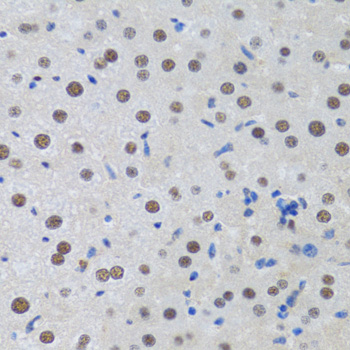
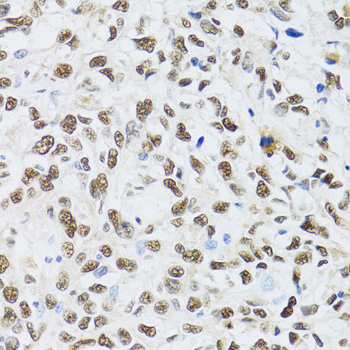
A05612 has real IHC images of paraffin-embedded rat liver, human liver cancer, and mouse testis (catalog: A05612 IHC image captions). No IF image is supplied (catalog: A05612 IF images).

Real IHC data Immunohistochemistry of paraffin-embedded rat liver using MTA3 antibody at dilution of 1:100 .Perform microwave antigen retrieval with 10 mM PBS buffer pH 7.2 before commencing with IHC staining protocol.
Anti-MTA3 Antibody
Cat # A05612

The A05612 card lists IHC as a tested application and Human, Mouse, and Rat reactivity (catalog: A05612 applications/reactivity). Its images show paraffin-embedded rat liver, human liver cancer, and mouse testis stained at 1:100 (catalog: A05612 IHC image captions).

Which to pick: Choose A05612 for paraffin-section tissue IHC across Human, Mouse, and Rat; it is a rabbit polyclonal with listed IHC reactivity in those species (catalog: A05612 host/applications/reactivity and IHC image captions). Its captions specify microwave antigen retrieval in 10 mM PBS, pH 7.2, but do not report the fixative (catalog: A05612 IHC image captions). There is no IF/ICC pick because A05612 has no listed IF/ICC application or IF image (catalog: A05612 applications/IF images).

Each figure is that product's own IHC / IF validation image from its datasheet.

References

  1. UniProt Consortium. UniProt entry Q9BTC8 (MTA3_HUMAN, Metastasis-associated protein MTA3).
  2. Human Protein Atlas. MTA3 tissue IHC expression (reliability: Approved).
  3. Human Protein Atlas. MTA3 subcellular location (ICC-IF): Mainly localized to the nucleoplasm. In addition localized to vesicles..
  4. Human Protein Atlas. MTA3 antibody validation summary (1 antibodies).
  5. Metastasis-associated protein 3 in colorectal cancer determines tumor recurrence and prognosis. Oncotarget 2017 — PMC5514899.
  6. Immunohistochemical Expression of Metastasis-Associated Gene 3 (MTA3) and Epithelial-Mesenchymal Transition (EMT) Markers in Colorectal Carcinoma: A Cross-sectional Study. Iranian journal of pathology 2026 — PMC13351380.
  7. Analysis of MAT3 gene expression in NSCLC. Diagnostic pathology 2013 — PMC3853379.
  8. The metastasis-associated gene MTA3, a component of the Mi-2/NuRD transcriptional repression complex, predicts prognosis of gastroesophageal junction adenocarcinoma. PloS one 2013 — PMC3643958.
  9. PubMed PMID:10574462 — UniProt-cited evidence.
  10. PubMed PMID:14702039 — UniProt-cited evidence.
  11. PubMed PMID:15489334 — UniProt-cited evidence.