MTCH1 / Mitochondrial carrier homolog 1 · IHC design guide

Design Immunohistochemistry for MTCH1

MTCH1 is an outer mitochondrial membrane protein (UniProt), while tissue IHC shows granular cytoplasmic staining (HPA tissue IHC). This guide helps plan paraffin-section staining with the IHC-validated antibody and interpret results alongside the HPA tissue profile.

Evidence assembled Oct 2026 · For research use; verify linked source records and product datasheet before use
Immunohistochemistry protocol sheet for MTCH1 (IHC for MTCH1): expected localisation Granular cytoplasm in tissue sections (HPA tissue IHC), antibody A09592, validated IHC image, and IHC protocol steps
Printable MTCH1 IHC protocol sheet — expected localisation Granular cytoplasm in tissue sections (HPA tissue IHC), antibody A09592, controls and protocol steps. Open the full MTCH1 IHC guide →

MTCH1 Immunohistochemistry Experimental Design Guide

Expected localisation, validated protocols, controls and antibodies — the at-a-glance facts below, then the full design guide.

Must know before staining
Expected localisation Granular cytoplasm in tissue sections (HPA tissue IHC)
Staining pattern Granular cytoplasmic staining in glandular cells (HPA tissue IHC)
Antigen retrieval Citrate pH 6.0 HIER, 95–98 °C, 20 min (rule: cytoplasmic / membrane antigen)
Positive control ⓘ Adipose tissue+4 more · see all
Negative control ⓘ None in HPA (detected in all 45 tissues); use no-primary + isotype controls
Important caveats
Reasons your staining may differ from the expected pattern.
Fixation Keep fixation consistent across sections (standard IHC practice; not target-specific). Selected-image fixative and duration unreported (datasheet A09592); verify before use.
Caveat Staining and RNA have medium consistency (HPA tissue IHC)
Regulation No specific regulation reported (UniProt)
Isoform / epitope 3 isoforms; variant-specific epitope map unavailable (UniProt)
Section 1

Recommended MTCH1 IHC & IF Protocols

The catalog antibody’s IHC-P protocol is accompanied by one published MTCH1 IHC protocol using human breast cancer tissue (PMC11877058).

Recommended immunohistochemistry (IHC-P) protocol parameters
SampleParaffin-embedded human breast cancer tissue; fixative not specified (datasheet A09592)
FixationImage fixative and duration unreported (datasheet A09592); verify before use.
Sectioning4–5 µm sections on charged slides (standard)
DeparaffinisationXylene, graded ethanol series to water (standard)
Antigen retrievalHeat-induced epitope retrieval in citrate buffer, pH 6.0, 20 min at 95–98 °C (standard rule: cytoplasmic / membrane antigen)
Peroxidase block3% H2O2, 10 min, room temperature (standard)
Blocking10% normal serum of the secondary host, 30 min, room temperature (standard)
Primary antibodyRabbit anti-MTCH1, 1:50-1:200 (datasheet A09592)
Primary incubationOvernight at 4 °C (standard)
DetectionHRP-polymer secondary, DAB chromogen 5–10 min (standard)
CounterstainHematoxylin, blue, dehydrate and mount (standard)
Expected resultMTCH1-positive staining in adipocytes of adipose tissue (HPA tissue IHC: High). HPA tissue profile: General granular cytoplasmic expression. No signal in the no-primary control.
💡Decision noteStart with citrate pH 6.0 heat retrieval: it matches this page’s retrieval setting and the published protocol (PMC11877058).
Section 2

What Is the Expected MTCH1 Staining Pattern?

MTCH1 is an outer mitochondrial membrane protein with six transmembrane segments (UniProt Q9NZJ7 topology). In paraffin sections, expect granular cytoplasmic staining in HPA high-staining cells, including adipocytes, bronchial respiratory epithelial cells and cerebellar granular-layer cells (HPA tissue IHC). HPA rates the tissue IHC pattern Approved, with medium consistency between staining and RNA expression (HPA tissue IHC).

What am I looking at on my slide?
Granular cytoplasmic staining in adipocytes, bronchial respiratory epithelial cells or cerebellar granular-layer cells.This matches the reported general granular cytoplasmic profile and High staining in these cells (HPA tissue IHC). Granules are compatible with mitochondrial localization (UniProt Q9NZJ7 subcellular location), but chromogenic IHC alone does not identify individual organelles (general IHC practice).
Predominantly nuclear or sharply plasma-membrane staining, without a granular cytoplasmic component.That distribution conflicts with the reported tissue pattern (HPA tissue IHC) and outer mitochondrial membrane location (UniProt Q9NZJ7). Treat it as suspect; inspect the counterstain and compare control sections before assigning MTCH1 specificity (general IHC practice).
Strong staining confined to an unexpected cell population while an HPA high-staining population is weak.HPA lists specific High-staining cell populations, not a universal positive or negative rule (HPA tissue IHC). Consider cross-reactivity or endogenous detection activity, and compare staining with appropriate reagent controls (general IHC practice). Cell identity and tissue context matter.
Diffuse color across cells and extracellular spaces obscures cytoplasmic granules.The expected profile is granular and cytoplasmic (HPA tissue IHC). Diffuse staining is difficult to score as MTCH1; nonspecific binding or detection background may contribute (general IHC practice). Reassess specificity after background is reduced.
No detectable signal in a section containing an HPA High-staining cell population.An absent signal warrants a workflow check, but HPA's High rating is an observed tissue pattern, not a guarantee for every specimen (HPA tissue IHC). Verify cell identity and controls before interpreting absence as loss of MTCH1 (general IHC practice).
💡Expected MTCH1 appearanceCall positive a discernible granular cytoplasmic signal in HPA High-staining cells (HPA tissue IHC); isolated nuclear or diffuse staining is suspect against the outer mitochondrial membrane assignment (UniProt Q9NZJ7).
How each factor affects the staining
Tissue and cell selectionHPA reports High staining in adipocytes, bronchial respiratory epithelial cells and cerebellar granular-layer cells; peripheral nerve in soft tissue is Low, not negative (HPA tissue IHC). Choose controls by the named cell population, since a tissue-wide label can hide variation (general IHC practice).
Antibody evidenceThe listed antibody HPA015971 has IHC status Approved; HPA describes medium consistency between staining and RNA expression (HPA antibody record; HPA tissue IHC). This supports a working reference pattern while leaving unexpected staining open to investigation.
Topology and epitope interpretationMTCH1 has six transmembrane segments and alternating cytoplasmic and mitochondrial intermembrane regions (UniProt Q9NZJ7 topology). The supplied evidence does not locate the IHC antibody's epitope, so topology cannot establish its accessibility after section processing.
Isoforms and processingUniProt lists three isoforms, a 1–389 chain, and no annotated signal peptide or propeptide (UniProt Q9NZJ7). The supplied evidence does not identify which isoforms the IHC antibody detects or support a cleavage-based staining prediction.
IF/ICC evidenceHPA's ICC-IF summary says Membrane, while its main location is unavailable and no cell-line ICC-IF images are listed (HPA subcellular record). That summary cannot confirm a mitochondrial pattern for this antibody; evaluate IF/ICC on its dedicated guide page.
Why is my staining missing, weak or wrong?
SituationLikely causeNext action
A known HPA High-staining cell population has no visible signal (HPA tissue IHC).The cells may be misidentified, or the IHC workflow may have failed; this result alone cannot distinguish them (general IHC practice).Confirm the cell population and inspect positive and detection controls; review retrieval, antibody dilution and detection against the chosen IHC instructions (general IHC practice).
Staining is chiefly nuclear or at the plasma membrane.This conflicts with HPA's granular cytoplasmic profile and UniProt's outer mitochondrial membrane assignment (HPA tissue IHC; UniProt Q9NZJ7).Check compartment assignment against the counterstain, then compare controls and another appropriate section before scoring it as MTCH1 (general IHC practice).
Unexpected cells stain strongly while the expected cells do not.Cross-reactivity or endogenous detection activity is possible; HPA's cell-specific High ratings do not validate every stained cell (HPA tissue IHC; general IHC practice).Verify cell identity and compare reagent controls; assess unexpected staining separately from the expected cell population (general IHC practice).
Diffuse color masks the granular cytoplasmic pattern (HPA tissue IHC).Nonspecific binding or detection background may obscure a localized signal (general IHC practice).Inspect controls and review blocking, washing and detection conditions; score only a resolvable cellular pattern (general IHC practice).
A low-staining peripheral nerve is used as a negative control.HPA calls peripheral nerve in soft tissue Low and supplies no negative tissue in this record (HPA tissue IHC).Interpret weak staining there cautiously; use reagent controls to assess background rather than treating this tissue as a proven MTCH1-negative control (general IHC practice).
Can IF/ICC staining confirm the IHC localization?HPA reports Membrane for ICC-IF but lists no main location or cell-line ICC-IF images (HPA subcellular record).Do not treat that summary as mitochondrial confirmation; consult the separate IF/ICC guide for assay design and evaluate localization with appropriate controls (general IF practice).

Sample controls for MTCH1 IHC & IF

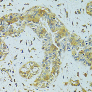
🧪Run breast first: glandular cells should stain (HPA: High in breast glandular cells), and the selected IHC image uses a paraffin-embedded breast cancer section (caption: SKU A09592). HPA detects MTCH1 in all 45 scored tissues, so there is no supported negative tissue; use no-primary and isotype controls, and treat cells without visible staining on the positive slide as background checks rather than proven MTCH1-negative cells (HPA: no negative tissue rows).
Positive control tissue: Adipose tissue (Adipocytes, HPA High)
Negative control tissue: None in HPA: MTCH1 is detected in all 45 scored tissues. Use a no-primary (secondary-only) and an isotype control instead.
ICC-IF cell lines (HPA subcellular resource): HPA carries no ICC-IF cell line for MTCH1; derive a cell-line control from the positive tissue's cell type (Adipocytes) and confirm it by RNA or western blot first.
Technical controls: Run a no-primary, secondary-only control and an isotype control matched to the primary antibody’s host species and clonality; use a matched MTCH1 knockout specimen, if available, as a biological specificity control (standard IHC practice). For chromogenic breast IHC, block endogenous peroxidase and check the control slide for residual background (standard IHC practice).
⚠️Feasibility: A target-specific fixation window or fixation effect is unreported, and the selected SKU’s paraffin-section caption does not state its fixative (caption: SKU A09592). The caption reports microwave antigen retrieval in 10 mM PBS, pH 7.2, followed by staining at 1:100; this provides a starting condition but does not establish that retrieval is required (caption: SKU A09592). No supplied comparison shows frozen sections or IF/ICC to be easier, and no breast-specific artefact is reported; assess IF background with a no-primary control if using that separate application (supplied application evidence; standard IF practice).

HPA tissue IHC evidence for MTCH1

Comprehensive Human Protein Atlas IHC scoring per tissue (reliability: Approved — Medium consistency between antibody staining and RNA expression data.). Rows are taken directly from the HPA tissue chart — click any row's HPA link to view the source.

Positive expression · recommended positive controls

TissueCell typeLevelEvidenceSource
Adipose tissue Adipocytes High Protein (IHC) HPA →
Adrenal gland Glandular cells High Protein (IHC) HPA →
Appendix Glandular cells High Protein (IHC) HPA →
Breast Glandular cells High Protein (IHC) HPA →
Bronchus Respiratory epithelial cells High Protein (IHC) HPA →

Undetected expression · recommended negative controls

TissueCell typeLevelEvidenceSource
None in HPA: MTCH1 is detected in all 45 scored tissues. Use a no-primary (secondary-only) and an isotype control instead.
Section 3

Advanced MTCH1 IHC Tips

Troubleshoot MTCH1 staining in paraffin sections using the documented tissue pattern, membrane topology, and antibody-specific IHC evidence.

Which retrieval conditions should I try when MTCH1 staining is weak?
Start with citrate buffer at pH 6.0, heated to 95–98 °C for 20 min (page retrieval specification). Keep section thickness, cooling, and detection conditions consistent while comparing retrieval runs, so a change in signal can be attributed to retrieval (standard IHC practice). If staining remains weak, test microwave retrieval in 10 mM PBS, pH 7.2, the condition reported for catalog antibody A09592 on a paraffin-embedded human breast cancer section (A09592 tissue-IHC caption). Compare granular cytoplasmic staining in the expected cells with background and a no-primary control; stronger diffuse staining alone does not establish improved specificity (HPA tissue IHC; standard IHC practice).
Could fixation explain weak or uneven MTCH1 staining?
Target-specific fixation sensitivity is unknown: the A09592 paraffin-section caption does not state a fixative, and the supplied tissue staining data do not establish one (A09592 tissue-IHC caption; HPA tissue IHC). Record the actual fixative, fixation duration, and processing history for each specimen before comparing staining intensity across sections (standard IHC practice). If fixation varies, compare sections processed together using the same pH 6.0, 95–98 °C, 20 min retrieval and detection run (page retrieval specification; standard IHC practice). Assess preservation and morphology alongside staining; a weak section alone cannot distinguish epitope masking from low expression or technical loss (standard IHC practice).
What staining pattern supports MTCH1 localisation in paraffin sections?
Expect a granular cytoplasmic pattern at tissue-IHC resolution, consistent with MTCH1 residing in the mitochondrial outer membrane (HPA tissue IHC; UniProt Q9NZJ7 subcellular location). Score staining within identifiable cells rather than treating diffuse extracellular color or nuclear-only signal as evidence of mitochondrial localisation (standard IHC practice; UniProt Q9NZJ7 subcellular location). Use tissue architecture and a counterstain to separate cellular granules from pigment, debris, and section folds (standard IHC practice). HPA reports high staining in breast glandular cells and cerebellar granular-layer cells, but its tissue-IHC assessment has medium agreement with RNA expression, so morphology and controls remain important (HPA tissue IHC).
How could epitope position or isoforms affect MTCH1 staining?
MTCH1 has 3 annotated isoforms and 6 transmembrane segments, so confirm which sequence the antibody immunogen represents before interpreting a negative result (UniProt Q9NZJ7 isoforms and topology; standard IHC practice). Cytoplasmic-facing intervals include residues 105–155, 230–254, and 343–371; other annotated intervals face the mitochondrial intermembrane space (UniProt Q9NZJ7 topology). Retrieval or fixation could affect access to a particular epitope, but the supplied A09592 caption does not identify its epitope or establish isoform coverage (A09592 tissue-IHC caption). Compare the immunogen sequence with isoform sequences and document that comparison before assigning different staining patterns to different isoforms (standard IHC practice).
How should I follow up an MTCH1 IHC pattern with immunofluorescence?
Use IF/ICC as a separate validation experiment; the supplied A09592 tissue image documents paraffin-section IHC, and the HPA subcellular record lists no ICC/IF image-bearing cell lines (A09592 tissue-IHC caption; HPA subcellular). Multiplex MTCH1 with a marker for the cell population being assessed, such as a glandular-cell marker when examining glandular staining, and include single-stain controls (HPA tissue IHC; standard IF practice). Choose a fluorophore channel after checking tissue autofluorescence in an unstained section (standard IF practice). Select permeabilisation according to the mapped antibody epitope: MTCH1 has cytoplasmic-facing and intermembrane-space-facing intervals, while the supplied caption does not map this antibody's epitope (UniProt Q9NZJ7 topology; A09592 tissue-IHC caption).
How can I reduce background without losing granular MTCH1 signal?
First distinguish granular cytoplasmic staining from diffuse haze, deposits, edge staining, or color over necrotic material by reviewing morphology and a no-primary control (HPA tissue IHC; standard IHC practice). Check deparaffinisation, washing, blocking, and the detection reagents in matched sections before changing antibody concentration (standard IHC practice). Include a peroxidase block when using enzyme-based chromogenic detection, and assess whether residual endogenous activity contributes to the signal (standard chromogenic IHC practice). Keep retrieval at pH 6.0, 95–98 °C for 20 min during initial comparisons; changing retrieval and detection together obscures the source of background (page retrieval specification; standard IHC practice).
How should I quantify MTCH1 staining across tissue sections? ⚠ ANSWER MARKED FOR VERIFICATION
Define the cell population and tissue area before scoring, then record the percentage of positive cells and staining intensity with a consistent threshold (standard IHC practice). An H-score can combine intensity with the percentage of cells at each intensity; alternatively, report positive-cell density per mm² when cell counts answer the study question (standard IHC practice). Normalise to evaluable tissue area or the number of relevant cells, and exclude folds, necrosis, and section edges using the same rules across samples (standard IHC practice). Report granular cytoplasmic staining separately from nonspecific diffuse color, because that is the tissue pattern described for MTCH1 (HPA tissue IHC).
When is a positive MTCH1 IHC result convincing?
A convincing result shows reproducible granular cytoplasmic staining in morphologically identifiable cells, consistent with the reported tissue pattern and mitochondrial outer-membrane localisation (HPA tissue IHC; UniProt Q9NZJ7 subcellular location). High staining in breast glandular cells or cerebellar granular-layer cells can guide comparison, but HPA rates agreement between staining and RNA expression as medium (HPA tissue IHC). Treat nuclear-only staining, isolated section-edge color, necrotic deposits, or signal reproduced in a no-primary control as possible artefacts requiring investigation (standard IHC practice; UniProt Q9NZJ7 subcellular location). In chromogenic assays, check endogenous peroxidase activity before attributing unexplained color to MTCH1 (standard chromogenic IHC practice).
Boster reagents

Best MTCH1 / Mitochondrial carrier homolog 1 IHC Antibodies

One anti-MTCH1 antibody has real IHC data from paraffin-embedded human breast cancer (A09592 image caption); the catalog lists Human, Mouse, and Rat reactivity and no IF/ICC application (A09592 catalog).

Real IHC data Immunohistochemistry of paraffin-embedded human breast cancer using MTCH1 antibody at dilution of 1:100 .Perform microwave antigen retrieval with 10 mM PBS buffer pH 7.2 before commencing with IHC staining protocol.
Anti-MTCH1 Antibody
Cat # A09592

A09592 is the only SKU shown; its IHC image depicts paraffin-embedded human breast cancer stained at 1:100 after microwave retrieval in 10 mM PBS, pH 7.2 (A09592 image caption). It is listed for IHC and WB with Human, Mouse, and Rat reactivity; IF/ICC is not listed (A09592 catalog).

Which to pick: Choose A09592 for paraffin-section tissue IHC because its own image documents that preparation and it is a rabbit polyclonal antibody listed for IHC (A09592 image caption; A09592 catalog). There is no IF/ICC-listed SKU to select (A09592 catalog). For mouse or rat samples, A09592 has catalog reactivity, but its IHC image documents only human tissue (A09592 catalog; A09592 image caption). The caption does not report the fixative (A09592 image caption).

Each figure is that product's own IHC / IF validation image from its datasheet.

References

  1. UniProt Consortium. UniProt entry Q9NZJ7 (MTCH1_HUMAN, Mitochondrial carrier homolog 1).
  2. Human Protein Atlas. MTCH1 tissue IHC expression (reliability: Approved).
  3. Human Protein Atlas. MTCH1 subcellular location (ICC-IF): Membrane.
  4. Human Protein Atlas. MTCH1 antibody validation summary (1 antibodies).
  5. Synergetic effect of doxorubicin and avenanthramide C on VDAC2/MTCH1 mitochondrial axis in breast cancer cells. International journal of health sciences 2025 — PMC11877058.
  6. Predictive model using four ferroptosis-related genes accurately predicts gastric cancer prognosis. World journal of gastrointestinal oncology 2024 — PMC11099433.
  7. PubMed PMID:12975309 — UniProt-cited evidence.
  8. PubMed PMID:14702039 — UniProt-cited evidence.
  9. PubMed PMID:14574404 — UniProt-cited evidence.