This website uses cookies to ensure you get the best experience on our website.


- Table of Contents
Plan chromogenic IHC for MTHFD1 in paraffin sections using the catalog antibody at 2–5 μg/mL (datasheet A02025-1). Expect cytoplasmic staining, strongest in hepatocytes, while interpreting specificity with care (HPA tissue IHC).

Expected localisation, validated protocols, controls and antibodies — the at-a-glance facts below, then the full design guide.
| Expected localisation | Cytoplasmic staining in tissue (HPA tissue IHC) | |
| Staining pattern | Hepatocytes show strong cytoplasmic staining (HPA tissue IHC) | |
| Antigen retrieval | EDTA pH 8.0 HIER, heat-mediated (datasheet A02025-1) | |
| Positive control | Liver+4 more · see all | |
| Negative control | None in HPA (detected in all 45 tissues); use no-primary + isotype controls |
| Fixation | Keep fixation consistent across sections (standard IHC practice; not target-specific) | |
| Caveat | Staining may reflect more than one gene (HPA tissue IHC) | |
| Regulation | Liver-enriched RNA expression (HPA tissue RNA) | |
| Isoform / epitope | No isoforms; loss of residue 1 may affect N-terminal epitopes (UniProt) |
The catalog antibody’s IHC-P protocol (datasheet: A02025-1) is followed by published MTHFD1 IHC methods from four articles (PMC11288495; PMC7947712; PMC11581708; PMC8264328).
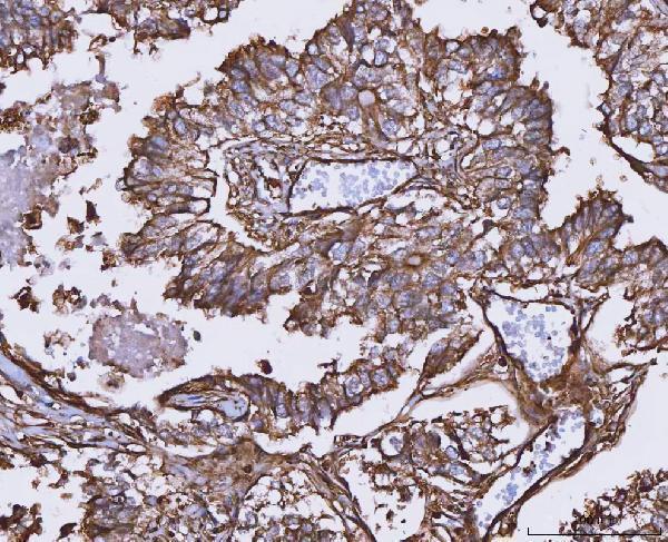
| Sample | Paraffin-embedded human adenocarcinoma of the lung tissue; fixative not specified (datasheet A02025-1) |
| Fixation | Image fixative and duration unreported (datasheet A02025-1); verify before use. |
| Sectioning | 4–5 µm sections on charged slides (standard) |
| Deparaffinisation | Xylene, graded ethanol series to water (standard) |
| Antigen retrieval | Heat retrieval: EDTA pH 8.0 (datasheet A02025-1); 20 min, 95–100 °C (standard) |
| Peroxidase block | 3% H2O2, 10 min, room temperature (standard) |
| Blocking | 10% goat serum (datasheet A02025-1) |
| Primary antibody | Rabbit anti-MTHFD1, 2-5 μg/ml (datasheet A02025-1) |
| Primary incubation | Overnight at 4 °C (datasheet A02025-1) |
| Detection | HRP-conjugated secondary, DAB chromogen (datasheet A02025-1) |
| Counterstain | Hematoxylin, blue, dehydrate and mount (standard) |
| Expected result | MTHFD1-positive staining in hepatocytes of liver (HPA tissue IHC: High). HPA tissue profile: General cytoplasmic expression, most abundant in liver. No signal in the no-primary control. |
MTHFD1 should show cytoplasmic staining across many tissues, with the strongest listed IHC signal in hepatocytes (UniProt P11586: cytoplasm and no transmembrane segment; HPA: general cytoplasmic expression, high in hepatocytes). HPA rates its tissue IHC evidence Approved but reports low agreement with RNA data and cautions that staining may include proteins from more than one gene (HPA: tissue IHC reliability).
| Hepatocytes show strong cytoplasmic chromogen; nearby tissue is less intense. | This fits the strongest listed tissue pattern: high hepatocyte staining and general cytoplasmic expression (HPA: tissue IHC). Compare cell compartments and tissue context before assigning a score. |
| A sharply nuclear, membranous or extracellular signal dominates the section. | The dominant compartment conflicts with cytoplasmic MTHFD1 localization (UniProt P11586: cytoplasm; HPA: tissue IHC). Consider an artifact or another detected protein; compartment alone cannot establish antibody specificity (HPA: tissue IHC reliability). |
| A low-staining cell group appears stronger than hepatocytes in the same run. | Check whether the signal follows tissue structures or the detection system. HPA lists cardiomyocytes and skeletal myocytes as low, versus high hepatocytes (HPA: tissue IHC). Cross-reactivity or endogenous chromogen-forming activity is possible (HPA: tissue IHC reliability; general IHC practice). |
| Chromogen forms a diffuse haze across cells, stroma and blank areas. | A haze without cell-shaped cytoplasmic contrast is difficult to score as MTHFD1 (HPA: general cytoplasmic expression; general IHC practice). Nonspecific antibody binding or detection background may contribute (general IHC practice). |
| A liver section shows no discernible hepatocyte signal. | This conflicts with the reported high hepatocyte staining (HPA: tissue IHC). Review run controls and tissue integrity before calling the sample negative (general IHC practice); HPA's Approved rating does not guarantee every antibody or run will reproduce its pattern (HPA: tissue IHC reliability). |
| Tissue and cell selection | Liver hepatocytes provide the strongest listed reference; HPA reports medium staining in several other listed cell types and low staining in selected muscle cells (HPA: tissue IHC). Low is not an established negative control. |
| Antibody interpretation | Four listed antibodies have Approved IHC status, while HPA reports low staining–RNA agreement and possible detection of protein from more than one gene (HPA: antibody list; tissue IHC reliability). Treat staining intensity as evidence to check, not proof of identity. |
| Compartment and protein form | MTHFD1 is cytoplasmic, lacks a transmembrane segment and has recorded 1–935 and N-terminally processed 2–935 chains (UniProt P11586). The supplied record gives no antibody epitope, so the processing entry cannot predict an antibody-specific staining difference. |
| Chromogenic detection | Endogenous enzyme activity or nonspecific reagent binding can add color independently of target staining (general IHC practice). Interpret chromogen against a suitable detection control, particularly when color lacks cytoplasmic cell boundaries (general IHC practice). |
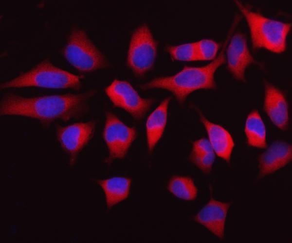
| IF/ICC Q: Where should MTHFD1 appear? | A: In the cytosol (HPA: subcellular ICC-IF, enhanced). That observation supports compartment interpretation here; IF/ICC setup belongs to its separate guide and does not establish a chromogenic IHC intensity threshold. |
| Situation | Likely cause | Next action |
|---|---|---|
| No hepatocyte staining in the liver reference section. | The run may have failed, tissue quality may be poor, or the antibody may not perform under the chosen conditions (general IHC practice). | Check the IHC-validated antibody's instructions, a known-positive section and detection controls; revisit antigen retrieval only as a general IHC optimization step, without assuming a documented MTHFD1 fixation effect (HPA: high in hepatocytes; general IHC practice). |
| Strong signal appears mainly in nuclei or along membranes. | That dominant pattern disagrees with the reported cytoplasmic location; off-target staining or an interpretation artifact is possible (UniProt P11586: cytoplasm; HPA: tissue IHC reliability). | Compare compartment boundaries with the counterstain and an appropriate control, then confirm the pattern with an independent antibody if available (general IHC practice; HPA: multiple listed IHC antibodies). |
| Low-listed muscle cells appear intensely positive. | The result departs from HPA's low staining in cardiomyocytes and skeletal myocytes; cross-reactivity or detection background may contribute (HPA: tissue IHC and reliability; general IHC practice). | Compare the cells with a liver reference and detection control in the same run; investigate reagent background before assigning a biological difference (HPA: high in hepatocytes; general IHC practice). |
| Diffuse chromogen obscures cell boundaries. | Nonspecific binding, incomplete blocking or endogenous detection activity can create broad background (general IHC practice). | Inspect the detection-only control, review blocking and antibody dilution, and score only interpretable cell-shaped cytoplasmic signal (general IHC practice; HPA: general cytoplasmic expression). |
| Staining is weak across every section. | A shared retrieval, antibody incubation or detection problem is possible (general IHC practice); HPA does not establish MTHFD1-specific fixation sensitivity (HPA: tissue IHC scope). | Compare a known-positive liver section and run controls, then adjust one general IHC step at a time according to the antibody instructions (HPA: high in hepatocytes; general IHC practice). |
| Two antibodies yield different cell patterns. | HPA reports low agreement with RNA data and cautions about detection of protein from more than one gene, despite Approved IHC ratings (HPA: tissue IHC reliability; antibody list). | Compare their cytoplasmic patterns in liver and the same tissue panel; document the discrepancy and avoid calling an antibody-specific pattern definitive without further validation (HPA: high in hepatocytes; general IHC practice). |


Comprehensive Human Protein Atlas IHC scoring per tissue (reliability: Approved — Low consistency between antibody staining and RNA expression data. Caution, targets protein from more than one gene.). Rows are taken directly from the HPA tissue chart — click any row's HPA link to view the source.
| Tissue | Cell type | Level | Evidence | Source |
|---|---|---|---|---|
| Liver | Hepatocytes | High | Protein (IHC) | HPA → |
| Adipose tissue | Adipocytes | Medium | Protein (IHC) | HPA → |
| Adrenal gland | Glandular cells | Medium | Protein (IHC) | HPA → |
| Appendix | Glandular cells | Medium | Protein (IHC) | HPA → |
| Bone marrow | Hematopoietic cells | Medium | Protein (IHC) | HPA → |
| Tissue | Cell type | Level | Evidence | Source |
|---|---|---|---|---|
| None in HPA: MTHFD1 is detected in all 45 scored tissues. Use a no-primary (secondary-only) and an isotype control instead. | ||||
Troubleshoot cytoplasmic MTHFD1 staining in paraffin section IHC using the catalog antibody’s documented conditions and compartment specific controls.
A02025-1 has human paraffin-section IHC images and HeLa-cell and human tonsil IF images (catalog image captions). Its listed reactivity covers human, mouse, and rat (catalog reactivity).
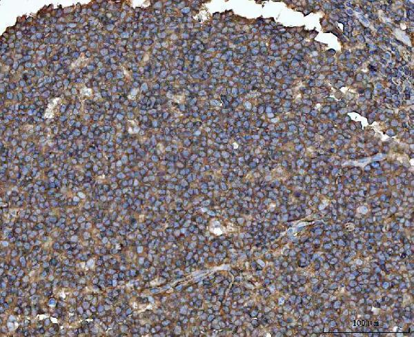
A02025-1 has IHC images from paraffin sections of human lung adenocarcinoma, chronic tonsillitis, diffuse large B-cell lymphoma, and liver cancer (catalog IHC captions). It also has IF images from HeLa cells and a paraffin section of human tonsil (catalog IF captions).
Which to pick: Choose A02025-1 for tissue IHC: its human paraffin-section captions document EDTA retrieval at pH 8.0 and primary antibody at 2 μg/ml (catalog IHC captions); the fixative is unreported (catalog IHC captions). The same SKU supports IF/ICC, with images from HeLa cells and human tonsil using 5 μg/ml primary antibody (catalog applications; catalog IF captions). For cross-species planning, A02025-1 lists human, mouse, and rat reactivity, while its supplied IHC and IF images show human samples only (catalog reactivity; catalog image captions).