This website uses cookies to ensure you get the best experience on our website.


- Table of Contents
Plan chromogenic MTHFD2 IHC in paraffin sections with the IHC-validated antibody A06465-1 (datasheet: IHC). Compare cytoplasmic staining in kidney tubule cells with the undetected signal reported for adipocytes (HPA tissue IHC).

Expected localisation, validated protocols, controls and antibodies — the at-a-glance facts below, then the full design guide.
| Expected localisation | Cytoplasmic tissue staining (HPA tissue IHC); mitochondrial protein (UniProt) | |
| Staining pattern | Cytoplasmic signal in several tissues, distinct in immune cells (HPA tissue IHC) | |
| Antigen retrieval | EDTA pH 8.0 HIER, heat-mediated (datasheet A06465-1) | |
| Positive control | Appendix+4 more · see all | |
| Negative control | Adipose tissue+4 more · see all |
| Fixation | Keep fixation consistent across compared paraffin sections. (selected-SKU IHC image A06465-1) | |
| Caveat | Immune-cell staining can complicate mixed-tissue scoring (HPA tissue IHC) | |
| Regulation | Staining intensity varies by cell type (HPA tissue IHC) | |
| Isoform / epitope | 2 isoforms; mature chain 36–350; epitope map unspecified (UniProt) |
Compare the catalog antibody’s IHC-P protocol (datasheet A06465-1) with published MTHFD2 IHC protocols for esophageal and lung cancer tissues (PMC7055195; PMC12009792; PMC6991687).
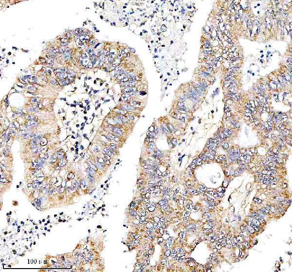
| Sample | Paraffin-embedded human colon cancer tissue; fixative not specified (datasheet A06465-1) |
| Fixation | Image fixative and duration unreported (datasheet A06465-1); verify before use. |
| Sectioning | 4–5 µm sections on charged slides (standard) |
| Deparaffinisation | Xylene, graded ethanol series to water (standard) |
| Antigen retrieval | Heat retrieval: EDTA pH 8.0 (datasheet A06465-1); 20 min, 95–100 °C (standard) |
| Peroxidase block | 3% H2O2, 10 min, room temperature (standard) |
| Blocking | 10% goat serum (datasheet A06465-1) |
| Primary antibody | Rabbit anti-MTHFD2, 1:50 recommended; image 1:100 (datasheet A06465-1) |
| Primary incubation | Overnight at 4 °C (datasheet A06465-1) |
| Detection | HRP-conjugated secondary, DAB chromogen (datasheet A06465-1) |
| Counterstain | Hematoxylin, blue, dehydrate and mount (standard) |
| Expected result | MTHFD2-positive staining in lymphoid tissue of appendix (HPA tissue IHC: High). HPA tissue profile: Cytoplasmic expression in several different tissues, distinct expression in immune cells. No signal in the no-primary control. |
MTHFD2 is mitochondrial and has no transmembrane segment (UniProt P13995 topology). In paraffin sections, expect cytoplasmic staining in selected cells, including kidney tubules, intestinal glands, and lymph node germinal centers (HPA tissue IHC). HPA rates its tissue staining Approved, with medium agreement between staining and RNA data and external verification pending (HPA tissue IHC). Interpret weak or unexpected staining with that limitation in mind.
| Cytoplasmic staining in kidney tubular cells, intestinal glandular cells, or lymph node germinal center cells. | This matches cell populations scored High by HPA tissue IHC. Mitochondrial localization supports a cytoplasmic location (UniProt P13995; HPA subcellular ICC-IF). A chromogenic section need not resolve individual mitochondria; assess the stained cells and compartment together. |
| Predominantly nuclear or cell-surface staining, with little corresponding cytoplasmic signal. | That distribution conflicts with mitochondrial localization (UniProt P13995; HPA subcellular ICC-IF). Treat it as a possible staining artefact or off-target signal, especially if the expected cytoplasmic pattern is absent; localization alone cannot identify its cause. |
| Strong staining in adipocytes or cardiomyocytes, particularly without staining in an expected positive cell population. | HPA reports MTHFD2 as Not detected in adipocytes of adipose tissue and cardiomyocytes of heart muscle (HPA tissue IHC). Investigate possible cross-reactivity or endogenous detection activity before calling these cells positive. The HPA result is a comparison point, not proof that every specimen must be negative. |
| Broad, even color across cells and surrounding tissue, obscuring cell boundaries. | This does not resemble HPA's cell-specific cytoplasmic pattern (HPA tissue IHC). Consider nonspecific antibody binding, incomplete blocking, or detection background as general IHC possibilities. Check a control without primary antibody and compare staining across the expected positive and negative cell populations. |
| No visible signal in kidney tubular cells or lymph node germinal center cells. | Both are scored High in HPA tissue IHC, so their absence calls for a technical check. Confirm that the section contains the named cells, then review the antibody's validated IHC-P conditions and detection controls (general IHC practice). A single unstained section does not establish biological absence. |
| Compartment and optical resolution | MTHFD2 is mitochondrial and lacks a transmembrane segment (UniProt P13995 topology). HPA tissue IHC describes cytoplasmic expression, while HPA ICC-IF supports mitochondrial localization. Score the cytoplasmic pattern in IHC without requiring visible mitochondrial puncta. |
| Cell population sampled | HPA scores duodenal and small-intestinal glandular cells, kidney tubular cells, and tonsil germinal center cells High; lung alveolar cells are Low (HPA tissue IHC). Name the cell population when interpreting a section, since a tissue-wide positive or negative label would hide that difference. |
| Strength of validation | The tissue IHC profile is Approved but has medium staining-to-RNA agreement and awaits external verification (HPA tissue IHC). HPA lists two antibodies with Approved IHC status, HPA049657 and CAB003684 (HPA antibodies). These ratings support comparison with the reported pattern without guaranteeing every specimen will match. |
| Isoforms and processing | UniProt lists two isoforms and a mature chain spanning residues 36–350 (UniProt P13995). No antibody epitope is supplied, so this record cannot establish whether either isoform or the processed region changes the observed IHC pattern. |
| Chromogenic detection | Endogenous enzyme activity can produce color when the matching enzyme-based detection system is used (general IHC practice). A control without primary antibody helps distinguish detection background from antibody-dependent staining; this is a workflow check, not an MTHFD2-specific effect. |
| Situation | Likely cause | Next action |
|---|---|---|
| Expected positive cells are unstained. | The named cells may be absent from the section, or the staining run may have failed (general IHC practice). | Confirm the cell population on the counterstained section; include a tissue and cell population scored High by HPA, then check the antibody's validated IHC-P conditions and run controls (HPA tissue IHC; general IHC practice). |
| Signal is mainly nuclear or along cell borders. | The compartment conflicts with mitochondrial MTHFD2 localization; artefact or off-target binding is possible (UniProt P13995; HPA subcellular ICC-IF). | Compare with a control lacking primary antibody and with a known positive cell population. Reassess specificity before scoring the unusual compartment as MTHFD2 (general IHC practice). |
| Adipocytes or cardiomyocytes stain strongly. | Those cell populations are reported Not detected (HPA tissue IHC); cross-reactivity or detection background is possible. | Inspect a control without primary antibody and compare the signal with HPA High cell populations on appropriately stained sections. Record the cell type and compartment rather than assigning positivity from color alone (HPA tissue IHC; general IHC practice). |
| Diffuse color prevents cell-by-cell scoring. | Nonspecific binding or background from the detection system may obscure the cell-specific pattern (general IHC practice; HPA tissue IHC). | Review blocking and antibody use against the validated IHC-P procedure; check a control without primary antibody. Score only when cytoplasmic signal can be assigned to identifiable cells (general IHC practice). |
| A negative comparison looks as dark as the expected positive cells. | The contrast does not follow HPA's reported High versus Not detected cell populations; background or an unsuitable comparison is possible (HPA tissue IHC). | Verify the exact cell types being compared and examine detection controls. Interpret differences cautiously because HPA rates tissue IHC agreement as medium and external verification is pending (HPA tissue IHC). |
| What should an IF/ICC image show? | HPA reports a supported mitochondrial location in ICC-IF, whereas its tissue IHC summary describes cytoplasmic staining (HPA subcellular ICC-IF; HPA tissue IHC). | For IF/ICC interpretation, look for mitochondrial localization and use the separate IF/ICC guide for experimental conditions. Do not require resolved mitochondrial puncta to score a chromogenic paraffin section (HPA subcellular ICC-IF; HPA tissue IHC). |


Comprehensive Human Protein Atlas IHC scoring per tissue (reliability: Approved — Medium consistency between antibody staining and RNA expression data. Pending external verification.). Rows are taken directly from the HPA tissue chart — click any row's HPA link to view the source.
| Tissue | Cell type | Level | Evidence | Source |
|---|---|---|---|---|
| Appendix | Lymphoid tissue | High | Protein (IHC) | HPA → |
| Duodenum | Glandular cells | High | Protein (IHC) | HPA → |
| Gallbladder | Glandular cells | High | Protein (IHC) | HPA → |
| Kidney | Cells in tubules | High | Protein (IHC) | HPA → |
| Lymph node | Germinal center cells | High | Protein (IHC) | HPA → |
| Tissue | Cell type | Level | Evidence | Source |
|---|---|---|---|---|
| Adipose tissue | Adipocytes | Not detected | Protein (IHC) | HPA → |
| Adrenal gland | Glandular cells | Not detected | Protein (IHC) | HPA → |
| Bone marrow | Hematopoietic cells | Not detected | Protein (IHC) | HPA → |
| Breast | Adipocytes | Not detected | Protein (IHC) | HPA → |
| Epididymis | Glandular cells | Not detected | Protein (IHC) | HPA → |
Troubleshoot MTHFD2 staining in paraffin sections by checking retrieval, compartment, cell identity, and assay controls together.
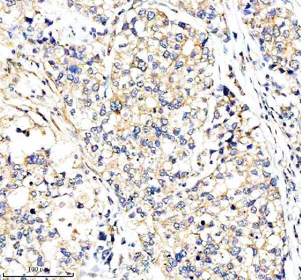
A06465-1 has IHC images from paraffin sections of human colon and lung cancers and mouse testis and epididymis; rat reactivity is listed without a tissue image (catalog: reactivity; IHC image captions).
A06465-1 is listed for IHC and reacts with human, mouse, and rat (catalog: applications and reactivity). Its IHC images show paraffin sections of human colon cancer, human lung cancer, mouse testis, and mouse epididymis (A06465-1 IHC image captions).
Which to pick: For chromogenic IHC on paraffin sections, choose A06465-1: its captions report EDTA retrieval at pH 8.0, a 1:100 primary dilution, and peroxidase/DAB detection; the catalog separately lists an IHC dilution of 1:50 (A06465-1 IHC image captions; catalog: IHC dilution). No SKU can be recommended for IF/ICC from this payload because A06465-1 has neither an IF/ICC application listing nor an IF image (catalog: applications and IF image alts). For cross-species IHC, A06465-1 is a rabbit polyclonal listed as human, mouse, and rat reactive, though its IHC images cover human and mouse only; the fixative used for those paraffin sections is unreported (catalog: host, dilution_raw, and reactivity; A06465-1 IHC image captions).