This website uses cookies to ensure you get the best experience on our website.
- Table of Contents
2 Citations 1 Q&As
Facts about Microsomal triglyceride transfer protein large subunit.

.
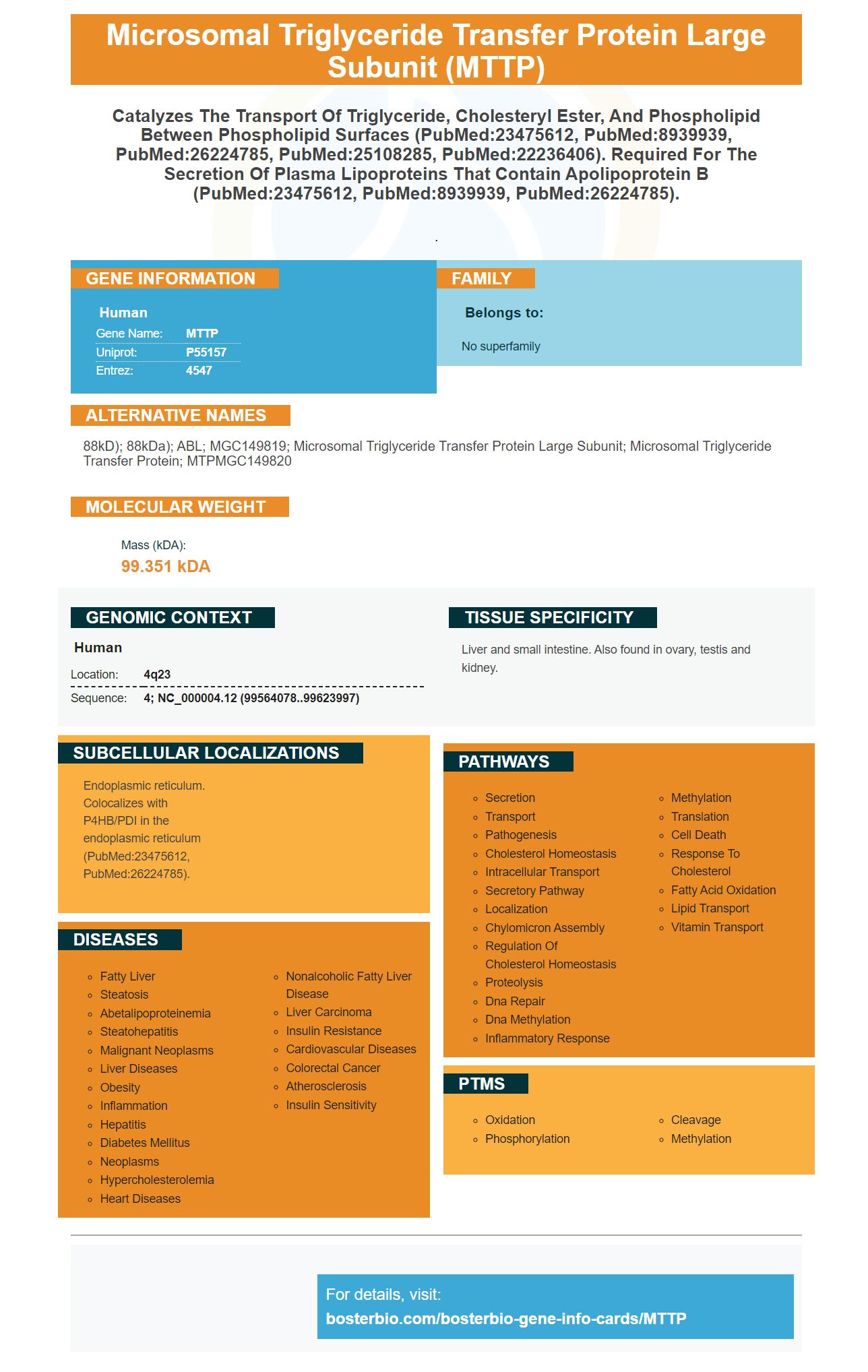
| Human | |
|---|---|
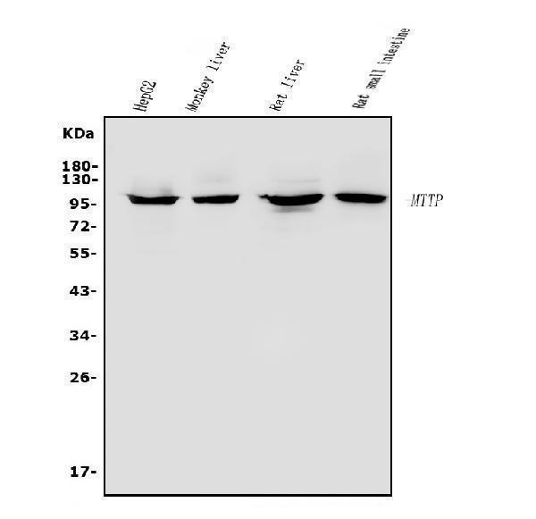
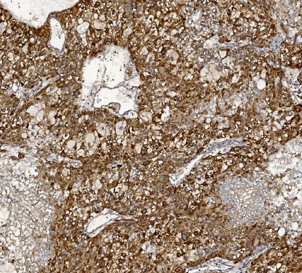
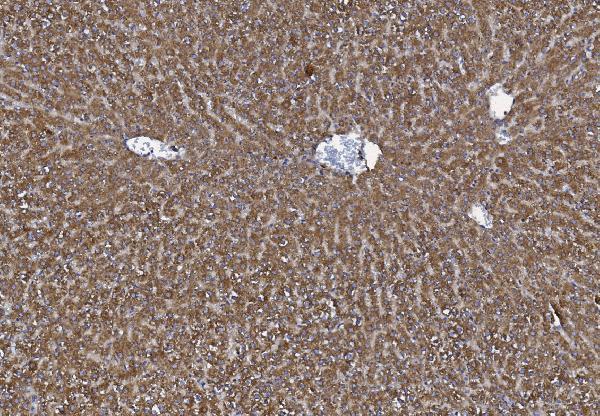
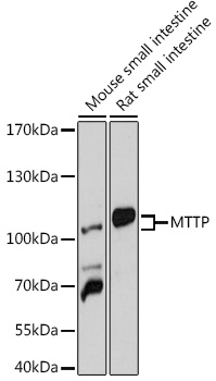
| Gene Name: | MTTP |
| Uniprot: | P55157 |
| Entrez: | 4547 |

| Belongs to: |
|---|
| No superfamily |

88kD); 88kDa); ABL; MGC149819; microsomal triglyceride transfer protein large subunit; microsomal triglyceride transfer protein; MTPMGC149820
Mass (kDA):
99.351 kDA

| Human | |
|---|---|
| Location: | 4q23 |
| Sequence: | 4; NC_000004.12 (99564078..99623997) |
Liver and small intestine. Also found in ovary, testis and kidney.
Endoplasmic reticulum. Colocalizes with P4HB/PDI in the endoplasmic reticulum (PubMed:23475612, PubMed:26224785).


PMID: 8111381 by Shoulders C.C., et al. Abetalipoproteinemia is caused by defects of the gene encoding the 97 kDa subunit of a microsomal triglyceride transfer protein.
PMID: 8361539 by Sharp D., et al. Cloning and gene defects in microsomal triglyceride transfer protein associated with abetalipoproteinaemia.