MVP / Major vault protein · IHC design guide

Design Immunohistochemistry for MVP

Plan chromogenic MVP IHC in paraffin sections using colon glandular cells and bronchial respiratory epithelium as positive tissue references (HPA tissue IHC). Compare cytoplasmic staining across consistently fixed sections, and assess unexpected signal in light of presumed off-target binding (HPA tissue IHC).

Evidence assembled Oct 2026 · For research use; verify linked source records and product datasheet before use
Immunohistochemistry protocol sheet for MVP (IHC for MVP): expected localisation Cytoplasmic, sometimes membranous in tissue (HPA tissue IHC), antibody M00642-1, validated IHC image, and IHC protocol steps
Printable MVP IHC protocol sheet — expected localisation Cytoplasmic, sometimes membranous in tissue (HPA tissue IHC), antibody M00642-1, controls and protocol steps. Open the full MVP IHC guide →

MVP Immunohistochemistry Experimental Design Guide

Expected localisation, validated protocols, controls and antibodies — the at-a-glance facts below, then the full design guide.

Must know before staining
Expected localisation Cytoplasmic, sometimes membranous in tissue (HPA tissue IHC)
Staining pattern Glandular/respiratory cells: cytoplasmic, sometimes membranous (HPA tissue IHC)
Antigen retrieval EDTA pH 8.0 HIER, heat-mediated (datasheet M00642-1)
Positive control ⓘ Appendix+4 more · see all
Negative control ⓘ Adipose tissue+4 more · see all
Important caveats
Reasons your staining may differ from the expected pattern.
Fixation Keep paraffin-section fixation consistent across samples. (standard IHC practice; not target-specific)
Caveat Presumed off-target binding can confound staining (HPA tissue IHC)
Regulation High in multidrug-resistant cancer cells (UniProt)
Isoform / epitope 0 isoforms; no transmembrane extracellular epitope (UniProt)
Section 1

Recommended MVP IHC & IF Protocols

The catalog antibody’s IHC-P protocol (datasheet: M00642-1) is paired with four published MVP staining workflows (PMC8210610; PMC6337862; PMC3493325; PMC8163044).

Recommended immunohistochemistry (IHC-P) protocol parameters
SampleParaffin-embedded mouse intestine tissue; fixative not specified (datasheet M00642-1)
FixationImage fixative and duration unreported (datasheet M00642-1); verify before use.
Sectioning4–5 µm sections on charged slides (standard)
DeparaffinisationXylene, graded ethanol series to water (standard)
Antigen retrievalHeat retrieval: EDTA pH 8.0 (datasheet M00642-1); 20 min, 95–100 °C (standard)
Peroxidase block3% H2O2, 10 min, room temperature (standard)
Blocking10% goat serum (datasheet M00642-1)
Primary antibodyMouse monoclonal (clone 8B12) anti-MVP, 0.5-1μg/ml (datasheet M00642-1)
Primary incubationOvernight at 4 °C (datasheet M00642-1)
DetectionStreptavidin-biotin complex (SABC), DAB chromogen (datasheet M00642-1)
CounterstainHematoxylin, blue, dehydrate and mount (standard)
Expected resultMVP-positive staining in glandular cells of appendix (HPA tissue IHC: High). HPA tissue profile: Cytoplasmic and cases of membranous expression in several tissues, most abundant in gastrointestinal and respiratory tract. No signal in the no-primary control.
💡Decision noteStart with heat-mediated EDTA pH 8.0 retrieval (datasheet: M00642-1); consider boiled citrate (PMC8210610) or CC1 retrieval (PMC8163044) when adapting those workflows.
Section 2

What Is the Expected MVP Staining Pattern?

In paraffin-section IHC, expect predominantly cytoplasmic MVP staining in gastrointestinal glandular and respiratory epithelial cells, with occasional membranous staining (HPA tissue IHC: Enhanced; medium staining–RNA consistency). MVP is also reported at the perinuclear region and nuclear pore complex; about 5% is found at nuclear pores (UniProt Q14764: subcellular location). It has no transmembrane segment (UniProt Q14764: topology).

What am I looking at on my slide?
Strong cytoplasmic staining in bronchial respiratory epithelium or colonic glandular cells.This matches a useful positive pattern: both cell populations are scored High by HPA (HPA tissue IHC: bronchus and colon). Compare staining within the expected cells, since intensity elsewhere in a section does not identify the cell producing the signal.
Predominantly nuclear staining, with little cytoplasmic signal in the expected positive cells.Treat this as a localisation mismatch and investigate staining artefact or nonspecific binding. UniProt places MVP mainly in the cytoplasm and reports a smaller nuclear-pore pool, about 5% (UniProt Q14764: subcellular location); HPA describes tissue staining as cytoplasmic with occasional membranous cases (HPA tissue IHC: profile).
Strong staining in adipocytes or skeletal myocytes while expected epithelial cells are weak.Question antibody specificity or endogenous detection activity before assigning this pattern to MVP. HPA reports MVP as Not detected in adipocytes and skeletal myocytes (HPA tissue IHC: adipose tissue and skeletal muscle). These comparisons are staining references, rather than proof that every such cell lacks MVP.
Diffuse colour across cells, stroma, or the section, without clear cell boundaries.The pattern cannot be scored confidently as MVP-positive. Check a no-primary control for nonspecific secondary or chromogen signal, assess blocking and washes, and compare expected cell populations (general IHC practice). HPA's reported tissue pattern is cell-associated, chiefly cytoplasmic (HPA tissue IHC: profile).
No signal in bronchial respiratory epithelium or intestinal glandular cells.First consider an assay failure or a sample-dependent loss of detectable staining, then review the antibody and detection workflow (general IHC practice). HPA scores bronchial respiratory epithelium and small-intestinal glandular cells High (HPA tissue IHC: bronchus and small intestine). One negative section cannot overturn those reference observations.
💡Expected MVP appearanceCall a result positive when clear, mainly cytoplasmic chromogenic signal is strong in bronchial respiratory epithelium or intestinal glandular cells (HPA tissue IHC: High); predominant nuclear staining or colour spread across unrelated cells warrants investigation (UniProt Q14764: subcellular location; general IHC practice).
How each factor affects the staining
Choice of positive and comparison cellsHPA scores glandular cells in appendix, colon, duodenum, rectum, and small intestine High, as well as respiratory epithelial cells in bronchus and nasopharynx (HPA tissue IHC). Adipocytes and skeletal myocytes are scored Not detected (HPA tissue IHC); use cell identity when interpreting either result.
Subcellular distributionPredominantly cytoplasmic signal fits both the tissue profile and UniProt location (HPA tissue IHC: profile; UniProt Q14764: subcellular location). HPA also describes cases of membranous staining, while UniProt reports perinuclear and minor nuclear-pore localisation; an isolated edge or nuclear signal needs context rather than automatic acceptance.
Evidence behind the tissue patternHPA labels tissue IHC reliability Enhanced but describes only medium consistency with RNA expression and says presumed off-target binding was disregarded (HPA tissue IHC: reliability). Its antibody table lists four antibodies with Enhanced IHC status (HPA antibodies). Those summaries support reference patterns, not specificity of an untested assay.
Topology and antigen processingMVP has no transmembrane segment, no signal peptide, no propeptide, and a reported major protein chain spanning residues 2–893 (UniProt Q14764: topology and processing). These annotations provide no basis to expect a shed extracellular staining pattern or to predict retrieval sensitivity; evaluate the observed cellular pattern directly.
Why is my staining missing, weak or wrong?
SituationLikely causeNext action
Expected positive epithelial cells remain blank.Primary antibody, retrieval, or chromogenic detection may be ineffective in this run (general IHC practice).Run a known-positive bronchus or intestinal section alongside the sample (HPA tissue IHC: High in bronchial respiratory epithelium and intestinal glandular cells). Check the catalog antibody's IHC-P instructions, retrieval step, and detection reagents before interpreting the sample as negative.
Colour covers the section with little cell-specific contrast.Nonspecific reagent binding, inadequate washing, or endogenous detection activity can obscure localisation (general IHC practice).Compare a no-primary control, strengthen appropriate blocking or washing, and review endogenous-activity blocking for the detection chemistry (general IHC practice). Score MVP only when signal resolves in the expected cells and cytoplasmic compartment (HPA tissue IHC: profile).
Staining is mainly nuclear.The pattern exceeds the limited nuclear-pore localisation reported for MVP (UniProt Q14764: about 5% at nuclear pores).Review counterstain, chromogen localisation, and no-primary control; then compare with a positive tissue (general IHC practice). Predominant cytoplasmic staining is the reference pattern (HPA tissue IHC: profile).
Adipocytes or skeletal myocytes stain as strongly as expected positive cells.Cross-reactivity or background is possible; HPA scores these cell types Not detected (HPA tissue IHC: adipose tissue and skeletal muscle).Check a no-primary control and compare staining across cell populations on the same run (general IHC practice). Do not use the negative-reference populations alone to establish antibody specificity.
A narrow cell-edge signal appears alongside cytoplasmic staining.Some membranous staining is reported in tissue, although MVP lacks a transmembrane segment (HPA tissue IHC: profile; UniProt Q14764: topology).Assess whether the expected epithelial cells also show cytoplasmic signal; investigate edge-only staining with controls before calling it a specific MVP pattern (general IHC practice).
Can ICC-IF localisation guide this IHC interpretation?HPA ICC-IF reports MVP in the cytosol with enhanced localisation evidence (HPA subcellular: Cytosol).Use that result as a localisation cross-check, while judging paraffin-section staining against HPA tissue IHC cell patterns (HPA tissue IHC: profile). Follow the separate IF/ICC guide for that application.

Sample controls for MVP IHC & IF

🧪Run colon first: glandular cells should stain (HPA: High in colon glandular cells). Use adipose tissue as a negative tissue, focusing on adipocytes (HPA: Not detected in adipocytes); on the colon slide, compare glandular staining with adjacent cells that lack visible signal, treating those cells as a background reference rather than assuming they lack MVP.
Positive control tissue: Appendix (Glandular cells, HPA High)
Negative control tissue: Adipose tissue (HPA Not detected)
ICC-IF cell lines (HPA subcellular resource): HPA ICC-IF images show MVP in A-431, U2OS, U-251MG, with annotated localisation: Cytosol (enhanced) (HPA subcellular).
Technical controls: Include a no-primary (secondary-only) control, an isotype-matched mouse IgG control, and an identically processed MVP-knockout specimen if available (selected IHC caption: mouse anti-MVP antibody). For the DAB assay, block endogenous peroxidase and check for endogenous biotin background because the selected IHC method uses a biotinylated secondary and streptavidin-biotin detection (selected IHC caption).
⚠️Feasibility: A target-specific fixation window or fixation effect is unreported, and the exact selected-SKU tissue-IHC caption does not state the fixative (selected IHC caption). Heat retrieval in EDTA at pH 8.0 was used for a mouse intestine paraffin section, but retrieval dependency is unreported (selected IHC caption). Neither frozen sections nor IF/ICC is established as easier by the supplied evidence; for intestinal tissue, check whether endogenous biotin contributes to background with the captioned detection method (selected IHC caption).

HPA tissue IHC evidence for MVP

Comprehensive Human Protein Atlas IHC scoring per tissue (reliability: Enhanced — Medium consistency between antibody staining and RNA expression data. Presumed off target binding observed and disregarded.). Rows are taken directly from the HPA tissue chart — click any row's HPA link to view the source.

Positive expression · recommended positive controls

TissueCell typeLevelEvidenceSource
Appendix Glandular cells High Protein (IHC) HPA →
Bronchus Respiratory epithelial cells High Protein (IHC) HPA →
Colon Glandular cells High Protein (IHC) HPA →
Duodenum Glandular cells High Protein (IHC) HPA →
Lung Alveolar cells type II High Protein (IHC) HPA →

Undetected expression · recommended negative controls

TissueCell typeLevelEvidenceSource
Adipose tissue Adipocytes Not detected Protein (IHC) HPA →
Breast Adipocytes Not detected Protein (IHC) HPA →
Caudate Glial cells Not detected Protein (IHC) HPA →
Esophagus Squamous epithelial cells Not detected Protein (IHC) HPA →
Heart muscle Cardiomyocytes Not detected Protein (IHC) HPA →
Section 3

Advanced MVP IHC Tips

Use the documented paraffin-section workflow as the starting point, then judge staining against MVP’s expected cell types and compartments.

How should I retrieve MVP in paraffin sections when staining is weak?
Start MVP paraffin-section IHC with heat-mediated retrieval in EDTA at pH 8.0 (datasheet M00642-1). The selected tissue image used this retrieval before 10% goat-serum blocking and 1 µg/ml primary antibody overnight at 4°C (datasheet M00642-1). If staining is weak, compare retrieval duration across adjacent sections while holding antibody concentration, detection and development constant (standard IHC practice). Include a known positive epithelial region and a primary-antibody omission control; compare cellular staining with background before increasing retrieval intensity, since excess heating can impair section morphology (HPA: high in small-intestinal glandular cells; standard IHC practice).
Could fixation explain inconsistent MVP staining between paraffin blocks?
The selected paraffin-section caption does not report a fixative, and no target-specific fixation comparison is supplied; MVP fixation sensitivity is therefore unknown (datasheet M00642-1). Record the fixative, fixation duration and processing history for each block before attributing a staining difference to biology (standard IHC practice). Compare sections from similarly processed blocks using the documented EDTA retrieval at pH 8.0 and the same antibody and detection conditions (datasheet M00642-1; standard IHC practice). Check morphology and a consistently stained reference section in each run, because uneven processing can complicate comparisons of staining intensity across blocks (standard IHC practice).
Where should MVP staining appear in a convincing IHC section?
Expect predominantly cytoplasmic staining, especially in epithelial populations of the gastrointestinal and respiratory tracts (HPA: tissue IHC profile). HPA reports high staining in small-intestinal glandular cells and bronchial respiratory epithelial cells, useful reference populations when assessing a run (HPA: tissue IHC). UniProt also places a minority of MVP at the nuclear pore complex, approximately 5%, so limited nuclear-rim signal is biologically plausible but should not dominate interpretation (UniProt Q14764: subcellular location). Score cytoplasmic and nuclear-rim patterns separately, and check any apparent membrane staining against morphology and background controls (HPA: cases of membranous expression; standard IHC practice).
Could an isoform or hidden epitope explain discordant MVP IHC?
The supplied record lists 0 MVP isoforms, so an annotated isoform difference does not explain discordant staining in this guide (UniProt Q14764: isoforms). MVP has no transmembrane segment and assembles into a large vault particle, which makes antibody access within fixed tissue worth evaluating (UniProt Q14764: topology and subunit). The catalog antibody’s binding epitope is unspecified here; do not assign its signal to the shoulder domain or to the reported phosphoserine at residue 445 (UniProt Q14764: domains and modified residues; supplied antibody evidence). Compare retrieval conditions and an independent antibody with a documented epitope, where available, before proposing an epitope-specific explanation (standard IHC practice).
How can IF help assess the cellular pattern seen by MVP IHC?
For the separate IF/ICC workflow, multiplex MVP with a marker identifying the epithelial cell population being examined, then assess signal within individual cells (HPA: high in gastrointestinal glandular and respiratory epithelial cells; standard IF practice). Select fluorophores after checking tissue autofluorescence in an unstained section; a longer-wavelength channel can help when shorter-wavelength background is prominent (standard IF practice). MVP is cytosolic and has no transmembrane segment, so permeabilisation should permit access to its intracellular epitope, whose precise position is unspecified here (HPA: cytosol; UniProt Q14764: topology; supplied antibody evidence). Include single-label controls for spectral bleed-through and interpret IF alongside the paraffin-section IHC pattern without treating the IHC caption as IF validation (standard IF practice; datasheet M00642-1).
How do I reduce diffuse or misleading MVP IHC background?
First compare the stained section with a primary-antibody omission control and inspect whether colour follows tissue structures rather than cells (standard IHC practice). The documented workflow blocked with 10% goat serum, used a biotinylated secondary antibody and developed with a streptavidin–biotin complex and DAB (datasheet M00642-1). Check peroxidase blocking and, for this biotin-based detection workflow, assess endogenous biotin as possible sources of unwanted colour (standard IHC practice). HPA reports presumed off-target binding in its tissue assessment, so require reproducible cytoplasmic staining in expected epithelial cells before calling a diffuse signal MVP-positive (HPA: reliability description and tissue IHC profile).
What should I measure when comparing MVP staining across sections? ⚠ ANSWER MARKED FOR VERIFICATION
Define the cell population and tissue region before scoring, then record the percentage of positive cells and staining intensity for a cell-based H-score (standard IHC practice). Keep scoring criteria, counterstain, imaging settings and DAB development consistent across the compared sections (standard IHC practice). Normalise positive-cell counts to the number of eligible cells, or positive area to the measured region area in mm², rather than to the whole section when epithelial content varies (standard IHC practice). Report cytoplasmic staining separately from rare nuclear-rim observations, and compare like cell types because HPA shows substantial differences among tissue populations (UniProt Q14764: subcellular location; HPA: tissue IHC).
When is apparent MVP positivity likely to be artefactual?
A convincing result shows cellular cytoplasmic staining in an appropriate population, such as intestinal glandular or bronchial respiratory epithelial cells (HPA: tissue IHC profile). Treat dominant nuclear staining with caution: UniProt places only about 5% of MVP at the nuclear pore complex, while HPA’s enhanced subcellular assignment is cytosolic (UniProt Q14764: subcellular location; HPA: subcellular). Check section edges, necrotic regions and the primary-antibody omission control for staining that tracks damage or persists without primary antibody (standard IHC practice). Also assess endogenous peroxidase or biotin signal in the documented DAB and streptavidin–biotin workflow before interpreting unexpected cells as MVP-positive (datasheet M00642-1; standard IHC practice).
Boster reagents

Best MVP / Major vault protein IHC Antibodies

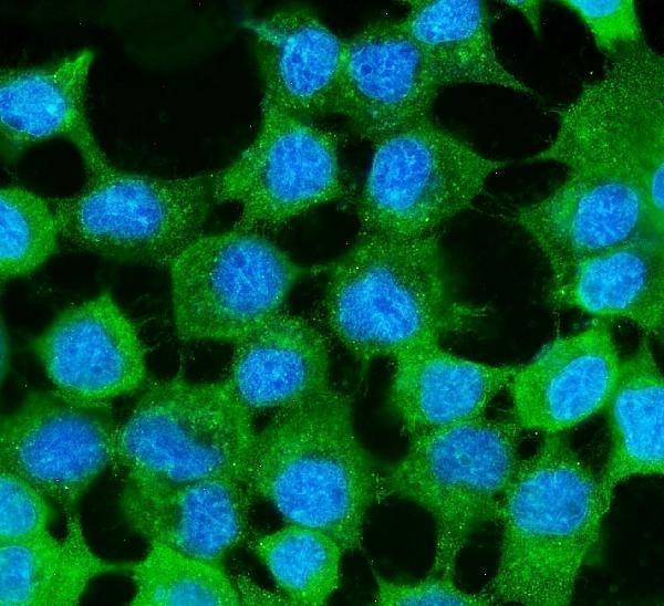
M00642-1 has IHC images from paraffin sections of human, mouse and rat tissue, plus an IF image from A431 cells (catalog image captions).

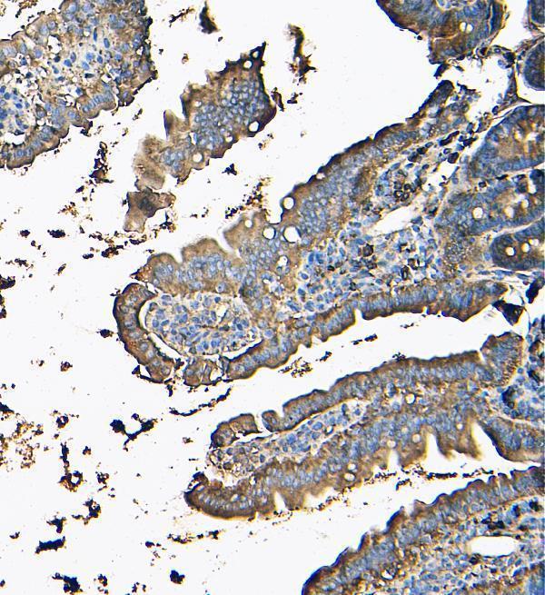
Real IHC data IHC analysis of MVP using anti-MVP antibody (M00642-1). MVP was detected in paraffin-embedded section of mouse intestine tissue. Heat mediated antigen retrieval was performed in EDTA buffer (pH8.0, epitope retrieval solution). The tissue section was blocked with 10% goat serum. The tissue section was then incubated with 1μg/ml mouse anti-MVP Antibody (M00642-1) overnight at 4°C. Biotinylated goat anti-mouse IgG was used as secondary antibody and incubated for 30 minutes at 37°C. The tissue section was developed using Strepavidin-Biotin-Complex (SABC) (Catalog # SA1021) with DAB as the chromogen.
Anti-MVP Antibody ® (monoclonal, 8B12)
Cat # M00642-1

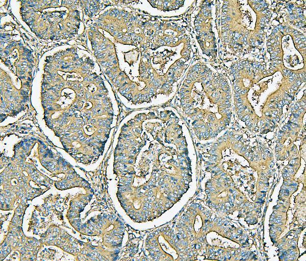
M00642-1 will render with an IHC figure from mouse intestine (catalog IHC caption). Its other IHC images show human colon and mammary cancer and rat intestine; its IF image shows A431 cells (catalog image captions).

Which to pick: For chromogenic IHC on paraffin sections, choose M00642-1: its IHC captions show EDTA pH 8 retrieval, 1 μg/ml primary antibody and DAB detection; the fixative is unreported (catalog IHC captions). For IF/ICC, M00642-1 has an A431 cell IF image and lists both applications (catalog IF caption; catalog applications). It is also the cross-species choice among the rendered cards, with human, mouse and rat reactivity and IHC images for all three species (catalog reactivity; catalog IHC captions).

Each figure is that product's own IHC / IF validation image from its datasheet.

References

  1. UniProt Consortium. UniProt entry Q14764 (MVP_HUMAN, Major vault protein).
  2. Human Protein Atlas. MVP tissue IHC expression (reliability: Enhanced).
  3. Human Protein Atlas. MVP subcellular location (ICC-IF): Localized to the cytosol..
  4. Human Protein Atlas. MVP antibody validation summary (4 antibodies).
  5. Major vault protein (MVP) negatively regulates osteoclastogenesis via calcineurin-NFATc1 pathway inhibition. Theranostics 2021 — PMC8210610.
  6. Adipocytes promote breast cancer resistance to chemotherapy, a process amplified by obesity: role of the major vault protein (MVP). Breast cancer research : BCR 2019 — PMC6337862.
  7. MVP expression in the prediction of clinical outcome of locally advanced oral squamous cell carcinoma patients treated with radiotherapy. Radiation oncology (London, England) 2012 — PMC3493325.
  8. Proteomic analyses identify major vault protein as a prognostic biomarker for fatal prostate cancer. Carcinogenesis 2021 — PMC8163044.
  9. PubMed PMID:7585126 — UniProt-cited evidence.
  10. PubMed PMID:15489334 — UniProt-cited evidence.
  11. PubMed PMID:11071864 — UniProt-cited evidence.