This website uses cookies to ensure you get the best experience on our website.


- Table of Contents
Plan MYBPC3 staining in paraffin sections using heart cardiomyocyte cytoplasmic staining as the expected pattern (HPA tissue IHC). This guide covers tissue controls and interpretation alongside the catalog antibody’s IHC dilution of 0.5–1 μg/ml (datasheet A01078-1).

Expected localisation, validated protocols, controls and antibodies — the at-a-glance facts below, then the full design guide.
| Expected localisation | Cardiomyocyte cytoplasm (HPA tissue IHC) | |
| Staining pattern | Heart cardiomyocytes show cytoplasmic staining (HPA tissue IHC) | |
| Antigen retrieval | Citrate pH 6 HIER, heat-mediated (datasheet A01078-1) | |
| Positive control | Heart muscle+1 more · see all | |
| Negative control | Adipose tissue+4 more · see all |
| Fixation | Keep fixation consistent across sections (standard IHC practice; not target-specific) | |
| Caveat | Testis spermatogonia also stain strongly (HPA tissue IHC) | |
| Regulation | Expression regulation not specified (UniProt) | |
| Isoform / epitope | 2 isoforms; check epitope coverage (UniProt) |
The catalog antibody's IHC-P protocol (datasheet: A01078-1) is followed by a published MYBPC3 IHC protocol using paraffin-embedded left ventricle (PMC8924128).
| Sample | Paraffin-embedded rat heart tissue; fixative not specified (datasheet A01078-1) |
| Fixation | Image fixative and duration unreported (datasheet A01078-1); verify before use. |
| Sectioning | 4–5 µm sections on charged slides (standard) |
| Deparaffinisation | Xylene, graded ethanol series to water (standard) |
| Antigen retrieval | Heat retrieval: Citrate pH 6, 20 min (datasheet A01078-1) |
| Peroxidase block | 3% H2O2, 10 min, room temperature (standard) |
| Blocking | 10% goat serum (datasheet A01078-1) |
| Primary antibody | Rabbit anti-MYBPC3, 0.5-1μg/ml (datasheet A01078-1) |
| Primary incubation | Overnight at 4 °C (datasheet A01078-1) |
| Detection | Streptavidin-biotin complex (SABC), DAB chromogen (datasheet A01078-1) |
| Counterstain | Hematoxylin, blue, dehydrate and mount (standard) |
| Expected result | MYBPC3-positive staining in cardiomyocytes of heart muscle (HPA tissue IHC: High). HPA tissue profile: Highly selective cytoplasmic expression in heart myocytes. No signal in the no-primary control. |
MYBPC3 is a thick filament associated protein of striated muscle A bands and has no transmembrane segment (UniProt Q14896: function, topology). In paraffin sections, expect strong cytoplasmic staining in heart cardiomyocytes (HPA: High in cardiomyocytes). HPA describes the tissue profile as highly selective for heart myocytes and rates its IHC evidence Enhanced because antibody staining agrees closely with RNA expression (HPA: tissue IHC reliability).
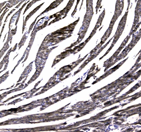
| Strong cytoplasmic staining follows the cardiomyocyte fibers in heart muscle. | This fits the reported high cardiomyocyte signal (HPA: High in cardiomyocytes) and MYBPC3’s association with striated muscle A bands (UniProt Q14896: function). Fine sarcomeric detail may depend on section orientation and image resolution (general IHC practice); its absence alone does not overturn a convincing cytoplasmic pattern. |
| Signal is concentrated in nuclei, along cell surfaces, or in extracellular material. | Those compartments do not fit the expected cardiomyocyte cytoplasmic profile (HPA: tissue IHC profile) or a protein with no transmembrane segment (UniProt Q14896: topology). Treat the pattern as possible artefact and compare it with the negative control before assigning MYBPC3 positivity (general IHC practice). |
| Prominent staining appears in an unexpected cell population or a reported negative tissue. | Consider cross-reactivity or endogenous detection activity (general IHC practice). HPA reports no detection in adipocytes of adipose tissue and glandular cells of adrenal gland, but reports high staining in testis spermatogonia (HPA: tissue IHC). Therefore, testis signal alone is not evidence of a false positive. |
| Brown precipitate covers many structures without a clear cardiomyocyte pattern. | Diffuse background prevents a reliable cell specific call (general IHC practice). Review the negative control and whether background follows tissue edges, damaged areas, or multiple compartments (general IHC practice). A widespread haze should not be scored as the selective heart myocyte staining reported by HPA (HPA: tissue IHC profile). |
| Heart cardiomyocytes show no detectable signal. | A negative result in the expected positive tissue conflicts with HPA’s high cardiomyocyte staining (HPA: High in cardiomyocytes). Check that cardiomyocytes are present and assess the control, antibody dilution, retrieval, and detection steps before interpreting the sample as MYBPC3 negative (general IHC practice). |
| Target location and topology | MYBPC3 is associated with thick filaments in striated muscle A bands and has no transmembrane segment (UniProt Q14896: function, topology). Interpret the expected signal within cardiomyocyte cytoplasm, rather than as a membrane outline (HPA: tissue IHC profile). |
| Tissue context | Heart muscle is RNA tissue enriched, and cardiomyocytes stain at a high level (HPA: tissue IHC). High staining is also listed for testis spermatogonia (HPA: tissue IHC); use the named cell population when interpreting that tissue. |
| Isoforms and modified residues | UniProt lists two isoforms and multiple modified residues, including phosphorylated serines (UniProt Q14896: isoforms, modified residues). The supplied sources do not map the IHC antibody epitopes, so they do not establish whether either feature changes staining. |
| Evidence for the IHC pattern | HPA rates the tissue IHC profile Enhanced and lists two rabbit antibodies with Enhanced IHC status (HPA: tissue IHC reliability; HPA: HPA040147, HPA043898). This supports the reported tissue pattern, without establishing a specific retrieval condition or fixation sensitivity. |
| IF/ICC evidence limit | HPA provides no main subcellular location or cell line ICC/IF images for this target (HPA: subcellular record). The cytoplasmic IHC expectation should not be presented as a validated IF/ICC pattern; consult the separate IF/ICC guide for that application. |
| Situation | Likely cause | Next action |
|---|---|---|
| Heart cardiomyocytes are blank while the negative control is clean. | The expected positive cell population has not been demonstrated (HPA: High in cardiomyocytes); a processing, retrieval, antibody, or detection step may have failed (general IHC practice). | Confirm cardiomyocytes are present, then check reagent activity, antibody dilution, retrieval, and detection against the catalog antibody’s IHC-P instructions (general IHC practice). Do not infer MYBPC3 absence from this slide alone. |
| Signal is mostly nuclear or outlines cardiomyocyte membranes. | This differs from the reported cytoplasmic profile (HPA: tissue IHC profile) and from the absence of a transmembrane segment (UniProt Q14896: topology); background or mislocalised detection is possible (general IHC practice). | Compare with a negative control and a heart positive control, then review blocking, washes, and detection background (general IHC practice). Score only a convincing cardiomyocyte cytoplasmic pattern. |
| A reported negative tissue stains strongly. | Cross-reactivity or endogenous detection activity may contribute (general IHC practice). HPA reports no detection in adipose tissue adipocytes and adrenal gland glandular cells (HPA: tissue IHC). | Check the stained cell identity against HPA’s cell level entries, inspect a negative control, and review detection blocking (general IHC practice). Do not use testis spermatogonia as a presumed negative control: HPA reports high staining there (HPA: tissue IHC). |
| Brown haze obscures cell boundaries across the section. | Diffuse reagent background or endogenous detection activity can reduce contrast (general IHC practice); such a pattern does not match selective cardiomyocyte cytoplasmic staining (HPA: tissue IHC profile). | Inspect negative controls, tissue condition, blocking, wash stringency, and detection development (general IHC practice). Reassess only after the background permits a clear cell level call. |
| Heart signal is weak or uneven despite visible cardiomyocytes. | Section quality, retrieval, antibody concentration, or detection may contribute (general IHC practice). The supplied sources give no MYBPC3 specific fixation effect or optimal retrieval setting. | Compare matched heart sections and controls, then follow the catalog antibody’s IHC-P retrieval and dilution instructions (general IHC practice). Avoid attributing the result to MYBPC3 epitope masking without supporting evidence. |
| An IF/ICC image is being used to validate the paraffin IHC result. | HPA lists no ICC/IF cell line images or main subcellular location for MYBPC3 (HPA: subcellular record); its Enhanced rating here concerns IHC tissue evidence (HPA: tissue IHC reliability). | Judge the paraffin section against the heart cardiomyocyte IHC pattern and its controls (HPA: tissue IHC; general IHC practice). Evaluate IF/ICC separately using application specific evidence. |


Comprehensive Human Protein Atlas IHC scoring per tissue (reliability: Enhanced — High consistency between antibody staining and RNA expression data.). Rows are taken directly from the HPA tissue chart — click any row's HPA link to view the source.
| Tissue | Cell type | Level | Evidence | Source |
|---|---|---|---|---|
| Heart muscle | Cardiomyocytes | High | Protein (IHC) | HPA → |
| Testis | Spermatogonia cells | High | Protein (IHC) | HPA → |
| Tissue | Cell type | Level | Evidence | Source |
|---|---|---|---|---|
| Adipose tissue | Adipocytes | Not detected | Protein (IHC) | HPA → |
| Adrenal gland | Glandular cells | Not detected | Protein (IHC) | HPA → |
| Appendix | Glandular cells | Not detected | Protein (IHC) | HPA → |
| Bone marrow | Hematopoietic cells | Not detected | Protein (IHC) | HPA → |
| Breast | Adipocytes | Not detected | Protein (IHC) | HPA → |
Troubleshoot MYBPC3 chromogenic IHC in paraffin sections using the catalog antibody’s tissue image, tissue expression data, and the protein’s known structure and function.
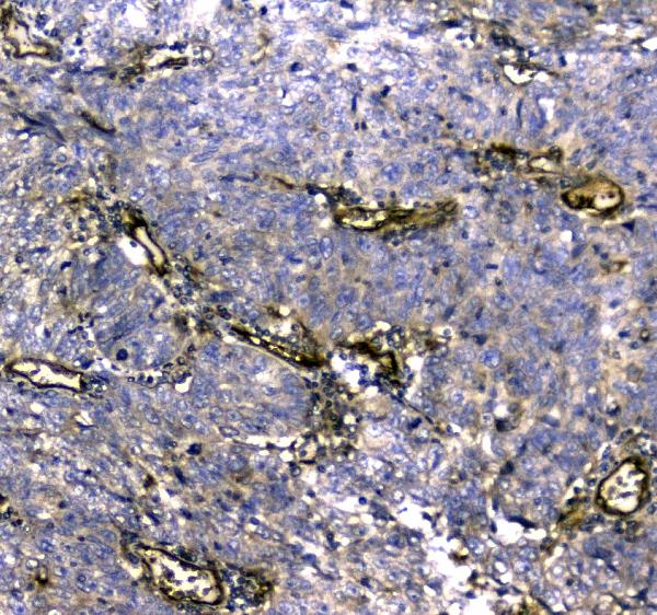
A01078-1 has real IHC images of paraffin sections from rat heart and human lung, mammary, and rectal cancer tissues (A01078-1 IHC image captions). No IF image is provided (catalog IF image alts).
A01078-1 is listed for IHC and has images from rat heart and human lung, mammary, and rectal cancer paraffin sections (catalog applications; A01078-1 IHC image captions). Its listed reactivity is human, mouse, and rat, while the supplied IHC images show human and rat tissue (catalog reactivity; A01078-1 IHC image captions).
Which to pick: For tissue IHC, choose A01078-1 for paraffin sections matching its image evidence; the captions do not report the fixative (A01078-1 IHC image captions). For IF/ICC, no listed SKU has an IF/ICC application or IF image (catalog applications; catalog IF image alts). For work across species, A01078-1 is a rabbit polyclonal with listed human, mouse, and rat reactivity; its supplied IHC images show human and rat tissue (catalog host and dilution_raw; catalog reactivity; A01078-1 IHC image captions).