MYDGF / Myeloid-derived growth factor · IHC design guide

Design Immunohistochemistry for MYDGF

Plan MYDGF chromogenic IHC on paraffin sections using the reported cytoplasmic tissue pattern to interpret staining (HPA tissue IHC). The catalog antibody has an IHC-P working range of 2–5 μg/mL (datasheet A08602).

Evidence assembled Oct 2026 · For research use; verify linked source records and product datasheet before use
Immunohistochemistry protocol sheet for MYDGF (IHC for MYDGF): expected localisation Cytoplasmic staining in most tissues (HPA tissue IHC), antibody A08602, validated IHC image, and IHC protocol steps
Printable MYDGF IHC protocol sheet — expected localisation Cytoplasmic staining in most tissues (HPA tissue IHC), antibody A08602, controls and protocol steps. Open the full MYDGF IHC guide →

MYDGF Immunohistochemistry Experimental Design Guide

Expected localisation, validated protocols, controls and antibodies — the at-a-glance facts below, then the full design guide.

Must know before staining
Expected localisation Cytoplasmic staining in most tissues (HPA tissue IHC)
Staining pattern Cytoplasmic staining in most tissues, including immune cells (HPA tissue IHC)
Antigen retrieval EDTA pH 8.0 HIER, heat-mediated (datasheet A08602)
Positive control ⓘ Placenta+4 more · see all
Negative control ⓘ Caudate+4 more · see all
Important caveats
Reasons your staining may differ from the expected pattern.
Fixation Keep fixation consistent across sections (standard IHC practice; not target-specific)
Caveat Secreted MYDGF may appear away from producing cells (UniProt)
Regulation Stimulus-linked changes unreported (UniProt)
Isoform / epitope No isoforms annotated; signal peptide 1–31 is cleaved (UniProt)
Section 1

Recommended MYDGF IHC & IF Protocols

The catalog antibody has an IHC-P protocol (datasheet A08602). The published options below cover fish retinal sections (PMC12346886) and human renal tissue (PMC12176760).

Recommended immunohistochemistry (IHC-P) protocol parameters
SampleParaffin-embedded human colorectal adenocarcinoma tissue; fixative not specified (datasheet A08602)
FixationImage fixative and duration unreported (datasheet A08602); verify before use.
Sectioning4–5 µm sections on charged slides (standard)
DeparaffinisationXylene, graded ethanol series to water (standard)
Antigen retrievalHeat retrieval: EDTA pH 8.0 (datasheet A08602); 20 min, 95–100 °C (standard)
Peroxidase block3% H2O2, 10 min, room temperature (standard)
Blocking10% goat serum (datasheet A08602)
Primary antibodyRabbit anti-MYDGF, 2-5 μg/ml (datasheet A08602)
Primary incubationOvernight at 4 °C (datasheet A08602)
DetectionHRP-conjugated secondary, DAB chromogen (datasheet A08602)
CounterstainHematoxylin, blue, dehydrate and mount (standard)
Expected resultMYDGF-positive staining in decidual cells of placenta (HPA tissue IHC: High). HPA tissue profile: Cytoplasmic expression in most tissues, including immune cells. No signal in the no-primary control.
💡Decision noteStart with EDTA pH 8.0 heat retrieval for the catalog antibody (datasheet A08602); the published protocols used citrate retrieval (PMC12346886; PMC12176760).
Section 2

What Is the Expected MYDGF Staining Pattern?

MYDGF should show predominantly cytoplasmic staining across many tissues, including immune cells (HPA tissue IHC: Approved; medium consistency). High staining is reported in placental decidual cells; medium staining occurs in bone marrow hematopoietic cells and several glandular cell populations (HPA tissue IHC). MYDGF is secreted, has no transmembrane segment, and also localizes to the ER and Golgi pathway (UniProt Q969H8).

What am I looking at on my slide?
Cytoplasmic signal in placental decidual cells, with weaker or variable staining elsewhere (HPA tissue IHC).This fits the reported High decidual cell signal and broad cytoplasmic profile (HPA tissue IHC). Compare staining within identified cell populations; HPA reports Medium staining in bone marrow hematopoietic cells and selected glandular cells (HPA tissue IHC).
Strong nuclear-only staining or a crisp, continuous plasma-membrane rim (UniProt Q969H8 topology).That pattern is difficult to reconcile with a secreted protein lacking a transmembrane segment (UniProt Q969H8). Review morphology and controls before calling it MYDGF; HPA tissue IHC supports cytoplasmic expression but does not establish a precise organelle pattern (HPA tissue IHC).
Apparent signal in cell populations reported as Not detected, such as glomerular cells or skeletal myocytes (HPA tissue IHC).Treat these as cell-specific discrepancies, not proof that an entire organ must be negative (HPA tissue IHC). Cross-reactivity or endogenous chromogen-generating activity are possible technical explanations (general IHC practice); assess the matched negative control and staining distribution.
Uniform haze over cells and extracellular spaces, obscuring cell boundaries (general IHC practice).Broad background can arise from nonspecific binding or detection chemistry (general IHC practice). MYDGF is secreted, so extracellular material alone cannot be ruled out biologically (UniProt Q969H8); require a convincing cellular pattern and clean controls before interpreting it.
No signal in placental decidual cells despite preserved tissue morphology (HPA tissue IHC: High).A negative known-positive population makes this run inconclusive for MYDGF (HPA tissue IHC; general IHC practice). Check antibody application, retrieval and detection against the validated IHC procedure (general IHC practice); absence here does not establish biological absence elsewhere.
💡Expected MYDGF appearanceCall positive when identifiable decidual cells show convincing cytoplasmic chromogen, potentially High in placenta (HPA tissue IHC); isolated nuclear staining, a sharp membrane rim, or uniform slide-wide haze warrants control review (UniProt Q969H8 topology; general IHC practice).
How each factor affects the staining
Secretory localization and topology (UniProt Q969H8).A signal peptide and absent transmembrane segment support intracellular secretory-pathway and possible extracellular distribution (UniProt Q969H8). Do not require a membrane outline; HPA's tissue-level IHC description is cytoplasmic (HPA tissue IHC).
Protein processing (UniProt Q969H8).The precursor has a signal peptide at residues 1–31; the annotated mature chain spans 32–173 (UniProt Q969H8). Interpret staining according to the antibody's documented epitope, if available; this payload provides none.
Tissue comparison strength (HPA tissue IHC).HPA rates tissue staining Approved with medium RNA–protein consistency and cautions that secretion can separate protein from RNA location (HPA tissue IHC). Use cell-level IHC observations as reference patterns, not a strict RNA-based pass/fail rule.
IHC antibody evidence (HPA antibodies).HPA041872 and HPA046744 each have Approved IHC status (HPA antibodies). The supplied status does not say IHC Enhanced; independent concordance or orthogonal confirmation should not be claimed from these entries alone.
IF/ICC interpretation question (HPA subcellular; HPA antibodies).Can IHC define an IF/ICC pattern? No: HPA lists MYDGF as secreted, gives no main ICC location or image-bearing cell line, and lists no ICC status for either antibody (HPA subcellular; HPA antibodies). Evaluate IF/ICC on its own guide page.
Why is my staining missing, weak or wrong?
SituationLikely causeNext action
Placental decidual cells stain weakly or remain blank (HPA tissue IHC: High).A run-level staining failure is possible when a reported positive population is absent (HPA tissue IHC; general IHC practice).Confirm tissue identity and morphology, then check the validated antibody dilution, retrieval, incubation and detection steps for this IHC run (general IHC practice).
Staining is mainly nuclear or forms a continuous membrane rim (UniProt Q969H8 topology).This conflicts with the reported cytoplasmic IHC profile and lacks support from a transmembrane segment (HPA tissue IHC; UniProt Q969H8).Inspect a matched negative control and repeat with the IHC-validated antibody; score only reproducible signal in morphologically identified cells (general IHC practice).
Glomerular cells or skeletal myocytes appear positive (HPA tissue IHC: Not detected).Cell identity, nonspecific binding or endogenous detection activity may account for the discrepancy (general IHC practice).Verify the cell population and compare a matched negative control; optimize blocking or endogenous activity quenching as appropriate for the detection system (general IHC practice).
Diffuse chromogen obscures the expected cell-level pattern (general IHC practice).Excess nonspecific staining or detection background is possible (general IHC practice); secretion also complicates interpretation of extracellular signal (UniProt Q969H8).Check negative controls, washing, antibody concentration and chromogen development; judge the result against identifiable cytoplasmic staining (general IHC practice; HPA tissue IHC).
A weak signal in lung macrophages is mistaken for a failed positive control (HPA tissue IHC: Low).HPA reports Low macrophage staining in lung, while placental decidual cells are High (HPA tissue IHC).Use the reported decidual cell pattern for a stronger tissue comparison; evaluate macrophages by cell identity and local background (HPA tissue IHC; general IHC practice).
A field labeled kidney is called wholly negative because glomerular cells lack stain (HPA tissue IHC).The Not detected entry applies specifically to cells in glomeruli, not every renal cell (HPA tissue IHC).Record the compartment and cell population scored; avoid extending a cell-specific HPA observation to the entire section (HPA tissue IHC).

Sample controls for MYDGF IHC & IF

🧪Run placenta first and look for MYDGF staining in decidual cells (HPA: High in placental decidual cells). Use caudate glial cells as a negative tissue control (HPA: Not detected in caudate glial cells); on the placenta slide, adjacent non-decidual areas should show only background-level signal, without assuming every non-decidual cell is MYDGF-negative (standard IHC interpretation).
Positive control tissue: Placenta (Decidual cells, HPA High)
Negative control tissue: Caudate (HPA Not detected)
ICC-IF cell lines (HPA subcellular resource): HPA carries no ICC-IF cell line for MYDGF; derive a cell-line control from the positive tissue's cell type (Decidual cells) and confirm it by RNA or western blot first.
Technical controls: Include a no-primary, secondary-only control, a concentration-matched nonimmune rabbit IgG control appropriate to the primary antibody’s clonality, and MYDGF-knockout material processed alongside the tissue as a biological negative (caption: rabbit primary; standard IHC control practice). Quench endogenous peroxidase and inspect blood-containing placental areas for background with the no-primary control (caption: HRP/DAB detection; standard IHC practice).
⚠️Feasibility: A target-specific fixation window or fixation effect is unreported, and the selected A08602 tissue-IHC caption does not state its fixative (caption: fixative unreported). The caption used heat retrieval in EDTA at pH 8.0, but it does not establish that retrieval is required (caption: EDTA pH 8.0). The supplied evidence does not show that frozen sections or IF are easier; cellular attribution may need care because MYDGF is secreted and is also reported in the ER–Golgi pathway (HPA: no ICC-IF images; UniProt Q969H8: subcellular locations).

HPA tissue IHC evidence for MYDGF

Comprehensive Human Protein Atlas IHC scoring per tissue (reliability: Approved — Medium consistency between antibody staining and RNA expression data. At least one protein variant secreted, tissue location of RNA and protein might differ and correlation is complex.). Rows are taken directly from the HPA tissue chart — click any row's HPA link to view the source.

Positive expression · recommended positive controls

TissueCell typeLevelEvidenceSource
Placenta Decidual cells High Protein (IHC) HPA →
Adrenal gland Glandular cells Medium Protein (IHC) HPA →
Appendix Glandular cells Medium Protein (IHC) HPA →
Bone marrow Hematopoietic cells Medium Protein (IHC) HPA →
Cerebral cortex Neuronal cells Medium Protein (IHC) HPA →

Undetected expression · recommended negative controls

TissueCell typeLevelEvidenceSource
Caudate Glial cells Not detected Protein (IHC) HPA →
Cerebellum Cells in granular layer Not detected Protein (IHC) HPA →
Hippocampus Glial cells Not detected Protein (IHC) HPA →
Kidney Cells in glomeruli Not detected Protein (IHC) HPA →
Ovary Ovarian stroma cells Not detected Protein (IHC) HPA →
Section 3

Advanced MYDGF IHC Tips

These IHC troubleshooting questions focus on MYDGF staining in paraffin sections, with one entry on adapting the assay for IF.

What retrieval should I use when MYDGF staining is weak?
Use heat-mediated antigen retrieval in EDTA at pH 8.0 for paraffin-section MYDGF IHC (datasheet A08602). That condition produced staining in a human colorectal adenocarcinoma paraffin section with the catalog antibody at 2 μg/ml, incubated overnight at 4°C (datasheet A08602). If signal remains weak, compare retrieved and unretrieved sections from the same specimen, keeping antibody concentration and detection conditions constant (standard IHC practice). Check that loss of signal is reproducible in cells expected to stain, such as colon glandular cells, before changing retrieval conditions (HPA: Medium in colon glandular cells; standard IHC practice).
Could fixation explain inconsistent MYDGF staining across paraffin sections?
Target-specific fixation sensitivity for MYDGF is unknown: the selected paraffin-section IHC caption does not state a fixative (datasheet A08602). Record each specimen’s fixative, fixation duration, processing history and section age, then compare specimens stained in the same run (standard IHC practice). Hold retrieval at EDTA pH 8.0 and antibody incubation at 2 μg/ml overnight at 4°C while assessing processing differences (datasheet A08602; standard IHC practice). Include a similarly processed reference section so a weak result is judged against run performance before assigning it to fixation (standard IHC practice).
How should I assess cytoplasmic and extracellular MYDGF staining?
Assess cytoplasmic staining in identifiable cells first, because tissue IHC shows cytoplasmic expression in most tissues, including immune cells (HPA: tissue IHC profile). MYDGF is secreted and also associated with the endoplasmic reticulum, ER–Golgi intermediate compartment and Golgi apparatus; its C-terminal RTEL motif may support ER retention (UniProt Q969H8: subcellular location). Thus, intracellular staining is plausible, but extracellular chromogen alone cannot identify which cells produced the protein (UniProt Q969H8: secreted; standard IHC interpretation). Score cellular and extracellular signal separately, and inspect morphology at higher magnification before assigning a positive cell type (standard IHC practice).
Could MYDGF processing or epitope position change the IHC pattern?
MYDGF has a signal peptide at residues 1–31 and a mature chain at 32–173, so epitope position matters when interpreting staining (UniProt Q969H8: processing). The supplied record lists no annotated isoforms, glycosylation sites, modified residues, domains or transmembrane segment (UniProt Q969H8: annotations). Because the selected IHC caption does not identify the antibody epitope, it cannot establish whether this reagent detects precursor, mature MYDGF or both (datasheet A08602; UniProt Q969H8: processing). If localisation is unexpected, check the reagent’s documented immunogen or epitope and assess signal in a validated control alongside the specimen (standard IHC practice).
How can I adapt this MYDGF IHC result for multiplex IF?
Treat IF as a separate assay: the selected antibody evidence is chromogenic IHC on a paraffin section, with no fixative reported (datasheet A08602). Multiplex MYDGF with a marker that identifies the cell population under study, then assess whether MYDGF signal falls within those cell boundaries (standard IF practice). Choose fluorophores after examining unstained tissue autofluorescence and reserve a cleaner spectral channel for the weaker signal (standard IF practice). Because MYDGF has no transmembrane segment and can occupy intracellular secretory compartments, optimise permeabilisation for an intracellular epitope while retaining a matched no-permeabilisation comparison (UniProt Q969H8: topology and subcellular location; standard IF practice).
What should I check when DAB staining is widespread?
Compare the stained section with a no-primary control to distinguish primary-antibody signal from detection-system background (standard IHC practice). The documented assay used 10% goat-serum block, 2 μg/ml primary antibody overnight at 4°C, a peroxidase-conjugated secondary for 30 minutes at 37°C, and DAB (datasheet A08602). If the no-primary section stains, review peroxidase blocking, secondary-antibody binding and DAB development as general chromogenic workflow steps (standard IHC practice). If background appears only with primary antibody, compare lower primary concentrations and inspect tissue edges and damaged areas separately from intact cells (standard IHC practice).
How should I quantify MYDGF across mixed cell populations? ⚠ ANSWER MARKED FOR VERIFICATION
Define the cell population and tissue area before scoring, since MYDGF IHC can show cytoplasmic staining in immune and other cells (HPA: tissue IHC profile; standard IHC practice). For a defined cell population, report an H-score or the percentage of positive cells using the same intensity thresholds across sections (standard IHC practice). For scattered positive cells, report density per mm² of viable tissue and exclude necrotic or technically damaged areas by a prespecified rule (standard IHC practice). Normalise counts to the corresponding cell population or viable area, and report extracellular staining separately because MYDGF is secreted (UniProt Q969H8: subcellular location; standard IHC practice).
How can I distinguish genuine MYDGF signal from staining artefact?
Look for reproducible cytoplasmic staining in morphologically identifiable cells, consistent with the reported tissue IHC profile (HPA: cytoplasmic expression in most tissues; standard IHC interpretation). Compare candidate positive cells with a suitable reference: decidual cells stain strongly, whereas glomerular cells were not detected in the supplied tissue profile (HPA: High in placental decidual cells; HPA: Not detected in kidney glomerular cells). MYDGF is secreted and occupies secretory compartments, so isolated extracellular staining or an unexpected compartment needs corroboration before assigning cellular expression (UniProt Q969H8: subcellular location; standard IHC interpretation). Review edge effects, necrosis and no-primary staining for artefact, including endogenous enzyme activity in chromogenic detection (standard IHC practice).
Boster reagents

Best MYDGF / Myeloid-derived growth factor IHC Antibodies

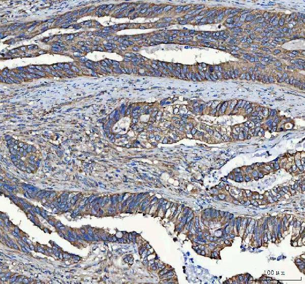
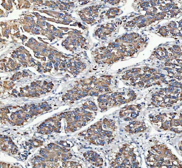
A08602 has IHC data from human paraffin sections of colorectal adenocarcinoma, liver cancer, lung cancer, and placenta (catalog image captions). Human, mouse, and rat reactivity is listed (catalog: reactivity).

Real IHC data IHC analysis of C19orf10/MYDGF using anti-C19orf10/MYDGF antibody (A08602). C19orf10/MYDGF was detected in a paraffin-embedded section of human colorectal adenocarcinoma tissue. Heat mediated antigen retrieval was performed in EDTA buffer (pH 8.0, epitope retrieval solution). The tissue section was blocked with 10% goat serum. The tissue section was then incubated with 2 μg/ml rabbit anti-C19orf10/MYDGF Antibody (A08602) overnight at 4°C. Peroxidase Conjugated Goat Anti-rabbit IgG was used as secondary antibody and incubated for 30 minutes at 37°C. The tissue section was developed using HRP Conjugated Rabbit IgG Super Vision Assay Kit (Catalog # SV0002) with DAB as the chromogen.
Anti-C19orf10/MYDGF Antibody ®
Cat # A08602

A08602 is the only SKU listed and has IHC images from human paraffin sections of colorectal adenocarcinoma, liver cancer, lung cancer, and placenta (catalog image captions). Its application list includes IHC but not IF/ICC (catalog: applications); the captions do not report a fixative (catalog image captions).

Which to pick: Choose A08602 for human paraffin-section IHC: its own captions show EDTA retrieval at pH 8.0 and primary antibody at 2 μg/ml (A08602 image captions). No SKU in this payload lists IF/ICC validation (catalog: applications), and A08602’s listed mouse and rat reactivity does not establish IHC validation in those species (catalog: reactivity; A08602 image captions). Clone information and the tissue fixative are unreported (catalog: clone field; A08602 image captions).

Each figure is that product's own IHC / IF validation image from its datasheet.

References

  1. UniProt Consortium. UniProt entry Q969H8 (MYDGF_HUMAN, Myeloid-derived growth factor).
  2. Human Protein Atlas. MYDGF tissue IHC expression (reliability: Approved).
  3. Human Protein Atlas. MYDGF subcellular location (ICC-IF): Secreted.
  4. Human Protein Atlas. MYDGF antibody validation summary (2 antibodies).
  5. The Rapid Activation of MYDGF Is Critical for Cell Survival in the Acute Phase of Retinal Regeneration in Fish. International journal of molecular sciences 2025 — PMC12346886.
  6. Single-Cell and Machine Learning Analyses Identify MYDGF as an Immune-Related Biomarker Associated With the Tumor Microenvironment in Clear Cell Renal Cell Carcinoma. Human mutation 2026 — PMC13401162.
  7. Myeloid-derived growth factor promotes M2 macrophage polarization and attenuates Sjögren's syndrome via suppression of the CX3CL1/CX3CR1 axis. Frontiers in immunology 2024 — PMC11532040.
  8. Cellular hierarchy framework based on single-cell and bulk RNA sequencing reveals fatty acid metabolic biomarker MYDGF as a therapeutic target for ccRCC. Frontiers in immunology 2025 — PMC12176760.
  9. PubMed PMID:15057824 — UniProt-cited evidence.
  10. PubMed PMID:15489334 — UniProt-cited evidence.
  11. PubMed PMID:19892738 — UniProt-cited evidence.