This website uses cookies to ensure you get the best experience on our website.


- Table of Contents
Plan MYEF2 paraffin IHC around its general nuclear staining pattern (HPA tissue IHC). Cerebral cortex glia stain strongly, while smooth muscle cells are reported as undetected; use these observations to guide control selection (HPA tissue IHC).

Expected localisation, validated protocols, controls and antibodies — the at-a-glance facts below, then the full design guide.
| Expected localisation | General nuclear staining in tissue (HPA tissue IHC) | |
| Staining pattern | Nuclear staining across multiple cell types (HPA tissue IHC) | |
| Antigen retrieval | Tris-EDTA pH 9.0 HIER, 95–98 °C, 20 min (rule: nuclear antigen) | |
| Positive control | Adrenal gland+4 more · see all | |
| Negative control | Smooth muscle |
| Fixation | Keep fixation consistent across sections (standard IHC practice; not target-specific) | |
| Caveat | Staining and RNA show medium concordance (HPA tissue IHC) | |
| Regulation | No specific expression regulator annotated (UniProt) | |
| Isoform / epitope | 4 isoforms; check antibody epitope coverage (UniProt) |
The catalog antibody’s IHC-P protocol is followed by one published chromogenic IHC protocol for MYEF2 in HCC tissues (PMC9854206).
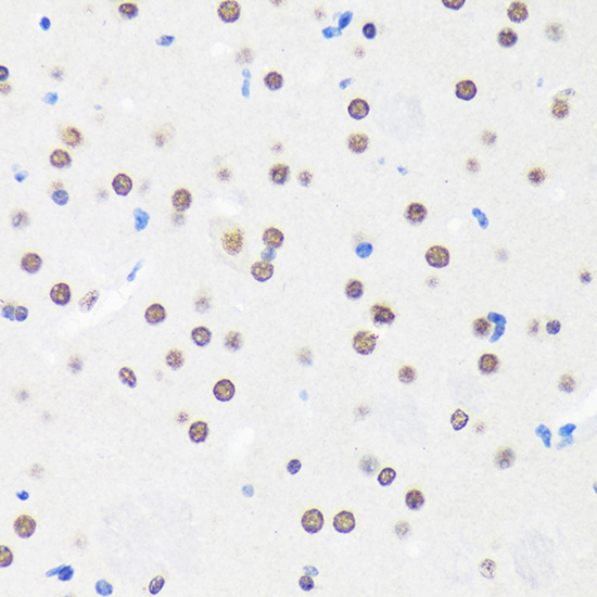
| Sample | Paraffin-embedded Mouse brain tissue; fixative not specified (datasheet A11666) |
| Fixation | Image fixative and duration unreported (datasheet A11666); verify before use. |
| Sectioning | 4–5 µm sections on charged slides (standard) |
| Deparaffinisation | Xylene, graded ethanol series to water (standard) |
| Antigen retrieval | Heat-induced epitope retrieval in Tris-EDTA buffer, pH 9.0, 20 min at 95–98 °C (standard rule: nuclear antigen) |
| Peroxidase block | 3% H2O2, 10 min, room temperature (standard) |
| Blocking | 10% normal serum of the secondary host, 30 min, room temperature (standard) |
| Primary antibody | Rabbit anti-MYEF2, 1:50-1:200 (datasheet A11666) |
| Primary incubation | Overnight at 4 °C (standard) |
| Detection | HRP-polymer secondary, DAB chromogen 5–10 min (standard) |
| Counterstain | Hematoxylin, blue, dehydrate and mount (standard) |
| Expected result | MYEF2-positive staining in glandular cells of adrenal gland (HPA tissue IHC: High). HPA tissue profile: General nuclear expression. No signal in the no-primary control. |
MYEF2 is a nuclear protein with no transmembrane segment (UniProt Q9P2K5 topology). In paraffin sections, expect predominantly nuclear staining in glandular, hematopoietic, respiratory epithelial and selected brain cells reported as high by HPA (HPA tissue IHC). The tissue pattern is rated Supported, with medium consistency between staining and RNA expression and external verification pending (HPA tissue IHC).
| Clear nuclear staining in adrenal glandular cells, bone marrow hematopoietic cells or cerebral cortex glial cells. | This matches the reported high staining in those cell populations and the general nuclear pattern (HPA tissue IHC). Judge the signal within the named cells; staining elsewhere in the section does not establish the same result. Compare signal with a negative control before scoring it as specific (general IHC practice). |
| Strong, predominantly cytoplasmic staining with little nuclear signal in an expected positive cell population. | This conflicts with nuclear localization (UniProt Q9P2K5; HPA tissue IHC) and warrants investigation of staining or detection artefact (general IHC practice). HPA reports additional cytosolic localization by ICC-IF, so a cytosolic component alone does not prove an artefact (HPA subcellular). |
| Strong staining of smooth muscle cells, especially when the expected positive cells stain weakly. | HPA reports MYEF2 as not detected in smooth muscle cells (HPA tissue IHC). Check for antibody cross-reactivity or endogenous detection activity before interpreting this as MYEF2; the supplied evidence does not identify which, if either, caused the signal (general IHC practice). |
| Broad color deposition across nuclei, cytoplasm and extracellular areas without clear cell boundaries. | A diffuse pattern cannot be scored confidently as the reported nuclear expression (HPA tissue IHC). Background from the staining or detection workflow is a possible explanation; use the negative control and tissue morphology to distinguish it from localized signal (general IHC practice). |
| No nuclear signal in an expected high-staining population, such as bronchial respiratory epithelial cells. | This is discordant with HPA's high result for those cells (HPA tissue IHC). First assess tissue preservation, controls and the IHC workflow (general IHC practice). Because HPA rates the tissue pattern Supported with external verification pending, one negative section cannot by itself settle MYEF2 expression (HPA tissue IHC). |
| Expected compartment | UniProt places MYEF2 in the nucleus, and HPA describes general nuclear tissue staining (UniProt Q9P2K5; HPA tissue IHC). Score the named cell populations and their nuclear signal together; intensity without the expected location is insufficient for a confident call (general IHC practice). |
| Tissue and cell population | HPA reports high staining in several glandular, hematopoietic and brain cell populations, low staining in vaginal squamous epithelial cells and skeletal myocytes, and no detection in smooth muscle cells (HPA tissue IHC). Choose controls by the reported cell population within a tissue, not by tissue name alone. |
| Evidence strength | The supplied antibody HPA004883 has Supported IHC status; HPA describes medium agreement between tissue staining and RNA and says external verification is pending (HPA antibodies; HPA tissue IHC). Treat an unexpected compartment or cell type as a finding to check, not as established MYEF2 biology. |
| Isoforms and epitope coverage | UniProt lists 4 MYEF2 isoforms (UniProt Q9P2K5). The supplied record does not map the IHC antibody epitope, so it cannot establish which isoforms contribute to staining. Do not explain a tissue difference by isoform recognition without separate evidence. |
| IF/ICC: where should signal appear? | HPA reports approved nucleoplasmic localization plus additional approved cytosolic localization in ICC-IF, with images from A-431, U-251MG, U2OS and KOLF2.1J (HPA subcellular). Its separate ICC antibody status is Approved (HPA antibodies); these observations do not define an IHC-P protocol. |
| Situation | Likely cause | Next action |
|---|---|---|
| An expected high-staining population has no visible nuclear signal. | The section or staining workflow may have failed; a biological absence is also possible. HPA's high call is Supported, with external verification pending (HPA tissue IHC). | Review morphology and the run's positive control, then check retrieval, primary-antibody incubation and detection steps against the chosen IHC-P procedure (general IHC practice). Avoid assigning a MYEF2-specific retrieval requirement from this record. |
| Signal appears only in cytoplasm in an IHC section. | The result diverges from general nuclear tissue staining (HPA tissue IHC; UniProt Q9P2K5). HPA's additional cytosolic ICC-IF location does not establish a cytoplasmic-only IHC pattern (HPA subcellular). | Inspect nuclear counterstain and cell boundaries, then compare with the positive and negative controls (general IHC practice). Record any mixed nuclear and cytoplasmic signal separately from a cytoplasmic-only result. |
| Smooth muscle cells stain strongly. | HPA lists smooth muscle cells as not detected (HPA tissue IHC). Cross-reactivity or endogenous detection activity could explain unexpected color, but this record does not distinguish them. | Compare an adjacent negative control and assess whether the color persists without primary antibody; review endogenous activity blocking where relevant to the detection chemistry (general IHC practice). |
| Weak color spreads across much of the section. | Diffuse background can obscure the nuclear pattern used for interpretation (HPA tissue IHC; general IHC practice). The payload provides no MYEF2-specific cause for background. | Check negative-control staining, blocking, washes and detection development, then assess nuclei in identifiable expected cells (general IHC practice). Avoid calling diffuse color positive solely because an HPA tissue is listed as high. |
| Low-staining cells look as dark as an expected high-staining population. | That contrast differs from HPA's low calls for vaginal squamous epithelial cells and skeletal myocytes versus its listed high populations (HPA tissue IHC). Section-to-section color intensity alone is not a validated expression measurement. | Compare the named cell populations within appropriately controlled sections and use consistent scoring criteria (general IHC practice). Keep HPA's Supported reliability and pending external verification in the interpretation (HPA tissue IHC). |
| A researcher wants to apply an IF/ICC image pattern directly to IHC-P scoring. | HPA reports nucleoplasm plus additional cytosol for ICC-IF, while its tissue IHC profile is general nuclear expression (HPA subcellular; HPA tissue IHC). The observations come from different applications. | Use nuclear staining in the reported tissue cell populations as the IHC-P reference (HPA tissue IHC). Consult the separate IF/ICC guide for that application; do not infer an IF protocol or an IHC-P cytoplasmic scoring rule from these records. |


Comprehensive Human Protein Atlas IHC scoring per tissue (reliability: Supported — Medium consistency between antibody staining and RNA expression data. Pending external verification.). Rows are taken directly from the HPA tissue chart — click any row's HPA link to view the source.
| Tissue | Cell type | Level | Evidence | Source |
|---|---|---|---|---|
| Adrenal gland | Glandular cells | High | Protein (IHC) | HPA → |
| Appendix | Glandular cells | High | Protein (IHC) | HPA → |
| Bone marrow | Hematopoietic cells | High | Protein (IHC) | HPA → |
| Breast | Glandular cells | High | Protein (IHC) | HPA → |
| Bronchus | Respiratory epithelial cells | High | Protein (IHC) | HPA → |
| Tissue | Cell type | Level | Evidence | Source |
|---|---|---|---|---|
| Smooth muscle | Smooth muscle cells | Not detected | Protein (IHC) | HPA → |
Troubleshoot MYEF2 staining by assessing retrieval, nuclear localisation, antibody epitope coverage and cell type before comparing chromogenic IHC results (UniProt Q9P2K5; HPA tissue IHC).
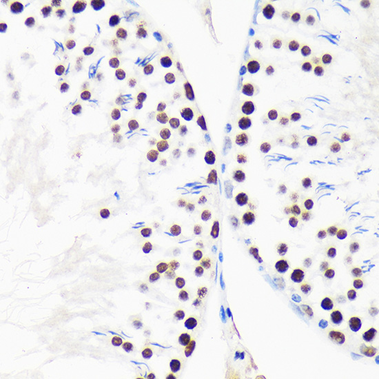
Anti-MYEF2 IHC images show paraffin-embedded mouse brain and rat testis (A11666 IHC captions). IF/ICC is listed for human, mouse and rat, but no IF image is supplied (A11666 catalog).
A11666 has IHC images of paraffin-embedded mouse brain and rat testis at 1:100 (A11666 IHC captions); IF/ICC is listed without an IF image (A11666 catalog). A11666-1 lists human paraffin-section IHC at 2–5 μg/ml and human IF/ICC at 5 μg/ml, but supplies no IHC or IF image (A11666-1 catalog).
Which to pick: For tissue IHC, pick A11666 when a paraffin-section example matters: its rabbit polyclonal antibody has mouse brain and rat testis IHC images, with microwave retrieval in 10 mM PBS, pH 7.2 (A11666 catalog and IHC captions). For human IF/ICC, either SKU is listed, while A11666-1 specifies 5 μg/ml for human samples (A11666 and A11666-1 catalogs). For cross-species work, A11666 lists human, mouse and rat reactivity and IHC/IF/ICC applications; its IHC captions document paraffin sections but do not report the fixative (A11666 catalog and IHC captions).