This website uses cookies to ensure you get the best experience on our website.


- Table of Contents
Plan chromogenic IHC on paraffin sections using skeletal muscle or heart muscle as positive controls (HPA tissue IHC). This guide covers cytoplasmic staining expectations (HPA tissue IHC) and the catalog antibody’s IHC dilution of 1:50–1:200 (datasheet A09420).

Expected localisation, validated protocols, controls and antibodies — the at-a-glance facts below, then the full design guide.
| Expected localisation | Cytoplasm in muscle; myofibrillar thick filaments (HPA tissue IHC; UniProt) | |
| Staining pattern | High cytoplasmic staining in myocytes and cardiomyocytes (HPA tissue IHC) | |
| Antigen retrieval | Citrate pH 6.0 HIER, heat-mediated (datasheet A09420) | |
| Positive control | Heart muscle+1 more · see all | |
| Negative control | Adipose tissue+4 more · see all |
| Fixation | Keep paraffin-section fixation consistent across samples. (standard IHC practice; not target-specific) | |
| Caveat | Staining may include proteins from more than one gene (HPA tissue IHC) | |
| Regulation | No expression regulation annotated (UniProt) | |
| Isoform / epitope | No annotated isoforms; one 1–1939 chain (UniProt) |
The catalog antibody’s IHC-P protocol is followed by four published MYH1 IHC protocols covering the samples and methods reported in their excerpts (datasheet A09420; PMC12331014; PMC13242725; PMC7807096; PMC9105468).
| Sample | Paraffin-embedded Human skeletal muscle tissue; fixative not specified (datasheet A09420) |
| Fixation | Image fixative and duration unreported (datasheet A09420); verify before use. |
| Sectioning | 4–5 µm sections on charged slides (standard) |
| Deparaffinisation | Xylene, graded ethanol series to water (standard) |
| Antigen retrieval | Heat retrieval: Citrate pH 6.0 (datasheet A09420); 20 min, 95–100 °C (standard) |
| Peroxidase block | 3% H2O2, 10 min, room temperature (standard) |
| Blocking | 10% normal serum of the secondary host, 30 min, room temperature (standard) |
| Primary antibody | Rabbit anti-MYH1, 1:50-1:200 (datasheet A09420) |
| Primary incubation | Overnight at 4 °C (standard) |
| Detection | HRP-polymer secondary, DAB chromogen 5–10 min (standard) |
| Counterstain | Hematoxylin, blue, dehydrate and mount (standard) |
| Expected result | MYH1-positive staining in cardiomyocytes of heart muscle (HPA tissue IHC: High). HPA tissue profile: Selective cytoplasmic expression in heart and skeletal muscle. No signal in the no-primary control. |
MYH1 should appear in the cytoplasm of muscle cells, consistent with its location in myofibrillar thick filaments and its lack of a transmembrane segment (UniProt P12882 localisation and topology). HPA reports high staining in skeletal myocytes and cardiomyocytes, with selective cytoplasmic expression in skeletal and heart muscle (HPA tissue IHC). Interpret that pattern with caution: HPA rates its tissue profile Enhanced but notes medium agreement with RNA data and antibody recognition of proteins from multiple genes (HPA tissue IHC).
| Strong cytoplasmic staining in skeletal myocytes, with myofibrillar distribution where structure is visible (HPA: High in skeletal myocytes; UniProt P12882 localisation). | This fits the expected positive pattern. Score signal within muscle cells rather than staining across the section; MYH1 is associated with myofibrillar thick filaments (UniProt P12882 localisation). HPA staining alone cannot establish that every positive fibre expresses MYH1 specifically because its profile carries a multiple-gene warning (HPA tissue IHC). |
| Strong cytoplasmic staining in cardiomyocytes (HPA: High in cardiomyocytes). | This matches the supplied HPA tissue observation, although HPA also describes skeletal muscle as RNA enriched (HPA tissue IHC). Treat heart staining as an observed antibody pattern, not independent proof of MYH1-specific protein abundance: HPA reports medium RNA agreement and possible recognition of proteins from multiple genes (HPA tissue IHC). |
| Predominantly nuclear, surface-rim or extracellular staining instead of muscle-cell cytoplasm. | That distribution conflicts with the documented myofibrillar cytoplasmic location and absence of a transmembrane segment (UniProt P12882 localisation and topology). First assess staining artefact and antibody specificity; the compartment mismatch alone does not identify its cause (general IHC interpretation). |
| Conspicuous staining in adipocytes, respiratory epithelial cells or marrow hematopoietic cells. | HPA reports these sampled cell types as not detected (HPA tissue IHC). A positive result there raises concern for cross-reactivity or endogenous detection activity; compare its location and intensity with a known-positive muscle section and the relevant detection controls (general IHC practice). |
| Diffuse section-wide colour, or no muscle-cell signal in a known-positive skeletal muscle section. | Diffuse colour makes cell-specific scoring unreliable; absent signal contradicts the reported high skeletal-myocyte staining (HPA tissue IHC). Use controls to distinguish a detection/background problem from a failed stain, then review routine IHC processing steps without assuming a MYH1-specific fixation effect (general IHC practice). |
| Tissue and cell context (HPA tissue IHC). | HPA reports high staining in skeletal myocytes and cardiomyocytes, while its listed adipocytes, respiratory epithelial cells and marrow hematopoietic cells are not detected (HPA tissue IHC). Use these observations to choose comparative sections, while retaining HPA's medium RNA agreement and multiple-gene caveat (HPA tissue IHC). |
| Antibody interpretation (HPA antibody validation; HPA tissue IHC). | The listed IHC ratings differ: HPA001349 is Supported and CAB010759 is Enhanced (HPA antibodies). An Enhanced rating supports reproducibility or orthogonal agreement under HPA's criteria; it does not remove the tissue profile's warning that staining may represent proteins from more than one gene (HPA tissue IHC). |
| Intracellular target location (UniProt P12882). | MYH1 is assigned to cytoplasmic myofibrillar thick filaments and has no transmembrane segment (UniProt P12882 localisation and topology). These annotations support judging muscle-cell cytoplasm as the relevant compartment; they do not supply a target-specific antigen retrieval condition or fixation sensitivity (UniProt P12882 record). |
| Q: Should focal-adhesion IF signal define the IHC positive pattern? (HPA subcellular ICC-IF). | A: No. HPA labels focal adhesion sites uncertain and warns that the ICC-IF antibodies target proteins from multiple genes (HPA subcellular ICC-IF). Keep that observation qualified on the separate IF/ICC page; judge paraffin IHC against the reported muscle-cell cytoplasmic pattern (HPA tissue IHC). |
| Situation | Likely cause | Next action |
|---|---|---|
| No signal in skeletal myocytes despite an intact known-positive section (HPA: High in skeletal myocytes). | A failed staining run or insufficient detection is possible; the supplied sources do not establish MYH1-specific fixation sensitivity (general IHC practice; UniProt P12882 record). | Check the positive control, antibody application, detection reagents and counterstain. Review the selected IHC antibody's validated procedure for antigen retrieval before changing conditions; do not infer a MYH1-specific retrieval requirement from HPA staining levels (general IHC practice). |
| Muscle cells stain mainly in nuclei or along cell borders (UniProt P12882 localisation and topology). | The compartment differs from the documented cytoplasmic myofibrillar location; staining artefact or cross-reactivity is possible, but location alone cannot distinguish them (UniProt P12882 localisation; general IHC interpretation). | Compare with a known-positive muscle section, inspect morphology and run an appropriate primary-omission control. Interpret persistent compartment mismatch cautiously because the HPA tissue profile warns of multiple-gene recognition (general IHC practice; HPA tissue IHC). |
| Adipocytes or respiratory epithelial cells appear positive (HPA: Not detected in those cells). | Cross-reactivity or endogenous chromogenic detection activity may account for unexpected staining; HPA's multiple-gene warning adds a specificity concern (general IHC practice; HPA tissue IHC). | Compare the signal with muscle-cell cytoplasm and check primary-omission and detection controls. Address endogenous enzyme activity using the detection system's standard controls before assigning unexpected cells MYH1 expression (general IHC practice). |
| Diffuse brown background obscures myocytes (general chromogenic IHC observation). | Nonspecific reagent binding, inadequate washing or endogenous detection activity can create background in chromogenic IHC (general IHC practice). | Check primary-omission and reagent controls, then review blocking, washes and antibody concentration within the chosen antibody's IHC procedure. Score only distinguishable cellular staining; HPA's high muscle staining does not validate diffuse colour (general IHC practice; HPA tissue IHC). |
| Heart staining seems stronger than expected from skeletal muscle RNA enrichment (HPA tissue IHC). | HPA reports high cardiomyocyte staining alongside skeletal-muscle-enriched RNA, medium RNA agreement and possible recognition of proteins from multiple genes (HPA tissue IHC). | Record heart staining as an observed IHC result and compare cell location and controls across sections. Avoid converting relative colour intensity into a MYH1 expression ranking without independent specificity evidence (HPA tissue IHC; general IHC interpretation). |
| Two antibodies give different muscle or off-target patterns (HPA antibody validation; HPA tissue IHC). | The listed IHC validation levels differ, and the HPA tissue profile cautions that antibodies can recognize proteins from multiple genes (HPA antibodies; HPA tissue IHC). | Record antibody identity and IHC validation status, compare matched controls and the cytoplasmic muscle-cell pattern, and report unresolved disagreement. Neither a matching location nor an Enhanced rating alone proves MYH1-specific staining (HPA antibodies; HPA tissue IHC; UniProt P12882 localisation). |
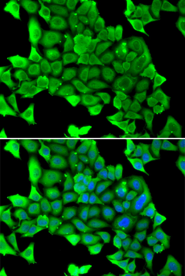


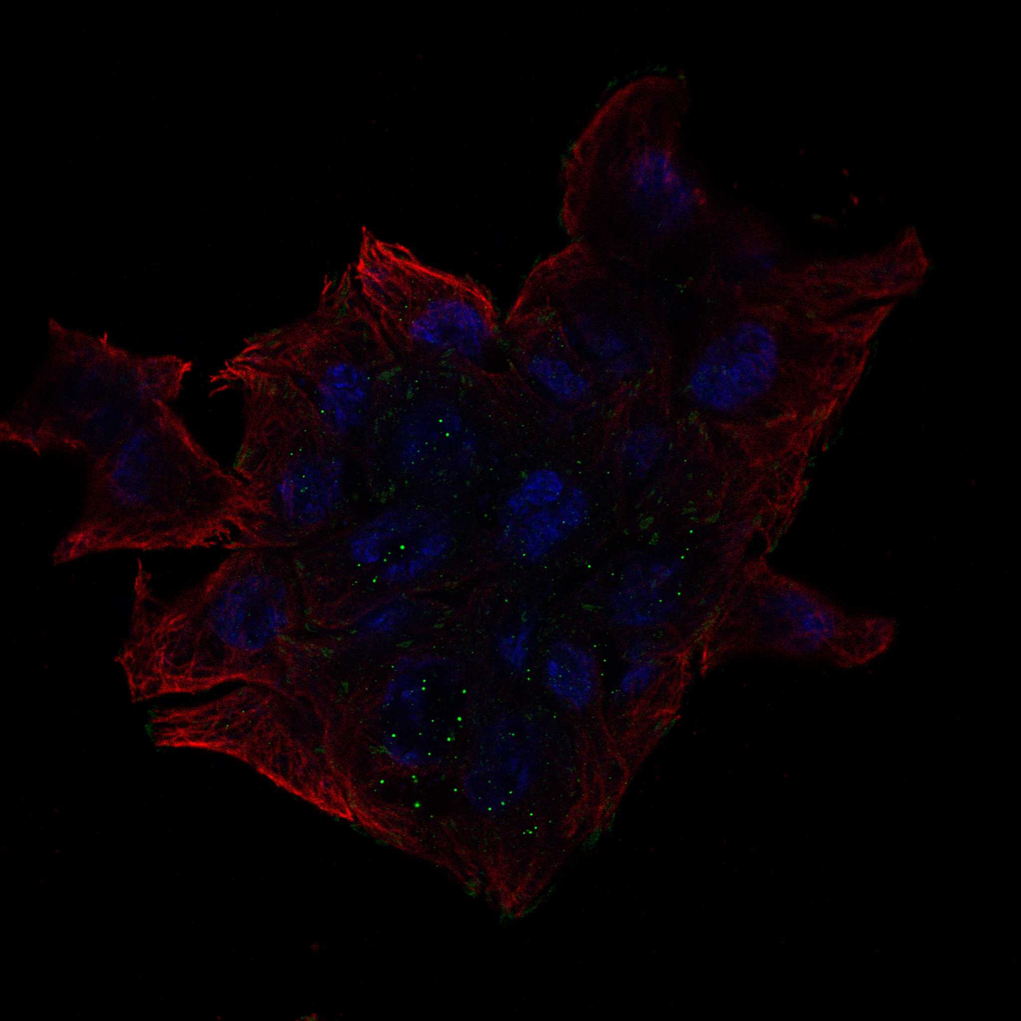

Comprehensive Human Protein Atlas IHC scoring per tissue (reliability: Enhanced — Medium consistency between antibody staining and RNA expression data. Caution, targets protein from more than one gene.). Rows are taken directly from the HPA tissue chart — click any row's HPA link to view the source.
| Tissue | Cell type | Level | Evidence | Source |
|---|---|---|---|---|
| Heart muscle | Cardiomyocytes | High | Protein (IHC) | HPA → |
| Skeletal muscle | Myocytes | High | Protein (IHC) | HPA → |
| Tissue | Cell type | Level | Evidence | Source |
|---|---|---|---|---|
| Adipose tissue | Adipocytes | Not detected | Protein (IHC) | HPA → |
| Adrenal gland | Glandular cells | Not detected | Protein (IHC) | HPA → |
| Appendix | Glandular cells | Not detected | Protein (IHC) | HPA → |
| Bone marrow | Hematopoietic cells | Not detected | Protein (IHC) | HPA → |
| Breast | Adipocytes | Not detected | Protein (IHC) | HPA → |
Troubleshoot MYH1 staining in paraffin sections by checking retrieval, cytoplasmic localization, antibody specificity, and matched controls.
A09420 has a human skeletal muscle paraffin-section IHC image (catalog IHC caption); IF/ICC is listed for the antibody, with human, mouse and rat reactivity (catalog applications and reactivity).
A09420 will render with an IHC image of human skeletal muscle paraffin sections stained at 1:50 after citrate pH 6.0 retrieval (catalog IHC caption). IF/ICC is listed, and human, mouse and rat reactivity is reported; no IF image is supplied (catalog applications, reactivity and image records).
Which to pick: Choose A09420 for paraffin-section IHC: its own image documents human skeletal muscle staining, and the listed IHC dilution range is 1:50–1:200 (catalog IHC caption and dilution). A09420 is also the listed IF/ICC option, with a 1:50–1:100 dilution range, and the listed cross-species option for human, mouse and rat; it is polyclonal (catalog applications, dilution, reactivity and antibody details). The IHC caption specifies paraffin embedding and citrate retrieval but does not report the fixative (catalog IHC caption).