MYH11 / Myosin-11 · IHC design guide

Design Immunohistochemistry for MYH11

Plan MYH11 chromogenic IHC in paraffin sections using smooth muscle and myoepithelial cell staining as the tissue reference (HPA tissue IHC). This guide covers fixation consistency, antibody selection and interpretation of staining outside those cells.

Evidence assembled Oct 2026 · For research use; verify linked source records and product datasheet before use
Immunohistochemistry protocol sheet for MYH11 (IHC for MYH11): expected localisation Thick filaments of myofibrils expected (UniProt), antibody A02422-2, validated IHC image, and IHC protocol steps
Printable MYH11 IHC protocol sheet — expected localisation Thick filaments of myofibrils expected (UniProt), antibody A02422-2, controls and protocol steps. Open the full MYH11 IHC guide →

MYH11 Immunohistochemistry Experimental Design Guide

Expected localisation, validated protocols, controls and antibodies — the at-a-glance facts below, then the full design guide.

Must know before staining
Expected localisation Thick filaments of myofibrils expected (UniProt)
Staining pattern Smooth muscle and myoepithelial cell staining; compartment unreported (HPA tissue IHC)
Antigen retrieval EDTA pH 8.0 HIER, heat-mediated (datasheet A02422-2)
Positive control ⓘ Breast+4 more · see all
Negative control ⓘ Adipose tissue+4 more · see all
Important caveats
Reasons your staining may differ from the expected pattern.
Fixation Keep paraffin-section fixation consistent (standard IHC practice; not target-specific); Matched tissue-IHC evidence does not establish the fixation claim. Validate the specimen-specific method before use. (selected-SKU IHC image A02422-2)
Caveat Enterocytes and duodenal endocrine cells may also stain (HPA tissue IHC)
Regulation Tissue-specific expression in smooth muscle (UniProt)
Isoform / epitope 4 isoforms; verify epitope coverage; no transmembrane segment (UniProt)
Section 1

Recommended MYH11 IHC & IF Protocols

The catalog antibody uses EDTA pH 8.0 retrieval (datasheet A02422-2). These published MYH11 IHC protocols cover gastric cancer, human aorta, and porcine jejunum (PMC8359574; PMC8213747; PMC13446750).

Recommended immunohistochemistry (IHC-P) protocol parameters
SampleParaffin-embedded human appendix tissue; fixative not specified (datasheet A02422-2)
FixationImage fixative and duration unreported (datasheet A02422-2); verify before use.
Sectioning4–5 µm sections on charged slides (standard)
DeparaffinisationXylene, graded ethanol series to water (standard)
Antigen retrievalHeat retrieval: EDTA pH 8.0 (datasheet A02422-2); 20 min, 95–100 °C (standard)
Peroxidase block3% H2O2, 10 min, room temperature (standard)
Blocking10% goat serum (datasheet A02422-2)
Primary antibodyRabbit anti-MYH11, 1:50 recommended; image 1:100 (datasheet A02422-2)
Primary incubationOvernight at 4 °C (datasheet A02422-2)
DetectionHRP-conjugated secondary, DAB chromogen (datasheet A02422-2)
CounterstainHematoxylin, blue, dehydrate and mount (standard)
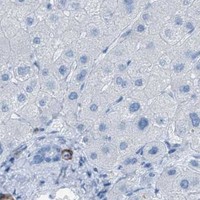
Expected resultMYH11-positive staining in myoepithelial cells of breast (HPA tissue IHC: High). HPA tissue profile: Distinct expression in myoepithelial and smooth muscle cells. No signal in the no-primary control.
💡Decision noteStart with heat-mediated EDTA pH 8.0 for the catalog antibody (datasheet A02422-2); the gastric cancer study used citrate pH 6.0 with a different antibody (PMC8359574).
Section 2

What Is the Expected MYH11 Staining Pattern?

MYH11 should stain smooth muscle cells and breast myoepithelial cells in paraffin sections (HPA tissue IHC: distinct expression; Enhanced reliability). Its thick-filament association and lack of a transmembrane segment support a predominantly intracellular interpretation (UniProt P35749). HPA also reports plasma membrane and cytosol localisation by ICC-IF; that result is context for interpreting localisation, not a paraffin IHC pattern (HPA ICC-IF).

What am I looking at on my slide?
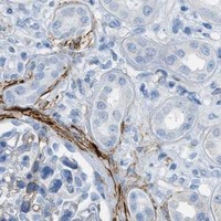
Strong staining outlines vessel walls or smooth muscle bundles, with signal in the muscle cells.This fits the expected cell pattern: HPA reports High staining in smooth muscle cells in smooth muscle and endometrium (HPA tissue IHC). Score the identified muscle cells; adjacent unstained cells do not make the result negative.
Breast myoepithelial cells stain strongly around glandular structures.This is another expected positive pattern (HPA tissue IHC: High in breast myoepithelial cells). Judge the staining by its cellular position, since the guide’s positive call depends on the stained cell type as well as intensity.
Signal is predominantly nuclear, away from identifiable smooth muscle or myoepithelial cells.Treat a nuclear-only pattern as suspect: HPA reports plasma membrane and cytosol localisation in ICC-IF, while UniProt places MYH11 in thick filaments (HPA ICC-IF; UniProt P35749). Check morphology and controls before assigning specificity.
Broad staining appears in cells expected to be negative, or across the whole section.Consider cross-reactivity or endogenous chromogen-generating activity (general IHC practice). HPA reports adipocytes and bronchial respiratory epithelial cells as Not detected; those findings apply to the named cells, not every cell in those tissues (HPA tissue IHC).
No staining appears in a known-positive smooth muscle area.A failed positive control leaves the negative result uninterpretable (general IHC practice). Smooth muscle cells are a strong expected positive reference (HPA tissue IHC: High); check control performance and the staining workflow before scoring the specimen negative.
💡Expected MYH11 appearanceCall positive when identifiable smooth muscle or breast myoepithelial cells show strong, predominantly nonnuclear staining (HPA tissue IHC: High; UniProt P35749 thick filaments); diffuse chromogen or isolated nuclear staining warrants investigation (general IHC practice; HPA ICC-IF).
How each factor affects the staining
Cell-specific tissue distributionUse morphology when choosing a control: High staining is reported in breast myoepithelial and smooth muscle cells, whereas duodenal endocrine cells, ovarian stroma cells and small-intestinal enterocytes have Medium staining (HPA tissue IHC). A tissue name alone cannot define every cell as positive.
Location evidence across assaysHPA’s plasma membrane and cytosol result comes from ICC-IF (HPA ICC-IF). UniProt lists thick filaments and no transmembrane segment (UniProt P35749). Use those findings to assess an unexpected compartment, while judging paraffin IHC chiefly against its observed cell pattern (HPA tissue IHC).
Isoforms and antibody coverageFour MYH11 isoforms are listed (UniProt P35749). The payload gives no epitope or isoform-coverage data for the catalog antibody, so an unexpected cell-specific difference cannot be assigned to an isoform from this evidence alone.
Antibody validationThree listed antibodies have Enhanced IHC status, and the tissue profile has Enhanced reliability (HPA antibodies; HPA tissue IHC). These support the reported pattern but do not establish that an uncharacterised antibody will behave identically.
Retrieval and detectionAntigen retrieval and chromogenic detection require appropriate run controls (general IHC practice). No MYH11-specific fixation sensitivity or retrieval condition is supplied; avoid attributing weak staining to epitope masking without experimental comparison.
Why is my staining missing, weak or wrong?
SituationLikely causeNext action
Known-positive smooth muscle is blank.The positive control or a staining step may have failed (general IHC practice).Verify the positive-control section, antibody application, retrieval and detection reagents before interpreting the specimen (general IHC practice; HPA tissue IHC: High in smooth muscle cells).
Only nuclei stain.The compartment conflicts with the reported ICC-IF localisation and thick-filament association (HPA ICC-IF; UniProt P35749).Inspect control sections and cellular morphology; reassess the signal as possible artefact before calling MYH11 positive (general IHC practice).
Respiratory epithelium stains broadly in a bronchus section.HPA reports bronchial respiratory epithelial cells as Not detected; the observation may reflect nonspecific or endogenous signal (HPA tissue IHC; general IHC practice).Compare an appropriate negative control and inspect whether staining is confined instead to identifiable smooth muscle cells (general IHC practice; HPA tissue IHC).
Chromogen covers many unrelated cells or tissue spaces.Diffuse background can arise from nonspecific binding or endogenous detection activity (general IHC practice).Check blocking and detection controls, then score only cell-associated staining with a credible MYH11 distribution (general IHC practice; HPA tissue IHC).
A Medium-level HPA cell population looks weaker than smooth muscle.Its reported level differs from the High smooth muscle reference (HPA tissue IHC: Medium versus High).Compare like cell types under the same run conditions; avoid treating weaker staining alone as assay failure (general IHC practice; HPA tissue IHC).
A specimen appears negative although its tissue has a positive HPA entry.HPA levels describe named cell populations, which may be absent from the area examined (HPA tissue IHC).Locate the relevant cells on the section and confirm that the run’s known-positive smooth muscle stained before issuing a negative call (general IHC practice; HPA tissue IHC).

Sample controls for MYH11 IHC & IF

🧪Run breast first: its myoepithelial cells should stain strongly (HPA: Breast myoepithelial cells, High). Use adipose tissue as the negative comparator for adipocytes (HPA: Adipocytes, Not detected); on the breast slide, adjacent luminal epithelial cells should lack MYH11 staining, while the myoepithelial layer stains (HPA: Breast myoepithelial cells, High; UniProt P35749: smooth-muscle expression).
Positive control tissue: Breast (Myoepithelial cells, HPA High)
Negative control tissue: Adipose tissue (HPA Not detected)
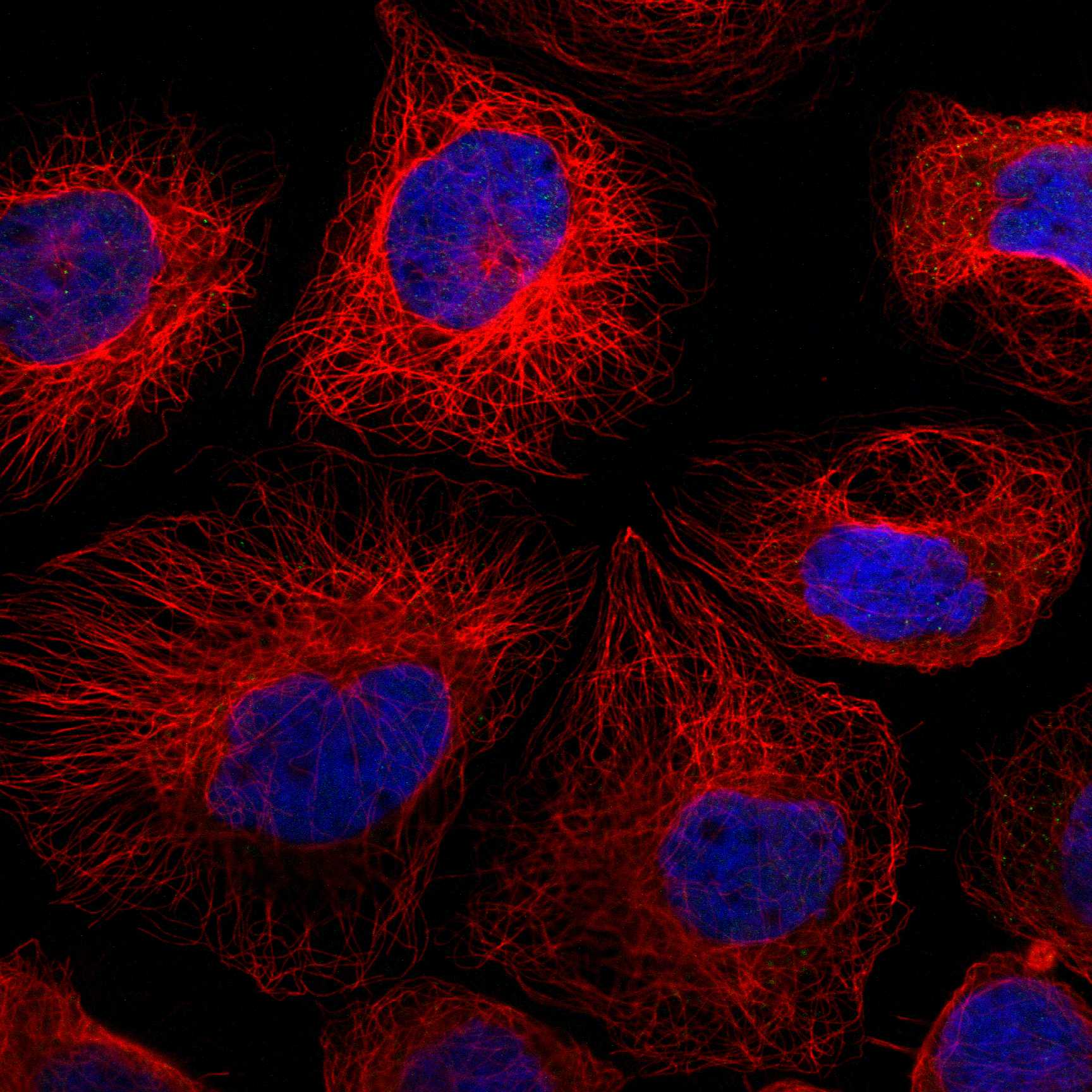
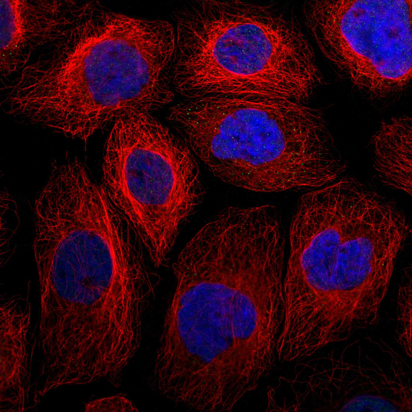
ICC-IF cell lines (HPA subcellular resource): HPA ICC-IF images show MYH11 in A-431, U-251MG, U2OS, HeLa BAC 5098, HeLa , with annotated localisation: Plasma membrane (enhanced), Cytosol (enhanced) (HPA subcellular).
Technical controls: Include a no-primary, secondary-only control, a nonimmune rabbit IgG control matched to the primary antibody’s clonality, and a MYH11 knockout sample or validated antigen-peptide competition control (selected-SKU caption: rabbit primary). Quench endogenous peroxidase before HRP–DAB detection, and score the adipose control by adipocytes rather than any vascular smooth muscle present (selected-SKU caption: HRP–DAB detection; HPA: Adipocytes, Not detected).
⚠️Feasibility: The selected-SKU paraffin-section caption reports heat-mediated EDTA retrieval at pH 8.0 and primary incubation at 1:100 overnight at 4°C; the fixative is unreported (selected-SKU tissue-IHC caption). No target-specific fixation window or fixation effect is reported, and the supplied evidence does not establish that frozen sections or IF are easier (selected-SKU tissue-IHC caption; HPA: ICC-IF images). Interpret adipose staining by cell type because vascular smooth muscle can be present alongside adipocytes (HPA: Adipocytes, Not detected; UniProt P35749: smooth-muscle expression).

HPA tissue IHC evidence for MYH11

Comprehensive Human Protein Atlas IHC scoring per tissue (reliability: Enhanced — High consistency between antibody staining and RNA expression data. Antibody staining in cells/structures not annotated, view images.). Rows are taken directly from the HPA tissue chart — click any row's HPA link to view the source.

Positive expression · recommended positive controls

TissueCell typeLevelEvidenceSource
Breast Myoepithelial cells High Protein (IHC) HPA →
Endometrium Smooth muscle cells High Protein (IHC) HPA →
Smooth muscle Smooth muscle cells High Protein (IHC) HPA →
Duodenum Endocrine cells Medium Protein (IHC) HPA →
Ovary Ovarian stroma cells Medium Protein (IHC) HPA →

Undetected expression · recommended negative controls

TissueCell typeLevelEvidenceSource
Adipose tissue Adipocytes Not detected Protein (IHC) HPA →
Adrenal gland Glandular cells Not detected Protein (IHC) HPA →
Appendix Endocrine cells Not detected Protein (IHC) HPA →
Bone marrow Hematopoietic cells Not detected Protein (IHC) HPA →
Bronchus Respiratory epithelial cells Not detected Protein (IHC) HPA →
Section 3

Advanced MYH11 IHC Tips

Troubleshoot MYH11 chromogenic IHC in paraffin sections by checking retrieval, cell identity, staining location and controls before comparing staining scores.

What retrieval should I try first when MYH11 staining is weak?
Start with heat-mediated retrieval in EDTA at pH 8.0 for paraffin sections stained with A02422-2 (datasheet A02422-2). The documented tissue-IHC workflow used this retrieval before an overnight incubation with primary antibody at 1:100 and 4°C (A02422-2 tissue-IHC caption). If staining is weak, check that sections reached the intended retrieval conditions and compare adjacent sections while changing only one heating variable at a time (standard IHC practice). Include a smooth muscle-rich region as a positive reference, since MYH11 is expressed in smooth muscle (UniProt P35749). Compare tissue morphology alongside signal; harsher retrieval can damage sections or increase nonspecific staining (standard IHC practice).
Could fixation explain inconsistent MYH11 staining between samples?
The A02422-2 image documents a paraffin-embedded appendix section but does not state its fixative (A02422-2 tissue-IHC caption). MYH11-specific sensitivity to fixative chemistry or fixation duration is therefore unknown from the supplied evidence; the HPA staining pattern does not establish either effect (A02422-2 tissue-IHC caption; HPA tissue IHC). Record each sample’s fixative, fixation duration and processing history before comparing staining intensity (standard IHC practice). Hold section preparation, EDTA retrieval at pH 8.0 and antibody incubation constant during that comparison (datasheet A02422-2; standard IHC practice). Assess preservation and staining in comparable smooth muscle structures on each slide before attributing a difference to MYH11 expression (UniProt P35749; standard IHC practice).
Where should convincing MYH11 staining appear in a tissue section?
Prioritize staining in morphologically identified smooth muscle cells and, where present, myoepithelial cells (HPA tissue IHC: distinct expression in both cell types). MYH11 is a myosin heavy chain associated with thick filaments, and its record lists no transmembrane segment (UniProt P35749). Cytoplasmic staining consistent with that role is plausible, while an exclusively sharp cell-border pattern needs careful review against tissue morphology (UniProt P35749; standard IHC interpretation). Cell-based imaging also reports plasma membrane and cytosol localization, so a membrane-associated appearance alone does not establish an extracellular epitope (HPA subcellular). Compare stained cells with neighboring structures and a matched negative control before assigning localisation (standard IHC practice).
How could epitope selection affect MYH11 staining across tissues?
MYH11 has 4 reported isoforms, and isoform 1 is mostly found in slowly contracting tonic muscles (UniProt P35749). Its annotated regions include a motor domain at residues 85–783 and an IQ region at 786–815 (UniProt P35749). The supplied A02422-2 caption does not map the antibody epitope, so staining differences cannot be assigned to a particular isoform or region from this evidence (A02422-2 tissue-IHC caption; UniProt P35749). When comparing tissues, keep retrieval and detection conditions constant and include a morphologically identified smooth muscle reference (standard IHC practice; HPA tissue IHC). Interpret unexpected loss cautiously until epitope information or an independent antibody supports an isoform-specific explanation (standard IHC practice).
How should I check MYH11 localisation in a multiplex IF experiment?
Treat IF as a separate validation exercise: the supplied A02422-2 tissue image documents chromogenic IHC, while the HPA cell-imaging record reports cytosol and plasma membrane localisation (A02422-2 tissue-IHC caption; HPA subcellular). Pair MYH11 with an independent marker of smooth muscle or myoepithelial cell identity, depending on the specimen, and inspect merged and single-channel images (HPA tissue IHC; standard IF practice). Choose a spectrally separated, preferably far-red MYH11 fluorophore when tissue autofluorescence obscures shorter wavelengths (standard IF practice). MYH11 has no transmembrane segment; for an intracellular epitope in fixed cells, assess mild permeabilisation and verify that cell boundaries remain interpretable (UniProt P35749; standard IF practice).
What should I change when DAB appears outside expected MYH11-positive cells?
First compare a no-primary control with the stained section to assess secondary-reagent binding and endogenous peroxidase signal (standard chromogenic IHC practice). The documented A02422-2 workflow used 10% goat serum blocking, a 1:100 primary dilution and peroxidase-linked detection with DAB (A02422-2 tissue-IHC caption). If diffuse background persists, check peroxidase blocking, washing and primary concentration on adjacent sections while preserving the documented EDTA pH 8.0 retrieval as the starting condition (standard IHC practice; datasheet A02422-2). Judge residual signal against morphology: smooth muscle and myoepithelial cells are supported expression sites, whereas staining throughout unrelated compartments needs investigation (HPA tissue IHC). Record DAB development consistently across comparison slides (standard IHC practice).
How should I quantify MYH11 staining when smooth muscle content varies? ⚠ ANSWER MARKED FOR VERIFICATION
Define the region and identify smooth muscle cells by morphology before scoring, because MYH11 expression is associated with smooth muscle and myoepithelial cells (UniProt P35749; HPA tissue IHC). Report the percentage of positive cells and staining intensity, or calculate an H-score from intensity categories 0–3 for a possible range of 0–300 (standard IHC scoring practice). Normalize counts to the number of eligible cells, or stained area to the measured smooth muscle area, rather than assuming equal smooth muscle content across sections (standard IHC practice). Keep thresholds, imaging settings and DAB development consistent across groups (standard IHC practice). Exclude folds, damaged edges and necrotic regions before scoring (standard IHC practice).
How can I distinguish true MYH11 staining from a tissue artefact?
Look for reproducible staining in morphologically recognizable smooth muscle cells or myoepithelial cells; these are supported MYH11-positive populations (HPA tissue IHC). A signal confined to an implausible compartment or unrelated cells warrants review, although cell imaging also reports cytosol and plasma membrane localisation (UniProt P35749; HPA subcellular). Compare serial sections, a no-primary control and the matched tissue morphology to identify edge staining, damaged or necrotic areas, and endogenous peroxidase signal (standard chromogenic IHC practice). The A02422-2 image used peroxidase detection and DAB, so brown deposits alone do not establish MYH11 identity (A02422-2 tissue-IHC caption; standard IHC interpretation). Resolve ambiguous patterns with an independent marker of the suspected cell population (standard IHC practice).
Boster reagents

Best MYH11 / Myosin-11 IHC Antibodies

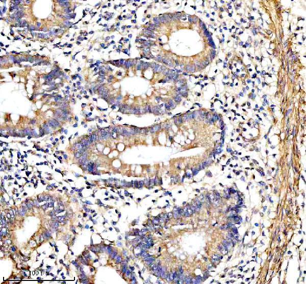
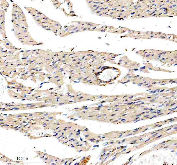
A02422-2 has pictured MYH11 IHC in paraffin sections of human appendix and rat heart; IF/ICC is also listed, without an IF image (catalog image captions; catalog applications).

Real IHC data IHC analysis of SMMHC/MYH11 using anti-SMMHC/MYH11 antibody (A02422-2). SMMHC/MYH11 was detected in a paraffin-embedded section of human appendix tissue. Heat mediated antigen retrieval was performed in EDTA buffer (pH 8.0, epitope retrieval solution). The tissue section was blocked with 10% goat serum. The tissue section was then incubated with 1:100 rabbit anti-SMMHC/MYH11 Antibody (A02422-2) overnight at 4°C. Peroxidase Conjugated Goat Anti-rabbit IgG was used as secondary antibody and incubated for 30 minutes at 37°C. The tissue section was developed using HRP Conjugated Rabbit IgG Super Vision Assay Kit (Catalog # SV0002) with DAB as the chromogen.
Anti-SMMHC/MYH11 Antibody
Cat # A02422-2

The rendered card is A02422-2, with pictured paraffin-section IHC in human appendix and rat heart (catalog image captions). M02422-1 lists human IHC and IF/ICC, while M02422-2 lists human and mouse IHC; neither has an IHC or IF image in the payload (catalog applications/reactivity; catalog image lists).

Which to pick: For tissue IHC, choose A02422-2 when a pictured paraffin-section example matters: its captions specify EDTA retrieval at pH 8.0, and the fixative is unreported (A02422-2 image captions). For IF/ICC, A02422-2 lists human, mouse and rat reactivity; M02422-1 is a human-reactive monoclonal alternative with IF/ICC listed, but neither has an IF image in the payload (catalog applications/reactivity; catalog clone; catalog image lists). For cross-species IHC, A02422-2 lists human, mouse and rat reactivity, while M02422-2 is a monoclonal option listed for human and mouse IHC without a pictured example (catalog applications/reactivity; catalog clone; catalog image lists).

Each figure is that product's own IHC / IF validation image from its datasheet.

References

  1. UniProt Consortium. UniProt entry P35749 (MYH11_HUMAN, Myosin-11).
  2. Human Protein Atlas. MYH11 tissue IHC expression (reliability: Enhanced).
  3. Human Protein Atlas. MYH11 subcellular location (ICC-IF): Mainly localized to the plasma membrane and cytosol..
  4. Human Protein Atlas. MYH11 antibody validation summary (3 antibodies).
  5. Interaction between DNMT3B and MYH11 via hypermethylation regulates gastric cancer progression. BMC cancer 2021 — PMC8359574.
  6. Basement membrane collagen IV deficiency promotes abdominal aortic aneurysm formation. Scientific reports 2021 — PMC8213747.
  7. Runx1 promotes scar deposition and inhibits myocardial proliferation and survival during zebrafish heart regeneration. Development (Cambridge, England) 2020 — PMC7197712.
  8. Repeated lengthening using distraction enterogenesis: A porcine model. Surgery 2026 — PMC13446750.
  9. PubMed PMID:16000639 — UniProt-cited evidence.
  10. PubMed PMID:10493829 — UniProt-cited evidence.
  11. PubMed PMID:10048485 — UniProt-cited evidence.