This website uses cookies to ensure you get the best experience on our website.


- Table of Contents
Plan chromogenic MYO1D IHC on paraffin sections with the catalog antibody’s 1:100–1:300 dilution range (datasheet: 1:100–1:300). Assess cytoplasmic staining and use controls to check specificity, given presumed off-target binding (HPA tissue IHC).

Expected localisation, validated protocols, controls and antibodies — the at-a-glance facts below, then the full design guide.
| Expected localisation | Cytoplasmic staining in most tissues (HPA tissue IHC) | |
| Staining pattern | Medium cytoplasmic staining in glandular and neuronal cells (HPA tissue IHC) | |
| Antigen retrieval | Citrate pH 6.0 HIER, 95–98 °C, 20 min (rule: cytoplasmic / membrane antigen) | |
| Positive control | Appendix+4 more · see all | |
| Negative control | Adipose tissue+4 more · see all |
| Fixation | Keep fixation consistent across paraffin sections. (standard IHC practice; not target-specific) | |
| Caveat | Presumed off-target binding; verify staining specificity (HPA tissue IHC) | |
| Regulation | Brain has the highest reported expression (UniProt) | |
| Isoform / epitope | 0 reported isoforms; chain spans residues 2–1006 (UniProt) |
The catalog antibody protocol is supplemented by published IHC methods for TNBC tissue microarrays (PMC10957690) and colorectal cancer sections (PMC8927999).
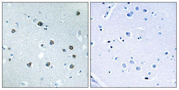
| Sample | Paraffin-embedded human brain tissue; fixative not specified (datasheet A09126-1) |
| Fixation | Image fixative and duration unreported (datasheet A09126-1); verify before use. |
| Sectioning | 4–5 µm sections on charged slides (standard) |
| Deparaffinisation | Xylene, graded ethanol series to water (standard) |
| Antigen retrieval | Heat-induced epitope retrieval in citrate buffer, pH 6.0, 20 min at 95–98 °C (standard rule: cytoplasmic / membrane antigen) |
| Peroxidase block | 3% H2O2, 10 min, room temperature (standard) |
| Blocking | 10% normal serum of the secondary host, 30 min, room temperature (standard) |
| Primary antibody | Rabbit anti-MYO1D, 1:100 - 1:300 (datasheet A09126-1) |
| Primary incubation | Overnight at 4 °C (standard) |
| Detection | HRP-polymer secondary, DAB chromogen 5–10 min (standard) |
| Counterstain | Hematoxylin, blue, dehydrate and mount (standard) |
| Expected result | MYO1D-positive staining in glandular cells of appendix (HPA tissue IHC: Medium). HPA tissue profile: Cytoplasmic expression in most tissues. No signal in the no-primary control. |
MYO1D should appear predominantly in the cytoplasm, with possible enrichment near the cell cortex and puncta resembling transport vesicles (UniProt O94832: subcellular location; HPA tissue IHC: cytoplasmic expression in most tissues). HPA reports medium staining in several glandular and neuronal cell populations (HPA tissue IHC). Its tissue IHC rating is Approved, but staining has low consistency with RNA data and remains pending external verification (HPA tissue IHC: reliability). MYO1D has no transmembrane segment (UniProt O94832: topology).
| Cytoplasmic chromogen in colon or duodenal glandular cells, with nuclei comparatively clear (HPA tissue IHC: Medium in these cells). | This fits the reported tissue pattern and cytoplasmic location (HPA tissue IHC; UniProt O94832: subcellular location). Compare the signal with an appropriate control on the same run; a matching compartment alone cannot establish antibody specificity because HPA flags presumed off-target binding (HPA tissue IHC: reliability). |
| Cytoplasmic staining in cerebellar Purkinje cells or cerebral cortex or hippocampal neurons (HPA tissue IHC: Medium in these cells). | These are reported positive cell populations and can help assess whether the run detects the expected pattern (HPA tissue IHC). Record the stained cell type as well as intensity: UniProt describes high expression in brain, whereas the HPA entries report staining at the level of specified cells (UniProt O94832: tissue specificity; HPA tissue IHC). |
| Predominantly nuclear staining, or a continuous cell-surface outline without appreciable cytoplasmic signal. | Neither is the principal reported location (HPA subcellular ICC-IF: cytosol; UniProt O94832: subcellular location and no transmembrane segment). Check the compartment against controls and staining quality before calling it MYO1D; cortex-associated signal can lie close to a membrane without indicating a transmembrane protein (UniProt O94832: subcellular location and topology). |
| Strong staining in adipocytes, esophageal squamous cells, or lung alveolar cells (HPA tissue IHC: Not detected in these cells). | The cell-type mismatch warrants scrutiny for cross-reactivity or endogenous detection activity (HPA tissue IHC: reliability; general IHC practice). Do not treat lung as uniformly negative: UniProt describes high lung expression at the tissue level, while HPA specifically reports no staining in alveolar cells (UniProt O94832: tissue specificity; HPA tissue IHC). |
| Diffuse chromogen across cells and extracellular spaces, or no signal in colon or duodenal glandular cells (HPA tissue IHC: Medium in these cells). | Diffuse staining obscures compartment assessment; an absent expected signal makes the run inconclusive (general IHC practice). Review background controls, detection, antigen retrieval and the catalog antibody's IHC-P instructions before assigning biological meaning (general IHC practice). HPA's Approved rating does not resolve a failed or noisy local run (HPA tissue IHC: reliability). |
| Which tissue and cell type are being scored? | HPA reports Medium staining in colon and duodenal glandular cells and in several neuronal populations, but Not detected in adipocytes and lung alveolar cells (HPA tissue IHC). Score the specified cells, not the entire section. UniProt's whole-tissue expression ranking cannot predict staining in every cell type (UniProt O94832: tissue specificity). |
| How certain is the antibody-based pattern? | The tissue IHC rating is Approved, with low staining–RNA consistency, external verification pending, and presumed off-target binding noted and disregarded in its assessment (HPA tissue IHC: reliability). The listed antibody HPA054744 has IHC Approved status; no Enhanced IHC status is supplied (HPA antibodies). Treat unexpected staining as provisional until controls support it. |
| Could a membrane-adjacent or punctate pattern be expected? | UniProt places MYO1D at the cell cortex near the apical membrane and in cytoplasmic puncta reminiscent of transport vesicles (UniProt O94832: subcellular location). Its lack of a transmembrane segment argues against interpreting a sharp, exclusive surface outline as its expected pattern (UniProt O94832: topology). |
| IF/ICC Q&A: where should fluorescence appear? | HPA's approved ICC-IF location is the cytosol (HPA subcellular ICC-IF). Assess cytosolic signal against the cell boundary and nucleus; UniProt also describes cortex-associated and vesicle-like punctate localization (UniProt O94832: subcellular location). This location guidance does not supply an IF/ICC protocol or validate an IHC-P result (HPA subcellular ICC-IF; general IHC practice). |
| Situation | Likely cause | Next action |
|---|---|---|
| No chromogen appears in colon or duodenal glandular cells (HPA tissue IHC: Medium). | A run-level detection or retrieval problem is possible; the tissue result alone cannot identify the failed step (general IHC practice). | Confirm tissue preservation and positive-control staining, then check the catalog antibody's IHC-P retrieval, dilution and detection instructions (general IHC practice). Do not infer MYO1D absence from this run. |
| The whole section develops a diffuse haze that masks cytoplasmic detail. | Nonspecific reagent binding, excess primary antibody or incomplete washing can raise background (general IHC practice). | Compare the no-primary control, review blocking and washes, and adjust primary antibody dilution using the validated IHC-P protocol (general IHC practice). Score only after cell boundaries and intracellular staining are distinguishable. |
| A brown precipitate appears in negative-control tissue or in cells reported Not detected (HPA tissue IHC). | Endogenous enzyme activity, if enzyme-based detection is used, or nonspecific binding can mimic chromogen signal (general IHC practice). | Inspect no-primary and detection-only controls and apply the detection system's endogenous-activity blocking procedure where relevant (general IHC practice). Reassess whether signal tracks the reported cytoplasmic cell pattern (HPA tissue IHC). |
| Staining is predominantly nuclear or forms only a crisp cell-surface rim. | The distribution conflicts with the reported cytosolic and cortex-associated locations; artefact or off-target binding is possible (HPA subcellular ICC-IF; UniProt O94832: subcellular location; HPA tissue IHC: reliability). | Check morphology, counterstain and controls, then compare a reported positive cell population on the same run (general IHC practice; HPA tissue IHC). Avoid scoring the atypical compartment as confirmed MYO1D. |
| Lung alveolar cells stain strongly despite an otherwise clean run (HPA tissue IHC: Not detected). | HPA's cell-specific result and UniProt's high whole-lung expression describe different levels of observation; neither establishes alveolar-cell positivity (HPA tissue IHC; UniProt O94832: tissue specificity). | Document the exact stained cell type and compare control sections before interpreting the signal (general IHC practice). HPA's low staining–RNA consistency and off-target caveat make this unexpected result provisional (HPA tissue IHC: reliability). |
| The run shows variable intensity among areas of a reported positive tissue (HPA tissue IHC). | Uneven section quality or reagent coverage can create an apparent gradient (general IHC practice); HPA's Medium category does not guarantee uniform staining in every cell (HPA tissue IHC). | Inspect morphology and within-run control staining, repeat the affected section if technical unevenness is evident, and record intensity by cell population rather than averaging the whole tissue (general IHC practice). |
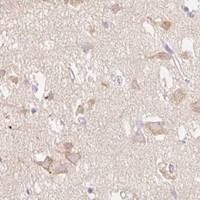


Comprehensive Human Protein Atlas IHC scoring per tissue (reliability: Approved — Low consistency between antibody staining and RNA expression data. Pending external verification. Presumed off target binding observed and disregarded.). Rows are taken directly from the HPA tissue chart — click any row's HPA link to view the source.
| Tissue | Cell type | Level | Evidence | Source |
|---|---|---|---|---|
| Appendix | Glandular cells | Medium | Protein (IHC) | HPA → |
| Cerebellum | Purkinje cells | Medium | Protein (IHC) | HPA → |
| Cerebral cortex | Neuronal cells | Medium | Protein (IHC) | HPA → |
| Colon | Glandular cells | Medium | Protein (IHC) | HPA → |
| Duodenum | Glandular cells | Medium | Protein (IHC) | HPA → |
| Tissue | Cell type | Level | Evidence | Source |
|---|---|---|---|---|
| Adipose tissue | Adipocytes | Not detected | Protein (IHC) | HPA → |
| Breast | Adipocytes | Not detected | Protein (IHC) | HPA → |
| Cervix | Glandular cells | Not detected | Protein (IHC) | HPA → |
| Endometrium | Cells in endometrial stroma | Not detected | Protein (IHC) | HPA → |
| Esophagus | Squamous epithelial cells | Not detected | Protein (IHC) | HPA → |
Troubleshoot MYO1D staining in paraffin sections by checking retrieval, compartment, controls and cell-specific signal before comparing samples.
A09126-1 has a real IHC image from paraffin-embedded human brain tissue and lists IF/ICC applications and human, mouse, and rat reactivity (catalog: A09126-1 image caption, applications, reactivity).
A09126-1 will render with an IHC image of paraffin-embedded human brain tissue and a peptide-blocked comparison (catalog: A09126-1 IHC image caption). It lists IHC, IF, ICC, and ELISA applications and human, mouse, and rat reactivity; no IF image is supplied (catalog: A09126-1 applications, reactivity, IF image alts).
Which to pick: For tissue IHC, choose A09126-1: its own image caption shows paraffin-embedded human brain tissue, but does not report the fixative (catalog: A09126-1 IHC image caption). For IF/ICC, A09126-1 is the listed option, although no IF image is supplied (catalog: A09126-1 applications, IF image alts). For cross-species work, A09126-1 is a rabbit polyclonal antibody listing human, mouse, and rat reactivity (catalog: A09126-1 host, dilution_raw, reactivity).