This website uses cookies to ensure you get the best experience on our website.


- Table of Contents
Plan MYO5A chromogenic IHC in paraffin sections using the cytoplasmic CNS staining pattern reported by HPA (HPA tissue IHC). This guide covers control selection, fixation consistency, and interpretation of tissue staining.

Expected localisation, validated protocols, controls and antibodies — the at-a-glance facts below, then the full design guide.
| Expected localisation | Cytoplasmic in several tissues, most abundant in CNS (HPA tissue IHC) | |
| Staining pattern | Cytoplasmic signal in cerebellar granular-layer cells and cortical neuropil (HPA tissue IHC) | |
| Antigen retrieval | Citrate pH 6.0 HIER, 95–98 °C, 20 min (rule: cytoplasmic / membrane antigen) | |
| Positive control | Cerebellum+4 more · see all | |
| Negative control | Adipose tissue+4 more · see all |
| Fixation | Matched tissue-IHC evidence does not establish the fixation claim. Validate the specimen-specific method before use. (selected-SKU IHC image A02216) | |
| Caveat | Parathyroid RNA is enhanced, but glandular staining is low (HPA tissue IHC) | |
| Regulation | Tissue-enhanced RNA: brain, parathyroid (HPA tissue RNA) | |
| Isoform / epitope | 3 isoforms; epitope coverage is unspecified (UniProt) |
Compare the catalog antibody’s IHC-P protocol with one published MYO5A IHC method (PMC10740236).
| Sample | Paraffin-embedded human breast cancer tissue; fixative not specified (datasheet A02216) |
| Fixation | Image fixative and duration unreported (datasheet A02216); verify before use. |
| Sectioning | 4–5 µm sections on charged slides (standard) |
| Deparaffinisation | Xylene, graded ethanol series to water (standard) |
| Antigen retrieval | Heat-induced epitope retrieval in citrate buffer, pH 6.0, 20 min at 95–98 °C (standard rule: cytoplasmic / membrane antigen) |
| Peroxidase block | 3% H2O2, 10 min, room temperature (standard) |
| Blocking | 10% normal serum of the secondary host, 30 min, room temperature (standard) |
| Primary antibody | Rabbit anti-MYO5A, 1:50-1:200 (datasheet A02216) |
| Primary incubation | Overnight at 4 °C (standard) |
| Detection | HRP-polymer secondary, DAB chromogen 5–10 min (standard) |
| Counterstain | Hematoxylin, blue, dehydrate and mount (standard) |
| Expected result | MYO5A-positive staining in cells in granular layer of cerebellum (HPA tissue IHC: High). HPA tissue profile: Cytoplasmic expression in several tissues, most abundant in CNS. No signal in the no-primary control. |
MYO5A should appear chiefly as cytoplasmic staining in several tissues, with the strongest reported signal in the CNS (HPA tissue IHC: Enhanced; medium consistency with RNA). Look for high staining in cerebellar granular-layer cells and cerebral-cortex neuropil, and medium staining in hippocampal neurons and several glandular cell populations (HPA tissue IHC). MYO5A has no transmembrane segment (UniProt Q9Y4I1 topology).
| Cytoplasmic staining in cerebellar granular-layer cells or cerebral-cortex neuropil. | This matches the reported high-signal sites (HPA tissue IHC: High). Judge the signal against local background and the tissue architecture; neuropil staining can extend between cell bodies, so a positive result need not outline individual cells (general IHC interpretation). |
| Predominantly nuclear, membranous, or extracellular staining in an otherwise positive region. | Treat the compartment mismatch as a warning of nonspecific signal or a detection artefact (HPA tissue IHC: cytoplasmic profile; general IHC interpretation). Check whether cytoplasmic signal remains after appropriate negative controls; UniProt does not annotate a MYO5A subcellular location (UniProt Q9Y4I1). |
| Strong staining in a cell population reported as undetected. | A signal in adipocytes, cardiomyocytes, or skeletal-muscle myocytes conflicts with those HPA observations (HPA tissue IHC: Not detected in these cells). Investigate antibody cross-reactivity or endogenous chromogenic activity before assigning it to MYO5A (general IHC practice). |
| Uniform color across tissue, lumina, and empty areas. | Diffuse staining that ignores cell boundaries is difficult to score as MYO5A (general IHC interpretation). In chromogenic IHC, inadequate blocking, excess primary antibody, or residual endogenous enzyme activity can contribute to background; compare a matched negative control (general IHC practice). |
| No signal in a cerebellar positive-control section. | Absence in granular-layer cells conflicts with the reported high staining there (HPA tissue IHC: High). First assess tissue preservation and the run controls, then review retrieval, primary-antibody dilution, and detection performance using the antibody's validated IHC-P procedure (general IHC practice). |
| Choice of reference tissue | Cerebellar granular-layer cells and cerebral-cortex neuropil are high-signal references; hippocampal neurons and breast, colon, endometrial, or fallopian-tube glandular cells have medium reported staining (HPA tissue IHC). A weak result in a medium-signal site alone is less informative than failure in a high-signal site (general IHC interpretation). |
| Antibody validation and interpretation limit | HPA lists HPA001356 as IHC Enhanced, meaning its pattern is supported by independent antibodies or orthogonal data (HPA antibody validation). The tissue profile still has medium consistency with RNA, so score the observed cells and compartment rather than treating every RNA-enriched tissue as a strong protein control (HPA tissue IHC). |
| Epitope and isoform coverage | MYO5A has three listed isoforms and defined motor, IQ, and Dilute regions (UniProt Q9Y4I1). The supplied evidence gives no antibody epitope or isoform coverage; staining cannot establish which isoform is present. Check the catalog antibody's documented epitope before making isoform-specific claims (general IHC interpretation). |
| IF/ICC Q&A: should puncta appear? | Yes, depending on the preparation: HPA reports approved IF/ICC localization at focal adhesion sites and centriolar satellites, with additional cytosolic and ciliary-associated sites (HPA subcellular). These cell-image observations do not require discrete puncta in chromogenic paraffin-section IHC, where HPA reports a cytoplasmic tissue profile (HPA tissue IHC). |
| Situation | Likely cause | Next action |
|---|---|---|
| Positive-control cerebellum is blank. | The result conflicts with high granular-layer staining (HPA tissue IHC); the failing step is not identified by that observation alone. | Confirm that the control tissue and detection run worked, then check retrieval, the primary-antibody dilution, and detection reagents against the validated IHC-P procedure (general IHC practice). |
| Only glandular tissue shows weak staining. | Several glandular populations have medium reported staining, while parathyroid glandular cells are low (HPA tissue IHC). | Compare the same run with a high-signal cerebellar or cortical section before calling the antibody negative; assess matched background and tissue preservation (HPA tissue IHC; general IHC practice). |
| Color appears in reported negative cell types. | Adipocytes, cardiomyocytes, and skeletal-muscle myocytes are reported as not detected; cross-reactivity or endogenous detection activity is possible (HPA tissue IHC; general IHC practice). | Review morphology and a no-primary control. If enzyme-based color remains without primary antibody, inspect the endogenous-enzyme blocking step (general IHC practice). |
| Nuclei dominate the staining. | The pattern conflicts with the reported cytoplasmic tissue profile (HPA tissue IHC); nonspecific staining or counterstain confusion may explain it (general IHC interpretation). | Compare with a no-primary section and inspect the chromogen separately from the nuclear counterstain; avoid scoring nuclear-only signal as MYO5A (general IHC practice). |
| Diffuse background hides cellular staining. | Excess primary antibody, inadequate blocking, or residual endogenous chromogenic activity can raise background (general IHC practice). | Use matched negative controls to locate the source, then review blocking, wash steps, primary-antibody dilution, and chromogen development within the validated IHC-P workflow (general IHC practice). |
| IF/ICC puncta and IHC sections look different. | HPA describes focal-adhesion and centriolar-satellite localization in IF/ICC images, but a cytoplasmic profile in tissue IHC (HPA subcellular; HPA tissue IHC). | Interpret each preparation against its own HPA pattern and controls. Do not reject a cytoplasmic paraffin-section result solely because it lacks resolvable IF/ICC puncta (HPA tissue IHC; general IHC interpretation). |
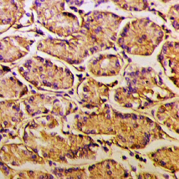
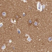
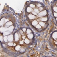
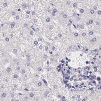
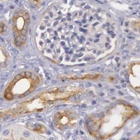


Comprehensive Human Protein Atlas IHC scoring per tissue (reliability: Enhanced — Medium consistency between antibody staining and RNA expression data.). Rows are taken directly from the HPA tissue chart — click any row's HPA link to view the source.
| Tissue | Cell type | Level | Evidence | Source |
|---|---|---|---|---|
| Cerebellum | Cells in granular layer | High | Protein (IHC) | HPA → |
| Cerebral cortex | Neuropil | High | Protein (IHC) | HPA → |
| Appendix | Lymphoid tissue | Medium | Protein (IHC) | HPA → |
| Breast | Glandular cells | Medium | Protein (IHC) | HPA → |
| Colon | Glandular cells | Medium | Protein (IHC) | HPA → |
| Tissue | Cell type | Level | Evidence | Source |
|---|---|---|---|---|
| Adipose tissue | Adipocytes | Not detected | Protein (IHC) | HPA → |
| Adrenal gland | Glandular cells | Not detected | Protein (IHC) | HPA → |
| Esophagus | Squamous epithelial cells | Not detected | Protein (IHC) | HPA → |
| Heart muscle | Cardiomyocytes | Not detected | Protein (IHC) | HPA → |
| Liver | Cholangiocytes | Not detected | Protein (IHC) | HPA → |
Troubleshoot MYO5A staining in paraffin section chromogenic IHC using the page retrieval conditions, cell specific controls, and cautious interpretation of cytoplasmic signal.
The catalog covers human tissue IHC and human, mouse, and rat IF/ICC; the supplied figure documents human paraffin-section IHC (catalog applications, reactivity, and A02216 image caption).
Only A02216 renders a card; it is listed for human IHC (catalog: A02216 applications and reactivity). Its own figure shows paraffin-embedded human breast cancer tissue at 1:50, with no fixative reported (catalog: A02216 image caption).
Which to pick: Choose A02216 for human tissue IHC: this rabbit polyclonal lists a 1:50–1:200 IHC dilution, and its own figure documents a paraffin section (catalog: A02216 host, dilution, and image caption). For IF/ICC, choose A02216-1, a rabbit polyclonal listed for human, mouse, and rat at 1:50–1:100 (catalog: A02216-1 applications, reactivity, host, and dilution). For broader species reactivity in an IHC-listed reagent, A02216-2 lists human, mouse, and rat, but its paraffin-section IHC entry specifies human at 2–5 μg/ml (catalog: A02216-2 reactivity and dilution).