This website uses cookies to ensure you get the best experience on our website.
- Table of Contents
Facts about Unconventional myosin-VIIa.
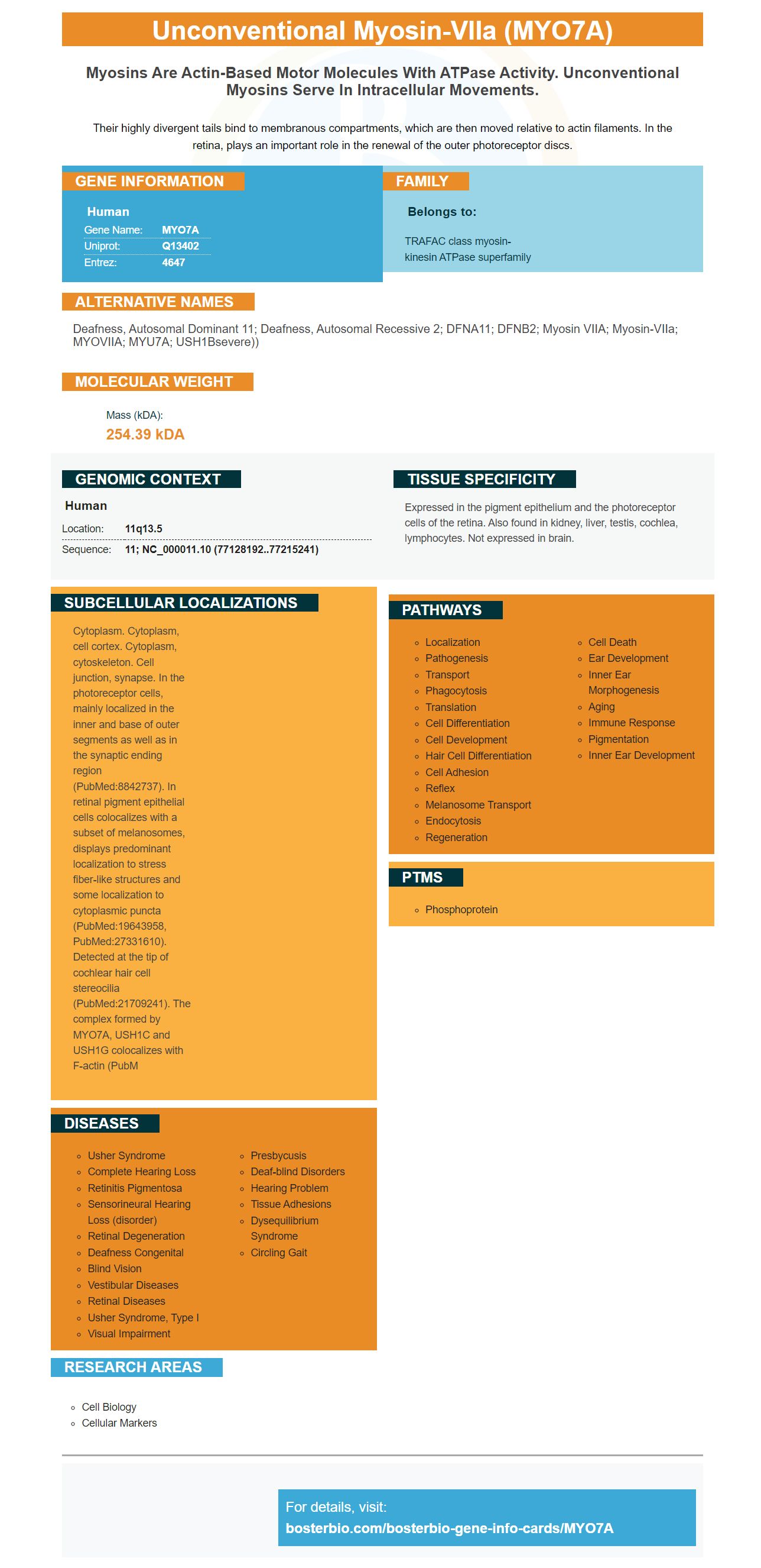
Their highly divergent tails bind to membranous compartments, which are then moved relative to actin filaments. In the retina, plays an important role in the renewal of the outer photoreceptor discs.
Human | |
---|---|
Gene Name: | MYO7A |
Uniprot: | Q13402 |
Entrez: | 4647 |
Belongs to: |
---|
TRAFAC class myosin-kinesin ATPase superfamily |
deafness, autosomal dominant 11; deafness, autosomal recessive 2; DFNA11; DFNB2; myosin VIIA; myosin-VIIa; MYOVIIA; MYU7A; USH1Bsevere))
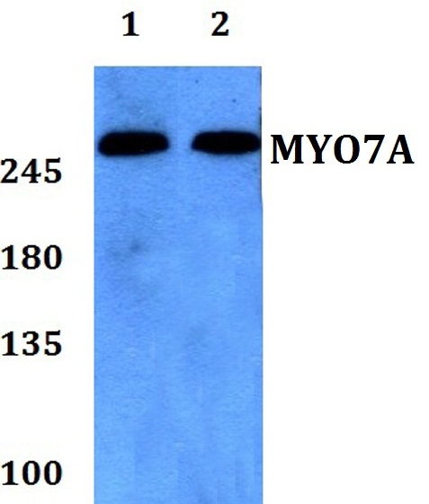
Mass (kDA):
254.39 kDA
Human | |
---|---|
Location: | 11q13.5 |
Sequence: | 11; NC_000011.10 (77128192..77215241) |
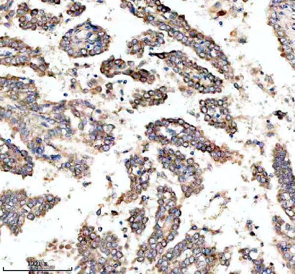
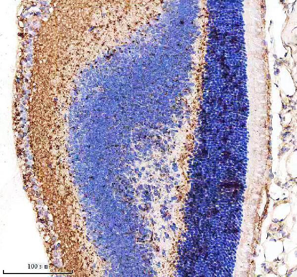
Expressed in the pigment epithelium and the photoreceptor cells of the retina. Also found in kidney, liver, testis, cochlea, lymphocytes. Not expressed in brain.
Cytoplasm. Cytoplasm, cell cortex. Cytoplasm, cytoskeleton. Cell junction, synapse. In the photoreceptor cells, mainly localized in the inner and base of outer segments as well as in the synaptic ending region (PubMed:8842737). In retinal pigment epithelial cells colocalizes with a subset of melanosomes, displays predominant localization to stress fiber-like structures and some localization to cytoplasmic puncta (PubMed:19643958, PubMed:27331610). Detected at the tip of cochlear hair cell stereocilia (PubMed:21709241). The complex formed by MYO7A, USH1C and USH1G colocalizes with F-actin (PubM
In this article, we will explore the Molecular pathogenesis, biological significance, and clinical applications of the MYO7A marker. We will be focusing on the high-affinity primary antibodies and their applications in veterinary diagnostics. Any scientist around the world can use the product credits. This article is not intended as a guide to the MYO7A mark. Anyone interested in obtaining the best data for their research can use this article.
Molecular pathogenesis is a study of host-pathogen interactions. It examines the role that microbial communities play in the spread of infectious diseases. Faculty members study the immune response of hosts to pathogens. The study of host resistance and interactions with pathogens is part of molecular pathogenesis. It also involves careful divisions of labor among the bacteria living on each chromosome. This information will provide more information about this field.
Recently, ETEC's virus-causing proteins were identified and are now implicated in disease severity. This discovery could be used to focus vaccine development efforts on broad antigens related to ETEC. ETEC's molecular pathogenesis is a subject of ongoing research. Vaccine development efforts continue to advance in tandem. But how will vaccine development proceed? It is important to develop an effective vaccine to protect against ETEC. Therefore, scientists are looking for new and alternative targets.
There are many mutations in the MYO7A gene. Some of these mutations, however, are conserved in many species. For example, D218N's mutation has a greater functional impact that the wild-type D218. In knockout mice mutations affecting DLNA11 are also found in the tectorial membrane. In addition, mutations in the POU4F3 transcription factor cause DFNA15 and DFNA17, two forms of progressive HL. Although the exact cause of DFNA17 has not been determined, mutations to these genes could cause HL-like symptoms.
The genetic association between MYO7A and the gene expression of HC was determined using a genetic linkage map. Twenty-three individuals from one family were genotyped. Genomic scanning took place at 10-cM intervals. This allowed the region to be identified and the rest of genome to be omitted. Further markers that were found in the region were identified. They yielded a maximum of 2.18 LOD scores at th=0. These markers were all within one cm of the MYO7A Gene.
The 11q13.5-located MYO7A genome encodes an uncommon myosin. Mutations in this gene can lead to Usher syndrome types I, II, and non-syndromic sensory deafness. MYO7A is composed of 49 exons. MYO7A's polypeptide contains 2215 amino acids. It is found in the retina and inner ear hair cells.
Mutations in the MYO7A gene affect the MYO7A protein's structure. Another disease-causing genetic is DFNA11. Mutations in the MYO7A genes are present in the DFNA11 gene. Mutations within DFNA11 affect the structure and function of the protein. Mutations in MYO7A are thought to cause disruptions in stereocilia structure, which is essential for auditory function.
The MYO7A genome encodes an uncommon myosin. Unconventional myosins are motor molecules that move along actin filaments. They can transport cargo and have tails that can be tethered with various macromolecular structure. This is why MYO7A is so important. It plays a major role in motor, sensory and cognitive development. Interestingly, the MYO7A gene has many functions in eukaryotic cells.
The MYO7A genes encode a protein that binds Actin. It has a motor-domain and a tail predicted to dimerize. Unconventional meosins can perform many functions in eukaryotic cellular cells, including movement and linking intracellular organelles and membranes. The protein's long tail, measuring 430 amino acids, is attached to a variety o macromolecules such as actin filaments or membrane-associated protein.
A study by Janecke et al. Janecke et al. The study also revealed that six intragenic polymorphisms were genotyped, indicating that nine mutation-bearing genes had been derived from the same ancestor. Another study conducted by Weston et al. Two probands in Sweden and the United States were found to be carrying the C31X genetic mutation. The mutation was considered a variant of unknown importance by the authors. However, it was not determined if it contributed to the syndrome.
These new findings allow the MYO7A genome to be used for the diagnosis and prognostication many diseases including cancer. The MYO7A gene is an excellent candidate for mutation screening in cancer patients. Researchers are currently studying a novel genetic variant that could be the cause of the disease. Based on genetic data, the study also suggests MYO7A as a promising candidate for cancer patients.
The MYO7A Gene is found in human eyes and photoreceptors. It is also implicated in Usher syndrome, which can cause blindness or deafness. Researchers have identified over 100 mutations to the human MYO7A genome. The MYO7A Gene is too large to allow for one AAV vector.
The D218N mutation was first discovered in MYO7A by a Chinese family that had DFNA11 mutated genes. This mutation replaces an aspartic acid negative charge with a neutral asparagine. Interchain interactions among neighboring B-strands were affected by the loss hydrogen bonds. MYO7A's G671 residue is located in the myosin head converter domain. The N-terminal globular globular domain interacts to actin filaments, and utilizes energy generated by ATP Hydrolysis. The central core is the motor of the myosin.
This product is a highly specific antisense antibody against the myosin VIIa/MYO7A marker, which has been identified in mouse tissues and recombinant and insect cell lysates. The company also offers knockout edited cell lines and gold standard validation. These high-affinity antibodies are available from multiple sources, including Abcam. Abcam's MYO7A antigen was developed with a mouse antibody as a control.
The TMTC4–c–Myc plasmid was used for immunoisolating a nonnuclear fraction HEK cells. The immunoisolated TMTC4–c–Myc protein proteins were purified with antibodies to c–Myc or Endo H. To stain this bound protein fraction, antibodies to ER proteins were used.
PMID: 8622919 by Weil D., et al. Human myosin VIIA responsible for the Usher 1B syndrome: a predicted membrane-associated motor protein expressed in developing sensory epithelia.
PMID: 8884267 by Chen Z.-Y., et al. Molecular cloning and domain structure of human myosin-VIIa, the gene product defective in usher syndrome 1B.