This website uses cookies to ensure you get the best experience on our website.
- Table of Contents
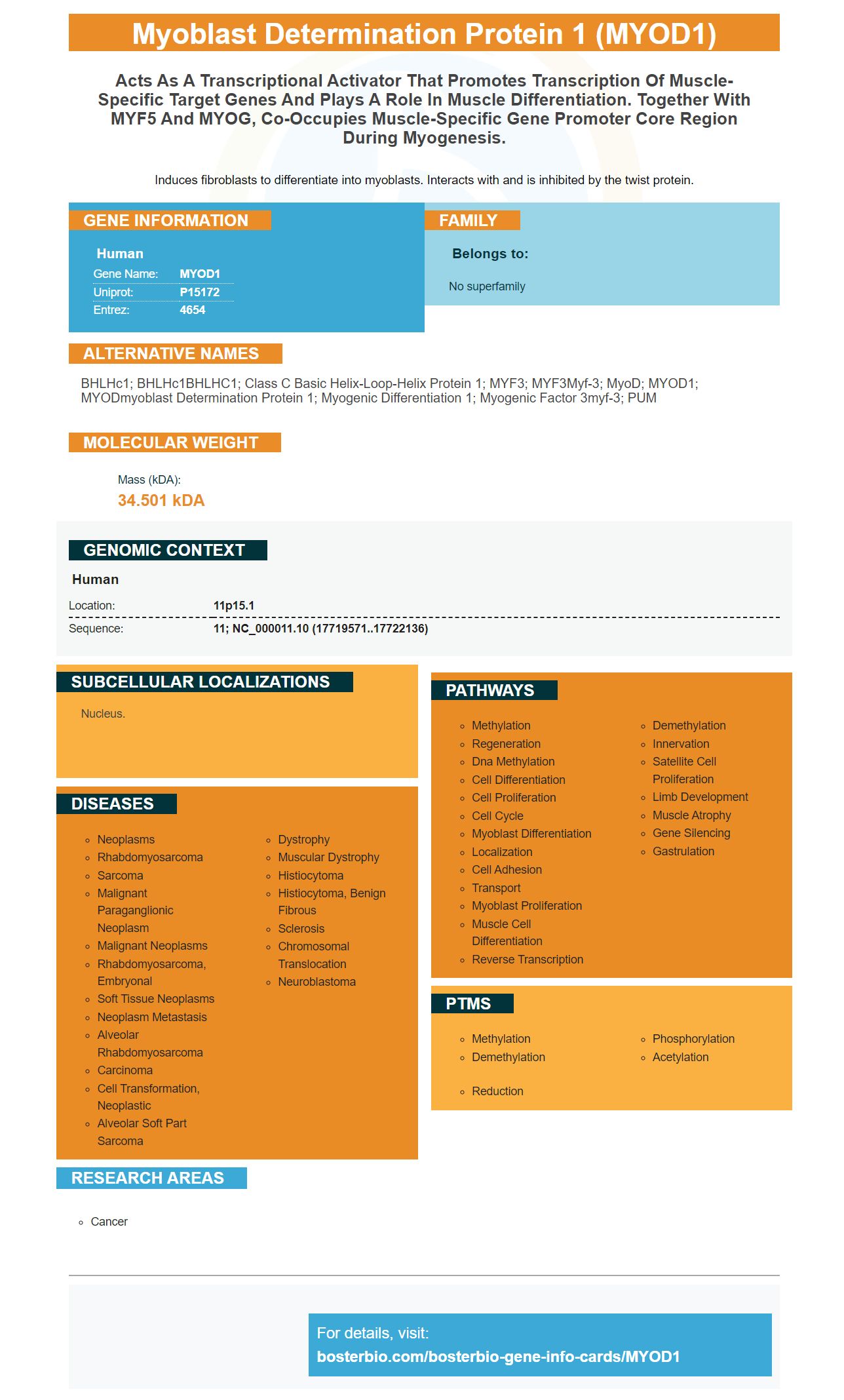
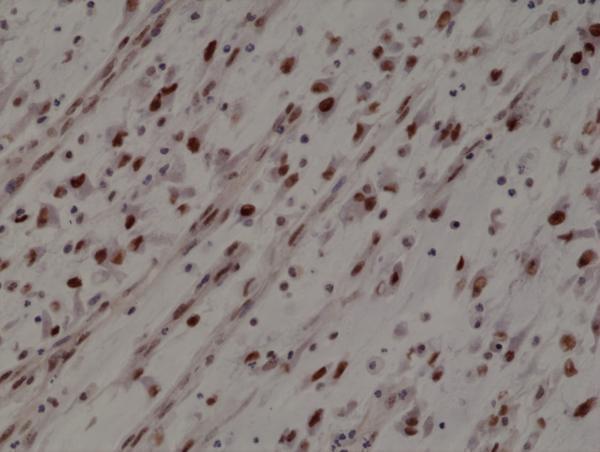
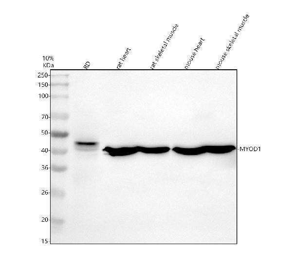
Facts about Myoblast determination protein 1.
Induces fibroblasts to differentiate into myoblasts. Interacts with and is inhibited by the twist protein.
Human | |
---|---|
Gene Name: | MYOD1 |
Uniprot: | P15172 |
Entrez: | 4654 |
Belongs to: |
---|
No superfamily |
bHLHc1; bHLHc1BHLHC1; Class C basic helix-loop-helix protein 1; MYF3; MYF3Myf-3; MyoD; MYOD1; MYODmyoblast determination protein 1; myogenic differentiation 1; Myogenic factor 3myf-3; PUM
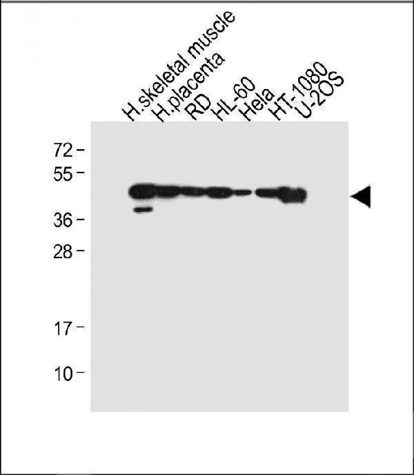
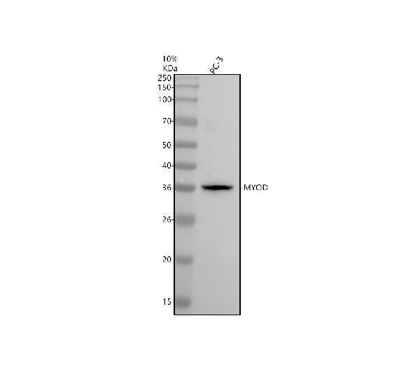
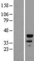
Mass (kDA):
34.501 kDA
Human | |
---|---|
Location: | 11p15.1 |
Sequence: | 11; NC_000011.10 (17719571..17722136) |
Nucleus.
You've likely noticed several helpful tips while researching the Boster Bio Anti MyoD MYOD1 marker. These tips include recommendations, incubation and applications. Continue reading to find out more. This article discusses some of the most useful information about this product. It's applicable for scientists worldwide and covers all the essential facts before you buy.
The Boster Bio Anti MyoD MyoD1Marker monoclonal antibody is used in IP or IF applications. It reacts with Rat, Mouse, and Human. Scientists who use the product are eligible for credit. It is available at a cost of $19 per ml. It is highly recommendable for clinical research.
The antibody is a monoclonal mouse antibody that specifically recognizes MYOD1 (the human gene). It can also be activated by other species, including chicken/birds, mice, and rats. It is suitable for use in immunohistochemistry, Western blot, and flow cytometry. The Boster Bio Anti-MyoD MyOD1 Marker can be used for quantifying the protein in tissue samples. This includes heart tissue, muscle, and liver tissue.
It has a large literature supporting its use in myogenic markers. MyoD1 is one of the earliest markers of myogenic commitment, and is also expressed in activated satellite cells, but not in quiescent cells. MyoD1 also plays a role in muscle repair. The MyoD1 Marker can be used to label tumor cells if the expression of this protein has been altered.
MYOD1 regulates the expressions of many genes, including myod which affects cell positioning and migration. This gene activates transcription elements to control muscle differentiate. The MYOD1 marker can be used in many research areas. The mechanism by which Myod works is still unknown. MYOD1 is one of the most frequently mutational genes in the genome. Its overexpression is often associated with muscle phenotypic differences.
Although Myod gene expression can be found in many cell types and tissues, it is most prominently expressed by primary fibroblasts. Both cell types contain transacting factors such as Msx1, that suppress Myod transcription. The PAX7 levels were higher in FSCHF and FGF than in Myod. But, the MYOD1 marker is still in its early stages of research.
The molecular analysis of myod expression has been a key tool in the differentiation of skeletal muscles. Myod was identified, which provided a framework that allowed us to study the function and regulation of chromatin-associated protein and tissue-specific transcription elements in the myogenic programme. Myod regulates genes throughout the myogenic programme, thereby achieving promoter-specific regulation. Furthermore, myod studies have also helped in integrating lineage-specific information with master regulatory factors.
In addition to Myod protein function, the other domains are highly conserved in myod. The acidicdomain acts as a transcriptional activation and binding domain by binding DNA close to E boxes. H/C plays an important part in chromatin modification. Myod1 has the ability in pigs to regulate the expression muscle-specific genes and prevent cells becoming non-muscle.
Many cell types have the MYOD1 genes. The differentiation of skeletal-muscle cells is affected by Myod gene expression. Myod, a transcription factor, has binding sites in the regulatory areas of several muscle gene genes. It forms heterodimers together with bHLH subfamily proteins and E-protein subfamily protein. Its DNA-binding domain is the basic region. It recognizes the CANNTG consensus sequence. Flanking sequences are a preferred choice.
Incubation requires the presence and function of a transcription factors, also known to be called a myogenic factor. This gene is involved myogenesis, which is the process by which stem cells are used to create skeletal muscle. It regulates cell fate and induces the arrest of the cell cell cycle. This is a prerequisite for myogenesis. The marker helps in the differentiation of cells that are in their pluripotent state, which may be a useful tool for studies of cell biology and myogenesis.
This marker can be used for assessing the activity level of myoblasts. MyoD, and MyoG are proteins that play a role in myogenesis. MyoD or MyoG recruit Brg1 at their target loci. Myoblasts with Arp5 gene silencing result in a higher amount of MyoD or MyoG. Myotube fusion is inhibited by Arp5 overexpression.
The amino acids sequence of MYOD1 are conserved by all species. The Myod1 basic region binds Ebox DNA. It also interacts to Pbx1/Meis1 o-factors. It also recognizes Ebox, a conserved sequence of DNA found throughout the genome. The HLH element of MYOD1 represents a highly conserved region.
In addition to its role in myogenesis, MYOD1 is also involved in the conversion of somatic cells into mature skeletal muscle. An established protocol includes the ectopic release of MYOD1 into pigfibroblasts. Signaling molecules associated with myogenesis, such as WNT activator and FGF, are applied in these modified conditions. These myotubes, once transdifferentiated have contractile potential and the characteristics of myoblasts.
In vitro studies have shown that MYOD lentivirus transduction of hAFS cell with MYOD lentivirus triggers muscle regeneration. It has also been demonstrated that MYOD lentivirus-hAFS cells can promote the differentiation and growth of skeletal myoblasts, both in vitro as well as in vivo. This method could help diagnose DMD. This allows us to develop an in vitro method to test MYOD1.
The cells were fixed with 4% Paraformaldehyde for 15 minutes, and then treated twice with Dulbecco’s phosphate-buffered saline. This was before the MYOD1 RTPCR. Incubation of the MYOD1 marker in Dulbecco's phosphate-buffered saline (DSBS) for transdifferentiation was determined. On day 8, multinucleated mobos were seen in both of the groups. qPCR analyses of cell culture conditions revealed an increase in the expression of MYF5, ExoMYOD1, MYOG and MYF5.
RMS patients can be helped by the MyoD1 gene. It is known that mutations in this gene cause poor responses to existing treatment regimens. The MYOD1 gene is expressed in high numbers in the myotome. Mutations can cause fatigue, muscle weakness, and other symptoms. Below are some recommendations for the use of the MYOD1 marker in RMS suffers.
PMID: 1850513 by Pearson-White S.H.; Human MyoD: cDNA and deduced amino acid sequence.
PMID: 9546368 by Chen B., et al. Methylation alterations of the MyoD1 upstream region are predictive of subclassification of human rhabdomyosarcomas.