MYSM1 / Deubiquitinase MYSM1 · IHC design guide

Design Immunohistochemistry for MYSM1

Plan MYSM1 staining in paraffin sections using the predominantly nuclear tissue pattern, with additional cytoplasmic staining, reported by HPA (HPA tissue IHC). The guide covers controls, interpretation and the catalog antibody’s IHC dilution of 1:100–1:300 (datasheet).

Evidence assembled Oct 2026 · For research use; verify linked source records and product datasheet before use
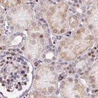
Immunohistochemistry protocol sheet for MYSM1 (IHC for MYSM1): expected localisation Predominantly nuclear, with cytoplasmic staining (HPA tissue IHC), antibody A06500, validated IHC image, and IHC protocol steps
Printable MYSM1 IHC protocol sheet — expected localisation Predominantly nuclear, with cytoplasmic staining (HPA tissue IHC), antibody A06500, controls and protocol steps. Open the full MYSM1 IHC guide →

MYSM1 Immunohistochemistry Experimental Design Guide

Expected localisation, validated protocols, controls and antibodies — the at-a-glance facts below, then the full design guide.

Must know before staining
Expected localisation Predominantly nuclear, with cytoplasmic staining (HPA tissue IHC)
Staining pattern General nuclear staining with additional cytoplasm (HPA tissue IHC)
Antigen retrieval Tris-EDTA pH 9.0 HIER, 95–98 °C, 20 min (rule: nuclear antigen)
Positive control ⓘ Appendix+4 more · see all
Negative control ⓘ None in HPA (detected in all 45 tissues); use no-primary + isotype controls
Important caveats
Reasons your staining may differ from the expected pattern.
Fixation Keep fixation consistent across sections (standard IHC practice; not target-specific)
Caveat Bacterial infection can shift MYSM1 into cytoplasm (UniProt)
Regulation Low tissue specificity; no defined inducer (HPA tissue IHC)
Isoform / epitope Three isoforms; check antibody epitope coverage (UniProt)
Section 1

Recommended MYSM1 IHC & IF Protocols

Compare the catalog antibody’s IHC-P protocol with three published MYSM1 staining protocols for paraffin sections (PMC5428969; PMC8881619; PMC12572397).

Recommended immunohistochemistry (IHC-P) protocol parameters
SampleParaffin-embedded human breast carcinoma tissue; fixative not specified (datasheet A06500)
FixationImage fixative and duration unreported (datasheet A06500); verify before use.
Sectioning4–5 µm sections on charged slides (standard)
DeparaffinisationXylene, graded ethanol series to water (standard)
Antigen retrievalHeat-induced epitope retrieval in Tris-EDTA buffer, pH 9.0, 20 min at 95–98 °C (standard rule: nuclear antigen)
Peroxidase block3% H2O2, 10 min, room temperature (standard)
Blocking10% normal serum of the secondary host, 30 min, room temperature (standard)
Primary antibodyRabbit anti-MYSM1, 1:100-1:300 (datasheet A06500)
Primary incubationOvernight at 4 °C (standard)
DetectionHRP-polymer secondary, DAB chromogen 5–10 min (standard)
CounterstainHematoxylin, blue, dehydrate and mount (standard)
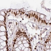
Expected resultMYSM1-positive staining in glandular cells of appendix (HPA tissue IHC: High). HPA tissue profile: General nuclear expression with additional cytoplasm. No signal in the no-primary control.
💡Decision noteStart with Tris-EDTA pH 9.0 at 95–98 °C for 20 min for nuclear MYSM1 (page retrieval; UniProt: nucleus). Use citrate retrieval when reproducing the cited protocols (PMC5428969; PMC8881619).
Section 2

What Is the Expected MYSM1 Staining Pattern?

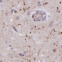
MYSM1 is nuclear and cytoplasmic, with cytoplasmic localization reported in response to bacterial infection (UniProt Q5VVJ2). In tissue IHC, expect predominantly nuclear staining with some cytoplasmic staining (HPA: general nuclear expression with additional cytoplasm). HPA reports high staining in bone marrow hematopoietic cells and several epithelial cell types, while overall tissue RNA specificity is low (HPA: tissue IHC). Its topology has no transmembrane segment (UniProt Q5VVJ2). HPA rates the tissue IHC pattern Approved, with medium staining–RNA consistency (HPA: tissue IHC).

What am I looking at on my slide?
Nuclear staining in bone marrow hematopoietic cells or reported glandular and epithelial cells.This fits the reported distribution (HPA: High in bone marrow hematopoietic cells; High in appendix, colon and duodenum glandular cells; High in bronchial, cervical and esophageal epithelial cells). Compare signal within each cell population; HPA's levels describe observed staining, not a required intensity for every specimen (HPA: tissue IHC).
Nuclear staining accompanied by cytoplasmic signal.This can fit MYSM1: HPA describes general nuclear expression with additional cytoplasm, and UniProt lists both compartments (HPA: tissue IHC; UniProt Q5VVJ2). Cytoplasmic localization during bacterial infection is a context-specific annotation, not evidence that a routine section is infected (UniProt Q5VVJ2).
Only membrane-edge or extracellular staining, without a convincing cellular nuclear pattern.Treat this as a possible artefact and check controls and morphology (general IHC practice). It does not match HPA's predominantly nuclear tissue pattern or the absence of a transmembrane segment (HPA: tissue IHC; UniProt Q5VVJ2 topology).
Strong staining in an unexpected cell population, especially without nuclear enrichment.Consider cross-reactivity or endogenous chromogen-generating activity, then assess controls (general IHC practice). Do not call a cell type MYSM1-negative solely because it is absent from HPA's examples: HPA reports low tissue RNA specificity and provides no negative tissue entries here (HPA: tissue IHC).
No signal in bone marrow hematopoietic cells or another reported high-staining population.First check whether tissue morphology and the staining run are interpretable (general IHC practice). Absence conflicts with HPA's reported high staining, but a single failed section cannot establish MYSM1 absence or a MYSM1-specific fixation effect (HPA: tissue IHC).
💡Expected MYSM1 appearanceA convincing positive shows predominantly nuclear staining, sometimes with cytoplasm, in reported hematopoietic or epithelial populations; HPA calls their staining High, whereas isolated membrane or extracellular color is suspect (HPA: tissue IHC; UniProt Q5VVJ2 topology; general IHC practice).
How each factor affects the staining
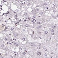
Tissue and cell populationHPA reports High staining in bone marrow hematopoietic cells and listed glandular and epithelial cells, but Low staining in hippocampal glia, hepatocytes, adipocytes and two other listed populations (HPA: tissue IHC). Low means a weaker reported pattern, not a negative control (HPA: tissue IHC).
Antibody evidenceHPA054291 is IHC Approved; HPA describes overall staining–RNA agreement as medium (HPA: antibody validation; tissue IHC). These ratings support comparison with the observed pattern but do not prove specificity in every specimen (general IHC interpretation).
Molecular form and locationUniProt lists 3 isoforms, no signal peptide or propeptide, and no transmembrane segment (UniProt Q5VVJ2). These annotations do not identify which isoforms an antibody detects or predict an epitope-specific retrieval condition; antibody epitope data are absent from the supplied record.
IF/ICC Q&A: Where should signal appear?Mainly in the nucleoplasm, with additional nucleolar staining (HPA: supported ICC-IF localization). HPA057306 has Supported ICC evidence, while HPA054291 has Approved IHC evidence (HPA: antibody validation). These are application-specific observations; this IHC section does not supply an IF/ICC protocol.
Why is my staining missing, weak or wrong?
SituationLikely causeNext action
No staining in a reported high-staining tissue.The run may have failed, or signal may be below detection (general IHC practice); the observation conflicts with HPA's reported High pattern (HPA: tissue IHC).Review tissue integrity, positive-control performance, retrieval and primary-antibody application against the chosen assay instructions (general IHC practice). Do not infer MYSM1-specific fixation sensitivity from this result.
Color appears broadly outside cells or across empty areas.Diffuse reagent or chromogen background can obscure cellular localization (general IHC practice).Inspect the no-primary control; review blocking, washes and detection exposure, then judge staining only where morphology identifies cells (general IHC practice).
Unexpected color concentrates in blood-rich or inflammatory areas.Endogenous peroxidase activity can contribute chromogenic signal in such areas (general IHC practice).Compare a no-primary control and review the peroxidase-blocking step for the detection system (general IHC practice). Do not equate color alone with MYSM1 positivity.
Staining appears only at cell borders.A border-only pattern conflicts with predominantly nuclear MYSM1 staining (HPA: tissue IHC); edge artefact is possible (general IHC practice).Check nuclear counterstain and morphology, compare controls, and repeat with the IHC-validated antibody if the pattern persists (general IHC practice; HPA: HPA054291 IHC Approved).
A low-staining tissue gives faint or variable color.HPA reports Low staining in selected cell populations; faint signal may be hard to distinguish from background (HPA: tissue IHC; general IHC practice).Compare the same run with a reported high-staining population and the no-primary control before scoring it as positive or absent (HPA: tissue IHC; general IHC practice).
Nuclear signal is accompanied by cytoplasmic color.Both compartments are documented; cytoplasmic signal alone does not establish bacterial infection (HPA: tissue IHC; UniProt Q5VVJ2).Score nuclear and cytoplasmic staining separately and compare cell morphology and controls before assigning a biological explanation (general IHC practice).

Sample controls for MYSM1 IHC & IF

🧪Start with bone marrow: hematopoietic cells should stain (HPA: High in bone marrow hematopoietic cells). HPA detects MYSM1 in all 45 scored tissues, so no tissue is established as negative; use no-primary and isotype controls, and assess any unstained cells on the positive slide for counterstain alone without specific chromogen signal, without assuming they are MYSM1-negative (HPA: no negative tissue rows; standard IHC practice).
Positive control tissue: Appendix (Glandular cells, HPA High)
Negative control tissue: None in HPA: MYSM1 is detected in all 45 scored tissues. Use a no-primary (secondary-only) and an isotype control instead.
ICC-IF cell lines (HPA subcellular resource): HPA ICC-IF images show MYSM1 in A-431, U-251MG, U2OS, with annotated localisation: Nucleoplasm (supported) (HPA subcellular).
Technical controls: Run a no-primary, secondary-only control and an isotype control matched to the primary antibody’s host species and clonality; use MYSM1 knockout material or peptide competition as a biological specificity control (standard IHC practice; A06500 tissue-IHC caption: peptide block). In bone marrow, block endogenous peroxidase and inspect residual pigment or background before scoring chromogenic staining (standard IHC practice).
⚠️Feasibility: Paraffin-section IHC is documented in a breast carcinoma caption, but the fixative is unreported; no target-specific fixation window or fixation effect is supplied (A06500 tissue-IHC caption). MYSM1-specific antigen retrieval dependence and whether frozen sections or IF are easier are unreported in the supplied evidence; optimize retrieval on control sections and assess nuclear localization during interpretation (HPA subcellular: nucleoplasm supported; standard IHC practice). Bone marrow pigment and endogenous peroxidase can complicate chromogenic scoring (standard IHC practice).

HPA tissue IHC evidence for MYSM1

Comprehensive Human Protein Atlas IHC scoring per tissue (reliability: Approved — Medium consistency between antibody staining and RNA expression data.). Rows are taken directly from the HPA tissue chart — click any row's HPA link to view the source.

Positive expression · recommended positive controls

TissueCell typeLevelEvidenceSource
Appendix Glandular cells High Protein (IHC) HPA →
Bone marrow Hematopoietic cells High Protein (IHC) HPA →
Bronchus Respiratory epithelial cells High Protein (IHC) HPA →
Cervix Squamous epithelial cells High Protein (IHC) HPA →
Colon Glandular cells High Protein (IHC) HPA →

Undetected expression · recommended negative controls

TissueCell typeLevelEvidenceSource
None in HPA: MYSM1 is detected in all 45 scored tissues. Use a no-primary (secondary-only) and an isotype control instead.
Section 3

Advanced MYSM1 IHC Tips

Troubleshoot MYSM1 staining by checking retrieval, controls and compartment-specific signal before comparing expression across specimens.

How should I optimize retrieval when MYSM1 staining is weak?
For MYSM1 paraffin-section IHC, start with Tris-EDTA pH 9.0 retrieval at 95–98 °C for 20 min (page protocol). Compare adjacent sections while keeping antibody incubation and chromogenic detection constant, and assess both signal and tissue morphology (standard IHC practice). MYSM1 can occupy the nucleus and cytoplasm, so judge retrieval against both compartments while expecting predominantly nuclear tissue staining (UniProt Q5VVJ2; HPA tissue IHC). If signal remains weak, test citrate pH 6.0 as a fallback on another section (standard IHC practice). Include a no-primary control to distinguish improved staining from retrieval-related background (standard IHC practice).
Could fixation explain weak or uneven MYSM1 staining?
Target-specific MYSM1 sensitivity to fixation is unknown from the supplied evidence; the selected paraffin-section caption does not state a fixative (A06500 caption). Record each specimen’s fixative, fixation interval and processing history before attributing a staining difference to biology (standard IHC practice). Compare sections processed together using the page’s Tris-EDTA pH 9.0, 95–98 °C, 20 min retrieval conditions (page protocol). Inspect preserved nuclear detail and section adhesion, then repeat staining on a comparably processed control if either is poor (standard IHC practice). Do not infer fixation tolerance from tissue expression patterns or MYSM1 topology and phosphorylation annotations (HPA tissue IHC; UniProt Q5VVJ2).
Should MYSM1 staining be nuclear, cytoplasmic or both?
Score nuclear and cytoplasmic chromogen separately: MYSM1 is annotated in both compartments, and tissue staining is generally nuclear with additional cytoplasm (UniProt Q5VVJ2; HPA tissue IHC). Nucleoplasmic localisation, with additional nucleolar localisation, is supported by cellular imaging and can help guide review of ambiguous nuclear signal (HPA subcellular). Cytoplasmic redistribution is reported in response to bacterial infection, but that context should not be assumed for an unstated specimen (UniProt Q5VVJ2). Check the same cell population across adjacent sections and use a no-primary control to assess diffuse cytoplasmic colour (standard IHC practice). Record compartment, intensity and positive-cell fraction independently before comparing specimens (standard IHC practice).
How can epitope choice affect MYSM1 IHC interpretation?
MYSM1 has 3 annotated isoforms, so check the antibody’s stated immunogen against the isoform sequence before treating absent staining as absent protein (UniProt Q5VVJ2). Its SANT, SWIRM and MPN domains span residues 116–167, 372–470 and 577–709, respectively (UniProt Q5VVJ2). Without an epitope map for the selected antibody, domain annotations cannot establish which isoforms or processed epitopes it detects (UniProt Q5VVJ2; A06500 caption). Compare retrieval conditions on adjacent sections and verify that staining follows plausible nuclear or cytoplasmic localisation (page protocol; UniProt Q5VVJ2). The selected paraffin-section image includes a peptide-blocked comparison, which supports assessing reagent-dependent staining without establishing isoform specificity (A06500 caption).
How should I investigate MYSM1 localisation with multiplex IF?
Treat IF as a separate assay: the selected antibody evidence describes paraffin-section chromogenic IHC, not IF validation (A06500 caption). Pair MYSM1 with a marker for the expected cell population, using the reported high staining in bone marrow hematopoietic cells or appendix glandular cells to guide specimen selection (HPA tissue IHC; standard IF practice). Place the dimmer signal in a fluorophore channel with low specimen autofluorescence, and check single-stain controls for spectral bleed-through (standard IF practice). Because MYSM1 has no transmembrane segment and is annotated in nuclear and cytoplasmic compartments, choose permeabilisation that admits antibody to the compartment being assessed (UniProt Q5VVJ2; standard IF practice). Compare nuclear and cytoplasmic signal with matched no-primary controls (standard IF practice).
What should I check when MYSM1 chromogen appears diffuse?
Inspect a no-primary section and the peptide-blocked comparison before calling diffuse chromogen MYSM1 signal; peptide blocking is shown for the selected paraffin-section image (standard IHC practice; A06500 caption). For peroxidase-based detection, confirm the endogenous peroxidase block and compare substrate development across matched sections (standard IHC practice). Review section edges, folds and damaged areas separately because staining there can obscure a cellular pattern (standard IHC practice). MYSM1 tissue staining is generally nuclear with additional cytoplasm, so diffuse extracellular colour warrants particular scrutiny (HPA tissue IHC; standard IHC practice). Recheck Tris-EDTA pH 9.0 retrieval at 95–98 °C for 20 min before changing other conditions (page protocol).
How should I score MYSM1 across mixed cell populations? ⚠ ANSWER MARKED FOR VERIFICATION
Define the cell population and nuclear versus cytoplasmic compartment before scoring, because MYSM1 can occupy both and tissue staining is generally nuclear (UniProt Q5VVJ2; HPA tissue IHC). For intensity, record an H-score as the sum of each intensity grade multiplied by its percentage of cells; also report the percentage positive (standard IHC practice). For sparse populations, report positive cells per mm² of viable tissue and state the area sampled (standard IHC practice). Normalize comparisons to the same eligible cell population, viable area, staining run and scoring threshold rather than to total mixed tissue (standard IHC practice). Exclude folds and necrotic regions by a prespecified rule (standard IHC practice).
When is a MYSM1-positive result convincing?
Look for cell-associated nuclear staining, allowing additional cytoplasmic signal, rather than relying on chromogen intensity alone (HPA tissue IHC; standard IHC practice). High staining has been reported in bone marrow hematopoietic cells and appendix glandular cells, providing useful tissue context without proving specificity in every specimen (HPA tissue IHC). Check suspicious signal at section edges or in necrotic regions against intact areas, a no-primary control and any peptide-blocked comparison (standard IHC practice; A06500 caption). Diffuse extracellular colour or signal reproduced without primary antibody argues for artefact, including endogenous enzyme activity in peroxidase-based IHC (standard IHC practice). Report the affected cell type and compartment when interpreting positive staining (standard IHC practice).
Boster reagents

Best MYSM1 / Deubiquitinase MYSM1 IHC Antibodies

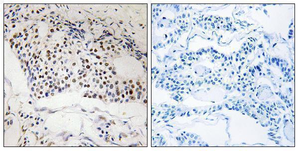
The catalog lists MYSM1 antibody A06500 for human and mouse IHC and IF; its supplied image shows paraffin-embedded human breast carcinoma by IHC (A06500 applications, reactivity, IHC caption).

Real IHC data Immunohistochemistry analysis of paraffin-embedded human breast carcinoma, using MYSM1 Antibody. The picture on the right is blocked with the synthesized peptide.
Anti-MYSM1 Antibody
Cat # A06500

A06500 lists IHC and IF applications with human and mouse reactivity (A06500 catalog). Its IHC figure shows paraffin-embedded human breast carcinoma alongside a peptide-blocked comparison; no IF image is supplied (A06500 image captions).

Which to pick: For tissue IHC, choose A06500 for paraffin sections based on its human breast carcinoma image; the fixative is unreported (A06500 IHC caption). For IF, A06500 is listed, but ICC use and an IF figure are undocumented (A06500 applications, image captions). For mouse samples, A06500 lists mouse reactivity, while its supplied IHC image shows human tissue (A06500 reactivity, IHC caption).

Each figure is that product's own IHC / IF validation image from its datasheet.

References

  1. UniProt Consortium. UniProt entry Q5VVJ2 (MYSM1_HUMAN, Deubiquitinase MYSM1).
  2. Human Protein Atlas. MYSM1 tissue IHC expression (reliability: Approved).
  3. Human Protein Atlas. MYSM1 subcellular location (ICC-IF): Mainly localized to the nucleoplasm. In addition localized to the nucleoli..
  4. Human Protein Atlas. MYSM1 antibody validation summary (2 antibodies).
  5. Expression of MYSM1 is associated with tumor progression in colorectal cancer. PloS one 2017 — PMC5428969.
  6. MYSM1 induces apoptosis and sensitizes TNBC cells to cisplatin via RSK3-phospho-BAD pathway. Cell death discovery 2022 — PMC8881619.
  7. MYSM1 promotes lung adenocarcinoma progression via TRAF2/3-mediated activation of MAPK and non-canonical NF-κB pathways. NPJ precision oncology 2025 — PMC12572397.
  8. Deubiquitinase Mysm1 regulates neural stem cell proliferation and differentiation by controlling Id4 expression. Cell death & disease 2024 — PMC10859383.
  9. PubMed PMID:11572484 — UniProt-cited evidence.
  10. PubMed PMID:14702039 — UniProt-cited evidence.
  11. PubMed PMID:17974005 — UniProt-cited evidence.