This website uses cookies to ensure you get the best experience on our website.


- Table of Contents
Start chromogenic MYZAP IHC-P with the catalog antibody at 5 μg/mL (datasheet: IHC-P). Assess cardiomyocyte intercalated discs and lung macrophages, and interpret staining cautiously because presumed off-target binding was observed (HPA tissue IHC).

Expected localisation, validated protocols, controls and antibodies — the at-a-glance facts below, then the full design guide.
| Expected localisation | Predominantly intercalated discs; low Z-disc signal (UniProt) | |
| Staining pattern | Cardiomyocyte intercalated discs; lung macrophage staining (HPA tissue IHC) | |
| Antigen retrieval | Citrate pH 6.0 HIER, 95–98 °C, 20 min (rule: cytoplasmic / membrane antigen) | |
| Positive control | Heart muscle+4 more · see all | |
| Negative control | Adipose tissue+4 more · see all |
| Fixation | Keep paraffin-section fixation consistent (standard IHC practice; not target-specific); assess effects empirically. | |
| Caveat | Presumed off-target binding may complicate scoring (HPA tissue IHC) | |
| Regulation | Heart muscle-enriched RNA (HPA tissue RNA) | |
| Isoform / epitope | 11 isoforms; mature chain 21–466; check epitope coverage (UniProt) |
The catalog antibody protocol uses citrate pH 6.0 retrieval (page retrieval rule). One published MYZAP IHC protocol reports low-pH retrieval for FFPE sections (PMC8656111).
| Sample | Tissue sections; selected-image fixative not specified (standard IHC workflow) |
| Fixation | Image fixative and duration unreported (datasheet A13349); verify before use. |
| Sectioning | 4–5 µm sections on charged slides (standard) |
| Deparaffinisation | Xylene, graded ethanol series to water (standard) |
| Antigen retrieval | Heat-induced epitope retrieval in citrate buffer, pH 6.0, 20 min at 95–98 °C (standard rule: cytoplasmic / membrane antigen) |
| Peroxidase block | 3% H2O2, 10 min, room temperature (standard) |
| Blocking | 10% normal serum of the secondary host, 30 min, room temperature (standard) |
| Primary antibody | Rabbit anti-MYZAP, 5 μg/mL (datasheet A13349) |
| Primary incubation | Overnight at 4 °C (standard) |
| Detection | HRP-polymer secondary, DAB chromogen 5–10 min (standard) |
| Counterstain | Hematoxylin, blue, dehydrate and mount (standard) |
| Expected result | MYZAP-positive staining in cardiomyocytes of heart muscle (HPA tissue IHC: High). HPA tissue profile: Most abundant expression in intercalated discs and lung macrophages. No signal in the no-primary control. |
MYZAP is predominantly at cardiomyocyte intercalated discs, with lower levels at sarcomeric Z discs; it is also associated with the cytoskeleton and cell junctions and has no transmembrane segment (UniProt P0CAP1). Expect strong staining in heart cardiomyocytes and lung macrophages (HPA: High in both). Interpret tissue staining cautiously: HPA rates its IHC evidence Approved, with medium RNA–staining consistency and presumed off-target binding (HPA: tissue IHC reliability).
| Strong, sharply defined staining at borders between cardiomyocytes. | This matches the predominant intercalated-disc localisation reported for MYZAP (UniProt P0CAP1: subcellular location) and the high cardiomyocyte staining observed in heart muscle (HPA: High in cardiomyocytes). Assess the pattern within intact heart architecture; intensity alone cannot establish that every stained structure contains MYZAP (HPA: tissue IHC reliability). |
| Prominent nuclear staining dominates cardiomyocytes. | A nuclear-dominant pattern conflicts with the reported junctional and cytoskeletal localisation (UniProt P0CAP1: subcellular location). Treat it as suspect and compare the antibody control and tissue morphology before scoring it as MYZAP; chromogen deposits or nonspecific binding can mislead localisation (general IHC practice). |
| Strong staining appears in cells outside the expected tissue pattern. | Check the cell identity before calling cross-reactivity: lung macrophages also stain strongly, while placental trophoblasts, cerebellar Purkinje cells and parathyroid glandular cells stain at medium levels (HPA: tissue IHC). An unexpected cell population may reflect nonspecific or endogenous detection activity; HPA also cautions that its tissue assay targets protein from more than one gene (HPA: tissue IHC reliability). |
| Broad, even chromogen covers cells and surrounding tissue. | Diffuse staining without a discernible cardiomyocyte junctional pattern is difficult to interpret against the predominant intercalated-disc localisation (UniProt P0CAP1: subcellular location). Review negative detection controls, blocking and washing for general background; do not assign widespread staining to MYZAP from colour alone (general IHC practice). |
| Heart cardiomyocytes show no discernible staining. | This conflicts with high cardiomyocyte staining in the HPA heart section (HPA: High in cardiomyocytes). First check tissue preservation, control performance and the IHC run; absent signal in one section does not establish absent MYZAP expression (general IHC practice). |
| Tissue and cell choice | Heart cardiomyocytes provide a high-staining reference, but lung macrophages are also high; adipocytes in adipose tissue and adrenal glandular cells are reported as not detected (HPA: tissue IHC). Score the specified cell population, since a whole-section positive or negative label can conceal which cells stained (general IHC practice). |
| Subcellular pattern | MYZAP is detected predominantly at cardiomyocyte intercalated discs and at low levels on sarcomeric Z discs (UniProt P0CAP1: subcellular location). Use compartment and tissue architecture alongside intensity when judging a result; membrane-associated staining should not be assumed to indicate a transmembrane protein (UniProt P0CAP1: topology). |
| Evidence and antibody specificity | HPA lists HPA039827 IHC as Approved, reports medium consistency with RNA expression, and warns of presumed off-target binding and detection of protein from more than one gene (HPA: antibody validation; HPA: tissue IHC reliability). A plausible staining pattern therefore supports interpretation but does not by itself prove molecular specificity. |
| Isoforms and antibody epitope | UniProt lists 11 MYZAP isoforms and a processed chain spanning residues 21–466 (UniProt P0CAP1: isoforms; processing). No epitope position is supplied here, so isoform coverage and any effect of processing on this antibody cannot be determined. Avoid attributing a missed stain to either mechanism without antibody-specific evidence. |
| Antigen retrieval and fixation | These are general IHC variables that can affect staining and should be assessed against run controls (general IHC practice). Neither source supplied here establishes a MYZAP-specific retrieval requirement or fixation sensitivity; do not infer one from the UniProt topology or HPA tissue staining levels. |
| IF/ICC expected pattern? | UniProt places MYZAP at cardiomyocyte intercalated discs and, more weakly, Z discs (UniProt P0CAP1: subcellular location). HPA supplies no ICC-IF images or main subcellular location for this record, and lists no ICC status for HPA039827 (HPA: subcellular; antibody validation). This evidence supports a localisation expectation, not an ICC-IF validation or protocol. |
| Situation | Likely cause | Next action |
|---|---|---|
| Heart positive control is blank. | The run may have failed, or the selected section may not show assessable cardiomyocytes (general IHC practice). High cardiomyocyte staining is expected in the HPA heart reference (HPA: tissue IHC). | Confirm cardiomyocyte-rich morphology and review the run's positive control, primary-antibody step, detection reagents and counterstain before interpreting a sample as negative (general IHC practice). |
| Signal is present but intercalated discs are indistinct. | Background or tissue detail may obscure the junctional pattern (general IHC practice); the expected predominant compartment is the intercalated disc (UniProt P0CAP1: subcellular location). | Compare intact neighbouring cardiomyocytes and the negative detection control; assess washing, blocking and chromogen development as general IHC variables before scoring localisation (general IHC practice). |
| Lung macrophages stain, but heart is negative. | HPA reports high staining in both populations, so the lung result alone does not resolve whether the heart result reflects tissue, run or antibody behaviour (HPA: tissue IHC; tissue IHC reliability). | Repeat assessment with an adequate heart section and run controls. Record each cell type separately, and avoid using macrophage staining alone to claim MYZAP-specific detection (HPA: tissue IHC reliability; general IHC practice). |
| Many unexpected cells stain strongly. | Cell misidentification, nonspecific binding or endogenous detection activity may contribute (general IHC practice); HPA flags presumed off-target binding and detection of protein from more than one gene (HPA: tissue IHC reliability). | Identify the stained cells by morphology, compare HPA's cell-specific tissue pattern and inspect negative detection controls before assigning the signal to MYZAP (HPA: tissue IHC; general IHC practice). |
| A proposed negative-control tissue shows staining. | HPA's ‘not detected’ calls apply to named cells, such as adipocytes in adipose tissue and adrenal glandular cells, rather than every cell in those sections (HPA: tissue IHC). | Score the named cell population and its compartment; then compare a negative detection control and the heart positive reference to judge whether the staining is interpretable (HPA: tissue IHC; general IHC practice). |
| Nuclei or the whole section appear uniformly brown. | That distribution does not match the reported predominant cardiomyocyte junctional pattern (UniProt P0CAP1: subcellular location) and may reflect background, endogenous detection activity or excessive chromogen development (general IHC practice). | Check the negative detection control, counterstain balance and chromogen development; score MYZAP only where cellular identity and a credible compartmental pattern can be resolved (general IHC practice). |
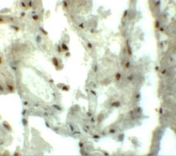
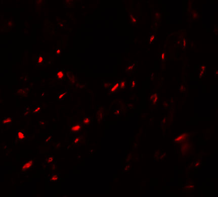


Comprehensive Human Protein Atlas IHC scoring per tissue (reliability: Approved — Medium consistency between antibody staining and RNA expression data. Caution, targets protein from more than one gene. Presumed off target binding observed and disregarded.). Rows are taken directly from the HPA tissue chart — click any row's HPA link to view the source.
| Tissue | Cell type | Level | Evidence | Source |
|---|---|---|---|---|
| Heart muscle | Cardiomyocytes | High | Protein (IHC) | HPA → |
| Lung | Macrophages | High | Protein (IHC) | HPA → |
| Cerebellum | Purkinje cells | Medium | Protein (IHC) | HPA → |
| Parathyroid gland | Glandular cells | Medium | Protein (IHC) | HPA → |
| Placenta | Trophoblastic cells | Medium | Protein (IHC) | HPA → |
| Tissue | Cell type | Level | Evidence | Source |
|---|---|---|---|---|
| Adipose tissue | Adipocytes | Not detected | Protein (IHC) | HPA → |
| Adrenal gland | Glandular cells | Not detected | Protein (IHC) | HPA → |
| Bone marrow | Hematopoietic cells | Not detected | Protein (IHC) | HPA → |
| Breast | Adipocytes | Not detected | Protein (IHC) | HPA → |
| Cervix | Glandular cells | Not detected | Protein (IHC) | HPA → |
Troubleshoot chromogenic MYZAP IHC by checking retrieval, controls, staining location and cell identity before comparing staining scores.
A13349 is listed for IHC-P and IF, with images of human lung tissue for both applications; catalog reactivity includes human, mouse, and rat (catalog: applications and reactivity; IHC and IF image captions).
A13349 has an IHC image of human lung tissue at 5 μg/mL (IHC image caption). A13349 also has an IF image of human lung tissue at 20 μg/mL; its listed reactivity is human, mouse, and rat (IF image caption; catalog: reactivity).
Which to pick: For tissue IHC, choose A13349: IHC-P is listed, and its own IHC image shows human lung tissue at 5 μg/mL; the fixative is unreported (catalog: applications; IHC image caption). For IF, choose A13349 at a starting concentration of 20 μg/mL; separate ICC validation and clone type are unreported (datasheet: IF starting concentration; catalog: applications and clone). For mouse or rat samples, A13349 lists reactivity with both species, while the supplied IHC and IF images show human tissue (catalog: reactivity; IHC and IF image captions).