MZB1 / Marginal zone B- and B1-cell-specific protein · IHC design guide

Design Immunohistochemistry for MZB1

Plan paraffin-section MZB1 IHC with spleen or tonsil as positive tissue controls (HPA tissue IHC; datasheet A08281-1). Assess cytoplasmic staining in immune-cell subsets, accounting for possible differences between protein staining and RNA location (HPA tissue IHC).

Evidence assembled Oct 2026 · For research use; verify linked source records and product datasheet before use
Immunohistochemistry protocol sheet for MZB1 (IHC for MZB1): expected localisation Cytoplasm in a subset of immune cells (HPA tissue IHC), antibody A08281-1, validated IHC image, and IHC protocol steps
Printable MZB1 IHC protocol sheet — expected localisation Cytoplasm in a subset of immune cells (HPA tissue IHC), antibody A08281-1, controls and protocol steps. Open the full MZB1 IHC guide →

MZB1 Immunohistochemistry Experimental Design Guide

Expected localisation, validated protocols, controls and antibodies — the at-a-glance facts below, then the full design guide.

Must know before staining
Expected localisation Cytoplasm in a subset of immune cells (HPA tissue IHC)
Staining pattern Subset of immune cells shows cytoplasmic staining (HPA tissue IHC)
Antigen retrieval EDTA pH 8.0 HIER, heat-mediated (datasheet A08281-1)
Positive control ⓘ Appendix+4 more · see all
Negative control ⓘ Adipose tissue+4 more · see all
Important caveats
Reasons your staining may differ from the expected pattern.
Fixation Keep tissue fixation consistent across samples. (standard IHC practice; not target-specific)
Caveat Secretion may separate protein staining from RNA location (HPA tissue IHC)
Regulation Higher expression in lymphoid tissue (UniProt)
Isoform / epitope 5 isoforms; signal peptide 1–22 is removed; check epitope (UniProt)
Section 1

Recommended MZB1 IHC & IF Protocols

The catalog antibody has an EDTA pH 8.0 IHC protocol (datasheet A08281-1). Published MZB1 IHC protocols below cover lymphoid and other tissues (PMC5791339; PMC7491119; PMC6008462).

Recommended immunohistochemistry (IHC-P) protocol parameters
SampleParaffin-embedded human spleen tissue; fixative not specified (datasheet A08281-1)
FixationImage fixative and duration unreported (datasheet A08281-1); verify before use.
Sectioning4–5 µm sections on charged slides (standard)
DeparaffinisationXylene, graded ethanol series to water (standard)
Antigen retrievalHeat retrieval: EDTA pH 8.0 (datasheet A08281-1); 20 min, 95–100 °C (standard)
Peroxidase block3% H2O2, 10 min, room temperature (standard)
Blocking10% goat serum (datasheet A08281-1)
Primary antibodyRabbit anti-MZB1, 1-2 μg/ml (datasheet A08281-1)
Primary incubationOvernight at 4 °C (datasheet A08281-1)
DetectionHRP-conjugated secondary, DAB chromogen (datasheet A08281-1)
CounterstainHematoxylin, blue, dehydrate and mount (standard)
Expected resultMZB1-positive staining in germinal center cells of appendix (HPA tissue IHC: Medium). HPA tissue profile: Cytoplasmic expression in a subset of immune cells in most tissues. No signal in the no-primary control.
💡Decision noteStart with EDTA pH 8.0 heat retrieval for the catalog antibody (datasheet A08281-1). Citrate pH 6.0 is a published alternative for gingival sections (PMC6008462).
Section 2

What Is the Expected MZB1 Staining Pattern?

MZB1 is an ER-lumen protein with no transmembrane segment; UniProt also reports secretion and granular cytoplasm around the nucleus (UniProt Q8WU39). In paraffin-section IHC, expect cytoplasmic staining in a subset of immune cells across many tissues (HPA tissue IHC: Enhanced). Interpret the stained cells alongside tissue architecture: a positive tissue does not imply that every cell should stain (HPA tissue IHC).

What am I looking at on my slide?
Discrete cytoplasmic staining in selected cells of a lymphoid tissue.This fits the reported IHC profile. HPA records medium staining in germinal center cells of appendix, lymph node and tonsil, and in spleen red-pulp cells (HPA tissue IHC). Score the proportion and intensity of the relevant cells; avoid assigning the tissue one uniform result when neighboring cells differ (general IHC practice).
Predominantly nuclear staining, with little convincing cytoplasmic signal.An isolated nuclear pattern lacks support from the reported ER-lumen and perinuclear cytoplasmic locations (UniProt Q8WU39) or the HPA tissue IHC profile (HPA tissue IHC). Treat it as suspect and compare control sections before calling it MZB1. A Golgi result reported by ICC-IF does not establish nuclear staining in paraffin IHC (HPA subcellular ICC-IF).
Strong staining throughout epithelial cells or adipocytes instead of a subset of immune cells.This differs from the reported IHC distribution (HPA tissue IHC). Adipocytes in adipose tissue and respiratory epithelial cells in bronchus are listed as not detected (HPA tissue IHC). Check for nonspecific antibody binding or endogenous detection activity using appropriate controls (general IHC practice); the image alone cannot distinguish those causes.
Uniform, low contrast color over cells and surrounding tissue.A widespread haze obscures the subset-of-immune-cells pattern reported by HPA (HPA tissue IHC). It cannot establish a positive cell population. Compare the no-primary control, review blocking and washes, and score only signal with a clear cellular boundary above background (general IHC practice). Secreted MZB1 alone does not justify calling uniform chromogen deposition specific (UniProt Q8WU39).
No detectable staining in a section expected to contain positive immune cells.Absence is inconclusive until tissue sampling and assay controls are checked. HPA reports medium staining in specified cells of appendix, bone marrow, lymph node, spleen, thymus and tonsil, not in every cell of those tissues (HPA tissue IHC). Confirm that the relevant compartment is present, then review antibody and detection performance (general IHC practice).
💡Expected MZB1 appearanceCall an IHC result positive when a subset of immune cells shows discernible cytoplasmic staining, potentially perinuclear and granular (HPA tissue IHC; UniProt Q8WU39); HPA's listed positive cell groups stain at medium level, while isolated nuclear color or uniform tissue haze warrants control review (HPA tissue IHC; general IHC practice).
How each factor affects the staining
Cellular compartmentMZB1 is reported in the ER lumen, secreted space and cytoplasm, with granular perinuclear cytoplasm described (UniProt Q8WU39). Its signal peptide spans residues 1–22, and the annotated mature chain spans 23–189; there is no transmembrane segment (UniProt Q8WU39). These annotations support cytoplasmic interpretation but do not specify an IHC retrieval condition.
Which cells are sampledHPA describes cytoplasmic expression in a subset of immune cells in most tissues, with medium staining in its listed positive cell groups (HPA tissue IHC). Evaluate the named cells within the section rather than treating an unstained neighboring population as assay failure (general IHC practice).
Secreted protein and RNA comparisonMZB1 is annotated as secreted (UniProt Q8WU39). HPA warns that tissue locations of RNA and protein may differ for secreted proteins, despite its Enhanced tissue IHC reliability assessment (HPA tissue IHC). Use observed cell-level staining and controls when interpreting a section; RNA distribution alone cannot assign a stained cell (general IHC practice).
Antibody validation and isoformsBoth listed antibodies, HPA043745 and HPA052694, have Enhanced IHC status; HPA052694 also has Approved ICC status (HPA antibodies). UniProt lists five MZB1 isoforms (UniProt Q8WU39). These records do not say which isoforms each antibody detects, so do not infer isoform-specific staining from the pattern.
IF/ICC Q&A: must its compartment match IHC?HPA reports Golgi apparatus localization in ICC-IF, with Approved status, while its tissue IHC summary is cytoplasmic (HPA subcellular ICC-IF; HPA tissue IHC). UniProt reports ER-lumen and perinuclear cytoplasmic localization (UniProt Q8WU39). Interpret each assay against its own evidence; the ICC-IF record supplies no paraffin-section protocol.
Why is my staining missing, weak or wrong?
SituationLikely causeNext action
A known-positive lymphoid section is blank.The relevant stained cells may be absent from the sampled area, or the IHC workflow may have failed (HPA tissue IHC; general IHC practice).Locate the expected cell population first; HPA lists medium staining in germinal center cells of lymph node and tonsil, among other sites (HPA tissue IHC). Then check a positive control and the antibody and detection steps (general IHC practice).
Color appears mainly in nuclei.That compartment is unsupported by the supplied MZB1 localization records (UniProt Q8WU39; HPA tissue IHC).Compare with a no-primary control, inspect cytoplasmic staining in an expected positive cell group, and review counterstain and detection conditions (general IHC practice). Do not score nuclear color alone as MZB1.
Adipocytes or bronchial epithelium stain strongly.HPA lists adipocytes in adipose tissue and respiratory epithelial cells in bronchus as not detected (HPA tissue IHC). Nonspecific binding or endogenous detection activity is possible (general IHC practice).Run an appropriate no-primary control and inspect the detection chemistry; confirm whether the color follows cells rather than tissue-wide background (general IHC practice).
A diffuse chromogenic haze covers the section.Background from blocking, washes or detection can hide the restricted cellular pattern (general IHC practice; HPA tissue IHC).Compare a no-primary control, review blocking and wash steps, and assess only clearly bounded cellular signal (general IHC practice). Do not use diffuse extracellular color alone to identify MZB1-positive cells.
Only scattered cells stain within a positive tissue.Restricted staining may be expected: HPA describes a subset of immune cells, and its listed positive cell groups have medium staining (HPA tissue IHC).Identify the stained cell population and record its fraction and intensity within the relevant area (general IHC practice). Compare with the HPA cell-level pattern before adjusting assay conditions (HPA tissue IHC).
IHC looks cytoplasmic while ICC-IF appears Golgi-localized.The supplied sources report cytoplasmic tissue IHC and Approved Golgi localization in ICC-IF (HPA tissue IHC; HPA subcellular ICC-IF).Judge the paraffin IHC section against its tissue cell pattern and controls (HPA tissue IHC; general IHC practice). Treat the ICC-IF result as assay-specific evidence, not as a paraffin IHC protocol or a reason by itself to reject cytoplasmic staining.

Sample controls for MZB1 IHC & IF

🧪Run spleen first and score cells in the red pulp for MZB1 staining (HPA: Medium in spleen red pulp cells). Use adipose tissue adipocytes as the biological negative (HPA: Not detected in adipocytes); on the spleen slide, cells outside the stained red pulp population should lack specific signal, although HPA does not designate a particular internal negative cell type (HPA: Medium in spleen red pulp cells).
Positive control tissue: Appendix (Germinal center cells, HPA Medium)
Negative control tissue: Adipose tissue (HPA Not detected)
ICC-IF cell lines (HPA subcellular resource): HPA ICC-IF images show MZB1 in HDLM-2, HaCaT, REH, with annotated localisation: Golgi apparatus (approved) (HPA subcellular).
Technical controls: Include a no-primary, secondary-only slide and a rabbit immunoglobulin-matched isotype control (hero caption: rabbit anti-MZB1 antibody); use MZB1-knockout material, if available, to test signal specificity (standard IHC practice). For the spleen DAB assay, quench endogenous peroxidase and check unstained tissue for pigment that could obscure the signal (hero caption: spleen, HRP/DAB; standard IHC practice).
⚠️Feasibility: A target-specific fixation window and fixation effect are unreported, and the selected-SKU paraffin-section caption does not state a fixative (hero caption: fixative not stated). Heat retrieval in EDTA at pH 8.0 preceded spleen staining, but its necessity was not tested against unretrieved sections (hero caption: heat-mediated EDTA retrieval). Frozen sections cannot be judged easier from the supplied evidence; ICC-IF images exist for HDLM-2, HaCaT and REH, while spleen pigment may complicate signal assessment (HPA subcellular: ICC-IF image cell lines; hero caption: spleen; standard IHC practice).

HPA tissue IHC evidence for MZB1

Comprehensive Human Protein Atlas IHC scoring per tissue (reliability: Enhanced — High consistency between antibody staining and RNA expression data. Secreted protein, tissue location of RNA and protein is expected to differ. Antibody staining in cells/structures not annotated, view images.). Rows are taken directly from the HPA tissue chart — click any row's HPA link to view the source.

Positive expression · recommended positive controls

TissueCell typeLevelEvidenceSource
Appendix Germinal center cells Medium Protein (IHC) HPA →
Bone marrow Hematopoietic cells Medium Protein (IHC) HPA →
Lymph node Germinal center cells Medium Protein (IHC) HPA →
Spleen Cells in red pulp Medium Protein (IHC) HPA →
Thymus Cortical cells Medium Protein (IHC) HPA →

Undetected expression · recommended negative controls

TissueCell typeLevelEvidenceSource
Adipose tissue Adipocytes Not detected Protein (IHC) HPA →
Adrenal gland Glandular cells Not detected Protein (IHC) HPA →
Breast Adipocytes Not detected Protein (IHC) HPA →
Bronchus Respiratory epithelial cells Not detected Protein (IHC) HPA →
Caudate Glial cells Not detected Protein (IHC) HPA →
Section 3

Advanced MZB1 IHC Tips

Use the paraffin-section IHC evidence for A08281-1 as the starting point, then assess staining by cell type, compartment and controls.

How should I retrieve MZB1 in paraffin sections when staining is weak?
Start with heat-mediated antigen retrieval in EDTA at pH 8.0 (datasheet A08281-1). The documented spleen stain used this retrieval before 2 μg/ml primary antibody overnight at 4°C, so keep those conditions together as a reference when troubleshooting (A08281-1 tissue-IHC caption). Check a spleen control on the same run and compare immune-cell staining before changing retrieval conditions (HPA tissue IHC; standard IHC practice). If signal remains weak, vary heating and cooling consistently across replicate sections, recording tissue damage as well as signal (standard IHC practice). Avoid judging retrieval from diffuse background alone; assess discrete cytoplasmic staining in the relevant cells (HPA tissue IHC).
Could fixation be causing weak or patchy MZB1 staining?
The A08281-1 spleen caption identifies a paraffin-embedded section but does not state its fixative, so target-specific fixation sensitivity is unknown (A08281-1 tissue-IHC caption). Record the fixative, fixation duration and processing history for each specimen, and compare similarly handled sections before attributing a difference to MZB1 biology (standard IHC practice). Use the documented EDTA retrieval and antibody conditions as a consistent starting point while assessing fixation effects (datasheet A08281-1; A08281-1 tissue-IHC caption). Examine morphology and staining across the section, because poor preservation or uneven processing can complicate interpretation (standard IHC practice). Neither reported tissue staining nor MZB1 topology establishes a target-specific fixation effect (HPA tissue IHC; UniProt Q8WU39 topology).
Where should convincing MZB1 chromogenic staining appear?
Look first for cytoplasmic staining in a subset of immune cells, the pattern reported across tissues by IHC (HPA tissue IHC). Perinuclear granular staining is compatible with the reported cytoplasmic distribution around the nucleus and with MZB1 in the endoplasmic reticulum lumen (UniProt Q8WU39 subcellular location). A separate ICC/IF assessment reports Golgi localisation, so compare compartment patterns with cell morphology instead of requiring one sharply bounded organelle pattern in chromogenic sections (HPA subcellular). MZB1 has no annotated transmembrane segment, making an exclusively crisp plasma-membrane outline a reason to investigate staining specificity (UniProt Q8WU39 topology). Assess the pattern in positive tissue alongside a matched negative control (HPA tissue IHC; standard IHC practice).
Can a negative stain exclude every MZB1 isoform?
No: MZB1 has 5 annotated isoforms, while the supplied antibody caption does not map the antibody epitope or establish recognition of each isoform (UniProt Q8WU39 isoforms; A08281-1 tissue-IHC caption). The precursor has a 1–22 signal peptide and a 23–189 mature chain, so epitope position and processing matter when interpreting a negative stain (UniProt Q8WU39 processing). There is no annotated transmembrane segment or glycosylation site to support a membrane-sided or glycan-masking explanation from this record alone (UniProt Q8WU39 topology and glycosylation). Check the antibody's epitope documentation and use an independently validated reagent or orthogonal expression evidence before making an all-isoform claim (standard IHC practice).
How can IF help assess an uncertain MZB1 IHC pattern?
Use IF as a separate, complementary observation: the reported ICC/IF subcellular location is Golgi, whereas tissue IHC reports cytoplasmic staining in a subset of immune cells (HPA subcellular; HPA tissue IHC). Multiplex MZB1 with a B-cell or plasma-cell marker appropriate to the question, and evaluate staining within identified cells rather than overlapping colours alone (UniProt Q8WU39 function; standard IF practice). Select a spectrally separated red or far-red fluorophore when tissue autofluorescence obscures shorter-wavelength signal, with single-label and no-primary controls (standard IF practice). Because MZB1 is reported in the endoplasmic reticulum lumen, check that permeabilisation permits antibody access to intracellular compartments; the supplied IHC caption does not establish IF fixation or permeabilisation conditions (UniProt Q8WU39 subcellular location; A08281-1 tissue-IHC caption).
What should I check when DAB stains broadly outside immune cells?
Compare the section with a no-primary control to distinguish primary-dependent staining from detection-system or tissue background (standard IHC practice). The documented spleen workflow used 10% goat serum blocking, peroxidase-conjugated goat anti-rabbit secondary antibody and DAB detection (A08281-1 tissue-IHC caption). Include an endogenous-peroxidase block in a chromogenic workflow, and inspect whether residual pigment or enzyme activity persists in controls (standard IHC practice). If background is primary-dependent, review antibody concentration, incubation, washing and blocking while retaining a positive tissue control; the documented primary condition was 2 μg/ml overnight at 4°C (A08281-1 tissue-IHC caption; standard IHC practice). Judge improvement by preservation of discrete cytoplasmic immune-cell signal (HPA tissue IHC).
How should I score MZB1 across heterogeneous tissue sections? ⚠ ANSWER MARKED FOR VERIFICATION
Define the region and cell population before scoring, because tissue IHC reports MZB1 in only a subset of immune cells (HPA tissue IHC). For an identified population, report percent positive cells and, where intensity is reproducible, an H-score on the conventional 0–300 scale (standard IHC practice). If spatial abundance matters, count positive cells per mm² of viable tissue and state how regions were selected (standard IHC practice). Normalise cell-level positivity to the number of eligible cells, and area-based density to analysed viable area rather than the whole slide (standard IHC practice). Keep retrieval, detection and scoring thresholds consistent across comparison groups (standard IHC practice).
How do I distinguish a true MZB1 signal from artefact?
A credible stain is cytoplasmic in an appropriate subset of immune cells and agrees with a positive control; spleen red-pulp cells and tonsil germinal-center cells have reported medium IHC staining (HPA tissue IHC). Perinuclear granularity can fit the reported distribution, whereas an exclusively crisp plasma-membrane pattern warrants scrutiny given the lack of an annotated transmembrane segment (UniProt Q8WU39 subcellular location and topology). Compare edge-heavy or necrotic regions with preserved interior tissue, and check no-primary controls for pigment or endogenous enzyme signal (standard IHC practice). A negative cell population does not negate expression elsewhere: HPA reports no detectable stain in adipocytes but cytoplasmic staining in immune-cell subsets (HPA tissue IHC).
Boster reagents

Best MZB1 / Marginal zone B- and B1-cell-specific protein IHC Antibodies

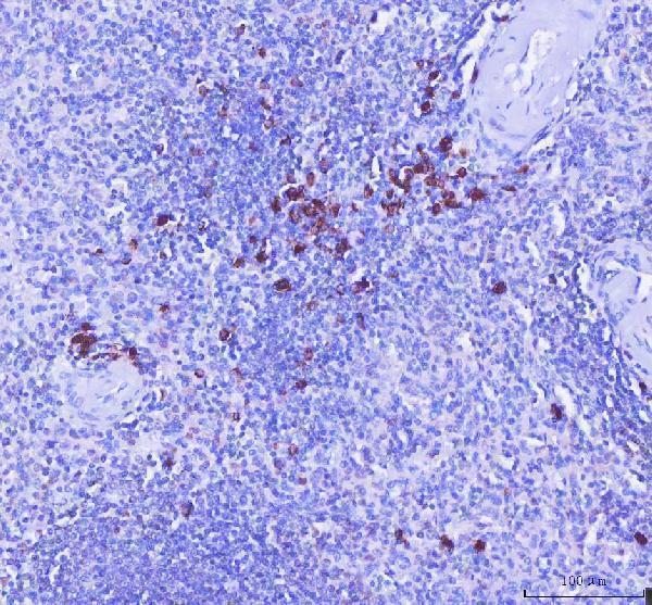
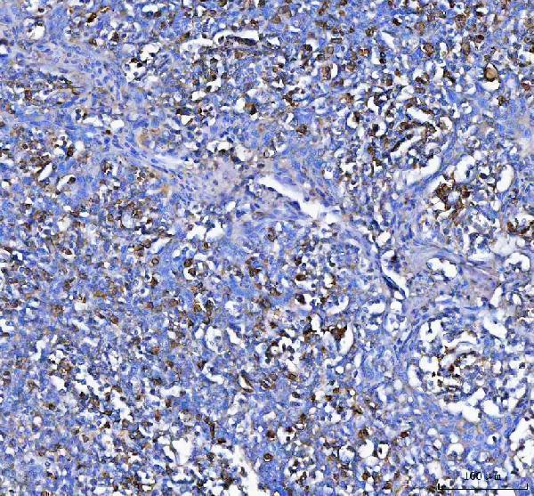
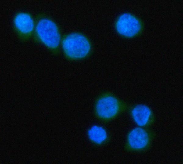
The IHC-validated anti-MZB1 antibody has paraffin-section images from human spleen and tonsil (A08281-1 IHC captions), plus IF data from Jurkat cells (A08281-1 IF caption).

Real IHC data IHC analysis of MZB1 using anti-MZB1 antibody (A08281-1). MZB1 was detected in a paraffin-embedded section of human spleen tissue. Heat mediated antigen retrieval was performed in EDTA buffer (pH 8.0, epitope retrieval solution). The tissue section was blocked with 10% goat serum. The tissue section was then incubated with 2 μg/ml rabbit anti-MZB1 Antibody (A08281-1) overnight at 4°C. Peroxidase Conjugated Goat Anti-rabbit IgG was used as secondary antibody and incubated for 30 minutes at 37°C. The tissue section was developed using HRP Conjugated Rabbit IgG Super Vision Assay Kit (Catalog # SV0002) with DAB as the chromogen.
Anti-MZB1 Antibody ®
Cat # A08281-1

A08281-1 will render with its human spleen IHC figure (A08281-1 card caption). Its additional captions show human tonsil IHC and Jurkat-cell IF (A08281-1 image captions).

Which to pick: Choose A08281-1 for human paraffin-section IHC: its spleen caption reports EDTA retrieval at pH 8.0 and 2 μg/ml primary antibody (A08281-1 IHC caption); the fixative is unreported (A08281-1 IHC captions). The same SKU is listed for IF and ICC, with a Jurkat-cell IF image at 5 μg/ml (A08281-1 applications and IF caption). No cross-species choice is supported because the catalog lists human reactivity only (A08281-1 reactivity).

Each figure is that product's own IHC / IF validation image from its datasheet.

References

  1. UniProt Consortium. UniProt entry Q8WU39 (MZB1_HUMAN, Marginal zone B- and B1-cell-specific protein).
  2. Human Protein Atlas. MZB1 tissue IHC expression (reliability: Enhanced).
  3. Human Protein Atlas. MZB1 subcellular location (ICC-IF): Localized to the Golgi apparatus..
  4. Human Protein Atlas. MZB1 antibody validation summary (2 antibodies).
  5. Increase of MZB1 in B cells in systemic lupus erythematosus: proteomic analysis of biopsied lymph nodes. Arthritis research & therapy 2018 — PMC5791339.
  6. MZB1 expression indicates poor prognosis in estrogen receptor-positive breast cancer. Oncology letters 2020 — PMC7491119.
  7. Proteomic profiling identifies novel diagnostic biomarkers and molecular subtypes for mucinous tubular and spindle cell carcinoma of the kidney. The Journal of pathology 2022 — PMC9311136.
  8. Gene expression profiling of periodontitis-affected gingival tissue by spatial transcriptomics. Scientific reports 2018 — PMC6008462.
  9. PubMed PMID:11350957 — UniProt-cited evidence.
  10. PubMed PMID:11042152 — UniProt-cited evidence.
  11. PubMed PMID:19805157 — UniProt-cited evidence.