This website uses cookies to ensure you get the best experience on our website.


- Table of Contents
Plan NACC1 paraffin-section IHC around the ubiquitous nuclear tissue pattern (HPA tissue IHC). This guide covers the catalog antibody’s 2–5 μg/ml IHC range (datasheet A08675-3) and scoring nuclear staining (HPA tissue IHC).

Expected localisation, validated protocols, controls and antibodies — the at-a-glance facts below, then the full design guide.
| Expected localisation | Nuclear tissue staining; cytoplasm is possible (HPA tissue IHC; UniProt) | |
| Staining pattern | Ubiquitous nuclear staining across tissue cell types (HPA tissue IHC) | |
| Antigen retrieval | EDTA pH 8.0 HIER, heat-mediated (datasheet A08675-3) | |
| Positive control | Adipose tissue+4 more · see all | |
| Negative control | None in HPA (detected in all 45 tissues); use no-primary + isotype controls |
| Fixation | Keep fixation conditions consistent across sections (standard IHC practice; not target-specific) | |
| Caveat | Phosphorylation may alter cytoplasmic distribution (UniProt) | |
| Regulation | Upregulated after chemotherapy in ovarian carcinoma (UniProt) | |
| Isoform / epitope | No annotated isoforms; one full-length chain (UniProt) |
The catalog antibody’s IHC-P protocol is accompanied by published NACC1 staining methods for urothelial carcinoma sections (PMC6210667) and mouse and human embryonic tissues (PMC3724875).
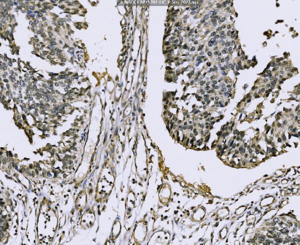
| Sample | Paraffin-embedded human bladder epithelial carcinoma tissue; fixative not specified (datasheet A08675-3) |
| Fixation | Image fixative and duration unreported (datasheet A08675-3); verify before use. |
| Sectioning | 4–5 µm sections on charged slides (standard) |
| Deparaffinisation | Xylene, graded ethanol series to water (standard) |
| Antigen retrieval | Heat retrieval: EDTA pH 8.0 (datasheet A08675-3); 20 min, 95–100 °C (standard) |
| Peroxidase block | 3% H2O2, 10 min, room temperature (standard) |
| Blocking | 10% goat serum (datasheet A08675-3) |
| Primary antibody | Rabbit anti-NACC1, 2-5 μg/ml (datasheet A08675-3) |
| Primary incubation | Overnight at 4 °C (datasheet A08675-3) |
| Detection | Streptavidin-biotin complex (SABC), DAB chromogen (datasheet A08675-3) |
| Counterstain | Hematoxylin, blue, dehydrate and mount (standard) |
| Expected result | NACC1-positive staining in adipocytes of adipose tissue (HPA tissue IHC: High). HPA tissue profile: Ubiquitous nuclear expression. No signal in the no-primary control. |
NACC1 should appear chiefly in nuclei across many cell types in paraffin-section IHC: HPA reports ubiquitous nuclear expression with Enhanced tissue-IHC reliability and low RNA tissue specificity (HPA: tissue IHC). Cytoplasmic distribution can occur and depends on phosphorylation (UniProt Q96RE7: subcellular location). NACC1 has no transmembrane segment, so a membrane-only pattern is unexpected (UniProt Q96RE7: topology). Interpret staining within the cell type being scored.
| Clear nuclear chromogen in adipocytes, adrenal or epididymal glandular cells, respiratory epithelium, neurons, glia, or decidual cells. | This fits the principal compartment and the listed high-staining cells (HPA: ubiquitous nuclear expression; High in the listed cells). Compare like cell types within a section: an organ-level judgment can hide a convincing nuclear result in its relevant cells (standard IHC interpretation). |
| Strong membrane-only staining, or a predominantly cytoplasmic pattern with little nuclear signal. | Recheck specificity and localization before calling this NACC1: HPA’s tissue profile is nuclear, and UniProt lists no transmembrane segment (HPA: tissue IHC; UniProt Q96RE7: topology). Cytoplasm alone is not proof of artifact, because phosphorylation-dependent cytoplasmic distribution is reported (UniProt Q96RE7: subcellular location). |
| Signal appears mainly in a different cell population from the one being assessed, while expected nuclei remain unstained. | Check cell identity, assay controls, and possible cross-reactivity or endogenous chromogenic activity (standard IHC interpretation). Broad NACC1 expression means an unexpected cell is not automatically negative; HPA provides cell-specific high and low observations, with no negative tissue listed (HPA: tissue IHC). |
| Hazy chromogen covers nuclei, cytoplasm, and spaces between cells without crisp cellular boundaries. | Treat this as unresolved background until the detection controls are reviewed (standard chromogenic IHC interpretation). A diffuse deposit cannot establish NACC1 localization, even where HPA reports high staining; the expected tissue pattern remains chiefly nuclear (HPA: tissue IHC). |
| A listed high-staining cell population shows no detectable nuclear signal. | First verify that the relevant cells are present and that the run’s positive control worked (standard IHC practice). Absence in one section cannot establish biological absence: HPA calls these populations High and rates its tissue-IHC evidence Enhanced (HPA: tissue IHC). |
| Cell-specific reference pattern | HPA lists High staining in adipocytes; adrenal and epididymal glandular cells; bronchial and nasopharyngeal respiratory epithelium; caudate neurons; cortical glia; and placental decidual cells (HPA: tissue IHC). Match the scored cell population to its reference; low RNA tissue specificity does not make every cell equally intense (HPA: RNA specificity and tissue IHC). |
| Low-staining comparators | HPA lists Low staining in parathyroid and prostate glandular cells, liver cholangiocytes, cardiomyocytes, and smooth muscle cells, but supplies no negative tissue (HPA: tissue IHC). These are weaker comparators, not validated zero-signal controls; an absent or faint result in one cannot validate a failed positive run (standard IHC interpretation). |
| Compartment and phosphorylation | HPA describes a chiefly nuclear tissue pattern and nucleoplasmic ICC-IF localization, with additional cytosol and vesicles (HPA: tissue IHC; HPA: subcellular ICC-IF). UniProt reports phosphorylation-dependent cytoplasmic distribution and phosphoserines at residues 188 and 259 (UniProt Q96RE7: localization and modified residues). Neither source establishes a fixation effect or a required cytoplasmic IHC intensity. |
| Antibody evidence | Two listed rabbit polyclonal antibodies have Enhanced IHC validation and Supported ICC status; the listed mouse monoclonal has Supported IHC status (HPA: antibody validation). This strengthens confidence in the reported nuclear reference pattern, while an individual assay still needs appropriate run controls (standard IHC practice). Validation status does not certify every tissue or staining condition. |
| Tumor context | UniProt reports NACC1 overexpression in several carcinomas, including ovarian serous carcinomas, and an association between intense primary-tumor immunoreactivity and early recurrence (UniProt Q96RE7: tissue specificity). Tumor intensity therefore needs comparison with the relevant cells and validated scoring context; staining alone is not a stand-alone clinical prediction (standard IHC interpretation). |
| Situation | Likely cause | Next action |
|---|---|---|
| No signal in an HPA-listed High population. | The relevant cells may be absent from the section, or the staining run may have failed (HPA: High cell list; standard IHC practice). | Confirm cell identity and morphology, then review the concurrent positive control and the catalog antibody’s IHC-P instructions for the run (standard IHC practice). |
| Only broad cytoplasmic staining is visible. | Cytoplasmic NACC1 is possible, but a nuclear-poor result departs from HPA’s dominant tissue pattern (UniProt Q96RE7: localization; HPA: tissue IHC). | Check nuclear counterstain, cell boundaries, and control sections before scoring; record cytoplasmic signal separately from convincing nuclear staining (standard IHC interpretation). |
| Strong membrane outlines dominate the slide. | A membrane-only pattern is unsupported by the reported localization and absence of a transmembrane segment (HPA: tissue IHC; UniProt Q96RE7: topology). | Review primary-antibody omission and detection controls for nonspecific or endogenous signal; reassess specificity if outlines persist (standard chromogenic IHC practice). |
| Every area has diffuse brown background. | Unresolved chromogen may reflect nonspecific binding or endogenous detection activity (standard chromogenic IHC practice). | Inspect the no-primary control, blocking and washing steps, and detection chemistry; score only staining with interpretable cellular boundaries (standard IHC practice). |
| A Low comparator stains more strongly than expected. | HPA’s Low category is an observed level, not a promise of no staining; cell composition and background can complicate comparison (HPA: tissue IHC; standard IHC interpretation). | Identify the exact scored cells and compare their nuclear signal with a concurrent High population; inspect detection controls if staining lacks cellular definition (HPA: High and Low cell lists; standard IHC practice). |
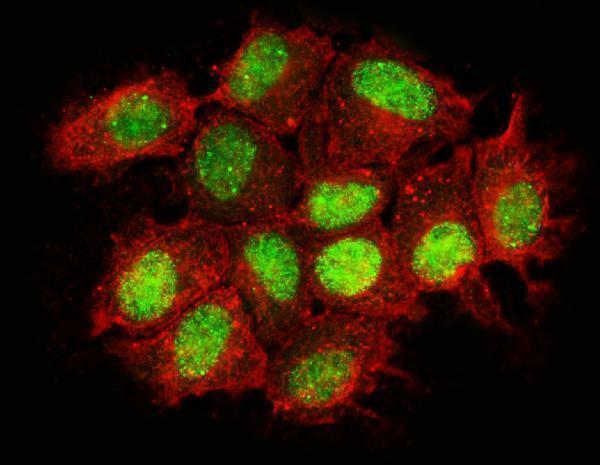
| Q: What if IF/ICC shows cytosolic NACC1? | HPA supports nucleoplasm as the main ICC-IF location and cytosol as an additional location; UniProt links cytoplasmic distribution to phosphorylation (HPA: subcellular ICC-IF; UniProt Q96RE7: localization). | A: Interpret it alongside the nuclear signal and appropriate controls; cytosol alone does not define the expected paraffin-section IHC pattern (HPA: tissue IHC; standard IHC interpretation). |
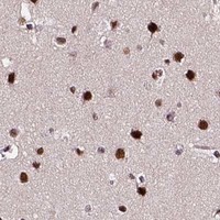


Comprehensive Human Protein Atlas IHC scoring per tissue (reliability: Enhanced — High consistency between antibody staining and RNA expression data.). Rows are taken directly from the HPA tissue chart — click any row's HPA link to view the source.
| Tissue | Cell type | Level | Evidence | Source |
|---|---|---|---|---|
| Adipose tissue | Adipocytes | High | Protein (IHC) | HPA → |
| Adrenal gland | Glandular cells | High | Protein (IHC) | HPA → |
| Bronchus | Respiratory epithelial cells | High | Protein (IHC) | HPA → |
| Caudate | Neuronal cells | High | Protein (IHC) | HPA → |
| Cerebral cortex | Glial cells | High | Protein (IHC) | HPA → |
| Tissue | Cell type | Level | Evidence | Source |
|---|---|---|---|---|
| None in HPA: NACC1 is detected in all 45 scored tissues. Use a no-primary (secondary-only) and an isotype control instead. | ||||
Troubleshoot NACC1 staining in paraffin sections by checking retrieval, nuclear localisation, detection background and how viable cells are scored.
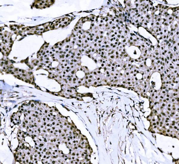
A08675-3 has real IHC data from human paraffin sections and IF data from MCF-7 cells (catalog IHC and IF image captions).
A08675-3 is listed for human IHC, with images from paraffin sections of bladder epithelial carcinoma, breast papillary carcinoma, and renal pelvis squamous metaplasia (catalog applications, reactivity, and IHC image captions). The same SKU is listed for IF/ICC, with an IF image from MCF-7 cells (catalog applications and IF image caption).
Which to pick: For tissue IHC, choose A08675-3: its paraffin-section captions report EDTA retrieval at pH 8.0, 2 μg/ml primary antibody, and DAB detection; the fixative is unreported (catalog IHC image captions). For IF/ICC, A08675-3 is listed for both applications and has an MCF-7 IF image using 5 μg/ml primary antibody; its host is rabbit and clonality is unreported (catalog applications, IF image caption, host, and clone). No listed SKU has reported reactivity beyond human for cross-species selection (catalog: reactivity Human).