This website uses cookies to ensure you get the best experience on our website.


- Table of Contents
Plan chromogenic IHC on paraffin sections for NCAN using staining in CNS processes as a reference (HPA tissue IHC). This guide covers fixation consistency, antibody concentration, detection and interpretation of a secreted protein (UniProt).

Expected localisation, validated protocols, controls and antibodies — the at-a-glance facts below, then the full design guide.
| Expected localisation | CNS processes (HPA tissue IHC); secreted protein (UniProt) | |
| Staining pattern | CNS processes: synaptic glomeruli capsules and cortical neuropil (HPA tissue IHC) | |
| Antigen retrieval | Citrate pH 6 HIER, heat-mediated (datasheet A06700-1) | |
| Positive control | Cerebellum+3 more · see all | |
| Negative control | Adipose tissue+4 more · see all |
| Fixation | Keep fixation consistent across paraffin sections (standard IHC practice; not target-specific) | |
| Caveat | Secreted NCAN may stain away from RNA-positive cells (HPA tissue IHC) | |
| Regulation | Brain-enriched RNA (HPA tissue RNA) | |
| Isoform / epitope | 0 isoforms annotated; mature chain begins at residue 23 (UniProt) |
The catalog antibody’s IHC-P protocol (datasheet A06700-1) is accompanied by published chromogenic NCAN protocols for liver tissue (PMC12571593) and Merkel cell carcinoma tissue (PMC10162530).
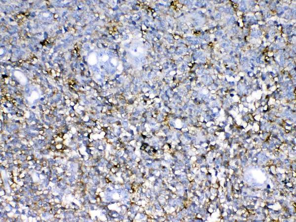
| Sample | Paraffin-embedded human glioma tissue; fixative not specified (datasheet A06700-1) |
| Fixation | Image fixative and duration unreported (datasheet A06700-1); verify before use. |
| Sectioning | 4–5 µm sections on charged slides (standard) |
| Deparaffinisation | Xylene, graded ethanol series to water (standard) |
| Antigen retrieval | Heat retrieval: Citrate pH 6, 20 min (datasheet A06700-1) |
| Peroxidase block | 3% H2O2, 10 min, room temperature (standard) |
| Blocking | 10% goat serum (datasheet A06700-1) |
| Primary antibody | Rabbit anti-NCAN, 1:100–1:500 starting range (standard) |
| Primary incubation | Overnight at 4 °C (standard) |
| Detection | Streptavidin-biotin complex (SABC), DAB chromogen (datasheet A06700-1) |
| Counterstain | Hematoxylin, blue, dehydrate and mount (standard) |
| Expected result | NCAN-positive staining in synaptic glomeruli - capsule of cerebellum (HPA tissue IHC: High). HPA tissue profile: Expression in processes in CNS. No signal in the no-primary control. |
NCAN is a secreted proteoglycan with no transmembrane segment (UniProt O14594 topology). In paraffin section IHC, expect staining associated with CNS processes: high signal in cerebellar synaptic glomeruli capsules and medium signal in cerebral cortical neuropil (HPA tissue IHC). HPA rates the tissue profile Enhanced, while noting medium consistency with RNA and disregarded presumed off target staining (HPA tissue IHC reliability).
| Structured staining outlines cerebellar synaptic glomeruli capsules; cortical neuropil also stains. | This matches the reported high cerebellar and medium cortical patterns (HPA tissue IHC). Assess the local process or extracellular pattern rather than counting positive neuronal cell bodies: NCAN is secreted and HPA describes expression in CNS processes (UniProt O14594 topology; HPA tissue IHC profile). |
| Strong staining concentrates in nuclei or forms a consistent intracellular pattern instead of following CNS processes. | Treat that compartment as suspect: NCAN is secreted, has no transmembrane segment, and HPA reports expression in processes (UniProt O14594 topology; HPA tissue IHC profile). Compare adjacent morphology and controls before scoring it as NCAN; the supplied sources do not establish a nuclear pattern. |
| Hematopoietic cells stain strongly, although the expected CNS pattern is absent. | HPA reports no detection in bone marrow hematopoietic cells (HPA tissue IHC). Consider antibody cross reactivity or endogenous chromogenic detection activity as general IHC possibilities. Staining of appendix or colon enterocyte microvilli alone is a different case: HPA reports medium signal there (HPA tissue IHC). |
| A uniform chromogenic haze obscures tissue structures and appears across expected positive and negative regions. | The haze cannot be assigned confidently to NCAN because the reported pattern is anatomically restricted (HPA tissue IHC profile). In general IHC practice, inspect secondary only and detection controls, then review antibody concentration, washing, and blocking; do not score diffuse color as extracellular NCAN solely because NCAN is secreted (UniProt O14594 topology). |
| Cerebellar synaptic glomeruli capsules show no signal in an otherwise interpretable section. | This conflicts with the reported high signal at that site (HPA tissue IHC). First verify that the region is present and that the IHC run worked; then review antibody application and retrieval using the antibody's IHC-P instructions. A negative slide by itself cannot establish absence of NCAN. |
| Secreted topology | NCAN has a signal peptide at residues 1–22 and no transmembrane segment (UniProt O14594 topology). Interpret staining in the context of CNS processes and extracellular distribution (HPA tissue IHC profile); a membrane rim on every cell is not an expected pattern established by these sources. |
| Tissue and structure | HPA reports high signal in cerebellar synaptic glomeruli capsules, medium in cortical neuropil, and no detection in caudate glial cells (HPA tissue IHC). Select and score the named structures: brain enrichment in RNA does not mean every brain cell should stain (HPA tissue IHC RNA specificity). |
| Antibody validation | The listed rabbit polyclonal antibody HPA036814 has an Enhanced IHC designation (HPA antibodies). The tissue profile also records medium staining versus RNA consistency and presumed off target binding that was disregarded (HPA tissue IHC reliability); interpret unexpected structures against the reported pattern and controls. |
| Core protein processing and glycans | UniProt annotates a mature core protein beginning at residue 23 and six glycosylation sites (UniProt O14594 processing and glycosylation). The payload gives no antibody epitope or target specific retrieval response, so these annotations cannot identify an optimal retrieval condition or explain a failed slide. |
| Reference negatives | HPA reports no detection in adipose tissue adipocytes and bone marrow hematopoietic cells (HPA tissue IHC). These offer pattern checks when present in a study, but the stated negative result applies to those cells in those tissues; it is not a claim that all non neural tissue is negative. |
| Situation | Likely cause | Next action |
|---|---|---|
| No cerebellar capsule signal. | The positive structure may be missing from the examined field, or the IHC run may have failed; HPA reports high staining in that structure (HPA tissue IHC). | Locate synaptic glomeruli capsules on the section, check run controls, and confirm antibody handling and the antibody's IHC-P retrieval instructions. Treat retrieval changes as general workflow optimization, since NCAN specific retrieval sensitivity is unreported. |
| Strong nuclear or widespread cell body color dominates the slide. | This conflicts with NCAN's secreted topology and the reported CNS process pattern (UniProt O14594 topology; HPA tissue IHC profile); nonspecific binding or detection artifact is possible in general IHC practice. | Recheck morphology at higher magnification and compare with a secondary only control. Score the reported structures separately; do not count nuclear color as NCAN without independent support. |
| Uniform haze masks the neuropil. | Broad background prevents assessment of the medium cortical neuropil pattern (HPA tissue IHC); excess detection signal or inadequate washing are general IHC possibilities. | Inspect a no primary control, review washing and blocking, and adjust the working antibody concentration within its documented IHC-P instructions. Reassess whether anatomical detail becomes visible. |
| Unexpected color appears in bone marrow hematopoietic cells. | HPA reports these cells as not detected (HPA tissue IHC). Cross reactivity or endogenous enzyme activity can mimic chromogenic staining in general IHC practice. | Compare no primary and secondary only controls, review the detection chemistry and endogenous enzyme blocking, and require the reported CNS pattern before interpreting the unexpected signal as NCAN. |
| Enterocyte microvilli stain in appendix or colon. | This can match the reported medium microvillar staining (HPA tissue IHC), even though NCAN RNA is tissue enriched in brain (HPA tissue IHC RNA specificity). Secreted protein location and local RNA expression need not align (HPA tissue IHC reliability). | Record the precise apical structure and intensity, check run controls, and assess the cerebellar or cortical pattern on an appropriate positive control before judging specificity. |
| Can an IF/ICC image establish the expected intracellular compartment? | HPA lists NCAN as secreted but supplies no main ICC-IF location or cell line images (HPA subcellular). The supplied evidence therefore does not define an intracellular IF pattern. | Use the CNS tissue IHC pattern as the supported localization reference (HPA tissue IHC). Consult the separate IF/ICC guide for that application; do not infer an IF protocol or a cell line pattern from the IHC evidence. |


Comprehensive Human Protein Atlas IHC scoring per tissue (reliability: Enhanced — Medium consistency between antibody staining and RNA expression data. At least one protein variant secreted, tissue location of RNA and protein might differ and correlation is complex. Presumed off target binding observed and disregarded.). Rows are taken directly from the HPA tissue chart — click any row's HPA link to view the source.
| Tissue | Cell type | Level | Evidence | Source |
|---|---|---|---|---|
| Cerebellum | Synaptic glomeruli - capsule | High | Protein (IHC) | HPA → |
| Appendix | Enterocytes - Microvilli | Medium | Protein (IHC) | HPA → |
| Cerebral cortex | Neuropil | Medium | Protein (IHC) | HPA → |
| Colon | Enterocytes - Microvilli | Medium | Protein (IHC) | HPA → |
| Tissue | Cell type | Level | Evidence | Source |
|---|---|---|---|---|
| Adipose tissue | Adipocytes | Not detected | Protein (IHC) | HPA → |
| Adrenal gland | Glandular cells | Not detected | Protein (IHC) | HPA → |
| Bone marrow | Hematopoietic cells | Not detected | Protein (IHC) | HPA → |
| Breast | Adipocytes | Not detected | Protein (IHC) | HPA → |
| Bronchus | Respiratory epithelial cells | Not detected | Protein (IHC) | HPA → |
Troubleshoot NCAN chromogenic IHC in paraffin sections using the documented antibody conditions and compartment-aware controls (selected IHC caption A06700-1; UniProt O14594).
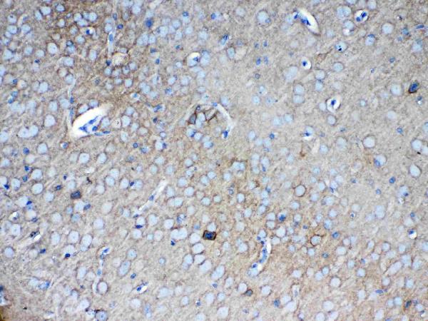
A06700-1 has IHC images of paraffin sections from human glioma and mouse brain; the payload supplies no IF images (A06700-1 IHC and IF image fields).
A06700-1 lists IHC for Human, Mouse, and Rat, with IHC images from human glioma and mouse brain paraffin sections (catalog applications and reactivity; A06700-1 IHC image captions). M06700 lists IHC and Human reactivity, but supplies no IHC image (catalog applications, reactivity, and IHC image field).
Which to pick: For tissue IHC, pick A06700-1 because its own captions document staining in paraffin sections; the fixative is unreported (A06700-1 IHC image captions). For work across species, A06700-1 lists Human, Mouse, and Rat reactivity, while M06700 is a rabbit monoclonal listed for Human only (catalog reactivity, host, and clone). Neither SKU lists IF/ICC as an application, so neither has catalog support for that choice (catalog applications).