NCOA1 / Nuclear receptor coactivator 1 · IHC design guide

Design Immunohistochemistry for NCOA1

Plan NCOA1 staining in paraffin sections using its widespread nuclear tissue pattern (HPA tissue IHC). The catalog antibody has a breast cancer IHC example (datasheet M00856); HPA cell type profiles can guide control selection (HPA tissue IHC).

Evidence assembled Oct 2026 · For research use; verify linked source records and product datasheet before use
Immunohistochemistry protocol sheet for NCOA1 (IHC for NCOA1): expected localisation Widespread nuclear staining (HPA tissue IHC), antibody M00856, validated IHC image, and IHC protocol steps
Printable NCOA1 IHC protocol sheet — expected localisation Widespread nuclear staining (HPA tissue IHC), antibody M00856, controls and protocol steps. Open the full NCOA1 IHC guide →

NCOA1 Immunohistochemistry Experimental Design Guide

Expected localisation, validated protocols, controls and antibodies — the at-a-glance facts below, then the full design guide.

Must know before staining
Expected localisation Widespread nuclear staining (HPA tissue IHC)
Staining pattern Nuclear staining across many cell types (HPA tissue IHC)
Antigen retrieval EDTA pH 8.0 HIER, heat-mediated (datasheet M00856)
Positive control ⓘ Adrenal gland+4 more · see all
Negative control ⓘ Adipose tissue
Important caveats
Reasons your staining may differ from the expected pattern.
Fixation Keep fixation consistent across sections (standard IHC practice; not target-specific)
Caveat Adipocytes may be unstained despite the broad tissue pattern (HPA tissue IHC)
Regulation Abundance regulation unreported (UniProt)
Isoform / epitope 3 isoforms; antibody epitope coverage is unknown (UniProt; catalog M00856)
Section 1

Recommended NCOA1 IHC & IF Protocols

The catalog antibody’s IHC-P protocol (datasheet M00856) is accompanied by published NCOA1 IHC methods for esophageal carcinoma (PMC6198200) and bladder urothelial carcinoma (PMC2674368).

Recommended immunohistochemistry (IHC-P) protocol parameters
SampleParaffin-embedded human breast cancer tissue; fixative not specified (datasheet M00856)
FixationImage fixative and duration unreported (datasheet M00856); verify before use.
Sectioning4–5 µm sections on charged slides (standard)
DeparaffinisationXylene, graded ethanol series to water (standard)
Antigen retrievalHeat retrieval: EDTA pH 8.0 (datasheet M00856); 20 min, 95–100 °C (standard)
Peroxidase block3% H2O2, 10 min, room temperature (standard)
Blocking10% goat serum (datasheet M00856)
Primary antibodyRabbit monoclonal (clone AEHD-14) anti-NCOA1, 1:50 (datasheet M00856)
Primary incubationOvernight at 4 °C (datasheet M00856)
DetectionHRP-conjugated secondary, DAB chromogen (datasheet M00856)
CounterstainHematoxylin, blue, dehydrate and mount (standard)
Expected resultNCOA1-positive staining in glandular cells of adrenal gland (HPA tissue IHC: High). HPA tissue profile: Ubiquitous nuclear expression. No signal in the no-primary control.
💡Decision noteStart with heat-mediated EDTA pH 8.0 retrieval (datasheet M00856); the bladder study also used EDTA pH 8 (PMC2674368).
Section 2

What Is the Expected NCOA1 Staining Pattern?

NCOA1 should show predominantly nuclear staining across many cell types (UniProt Q15788: nucleus; HPA tissue IHC: ubiquitous nuclear expression). High staining is reported in breast, colon and adrenal glandular cells, among others (HPA tissue IHC: High). HPA rates the tissue IHC pattern Supported, with medium consistency between antibody staining and RNA expression (HPA tissue IHC: reliability). NCOA1 has no transmembrane segment (UniProt Q15788: topology).

What am I looking at on my slide?
Distinct nuclear staining in glandular cells of breast, colon or adrenal gland, with interpretable tissue structure.This fits the expected compartment and reported High staining in those cells (UniProt Q15788: nucleus; HPA tissue IHC: High in breast, colon and adrenal glandular cells). Compare intensity within the same staining run; HPA levels describe observed tissue patterns, not a required intensity for every section (HPA tissue IHC: reliability Supported).
Strong cytoplasmic staining with little or no nuclear signal in an otherwise positive tissue.Recheck specificity and staining conditions before calling this a typical IHC result: HPA describes tissue expression as nuclear (HPA tissue IHC: ubiquitous nuclear expression). Cytosolic signal alone is not proof of an artefact, because ICC-IF also reports approved cytosolic localization (HPA ICC-IF: cytosol approved). The assays need separate interpretation.
Prominent staining of adipocytes where adjacent cells or a separate positive control stain as expected.Treat this as unexpected for the sampled cell type: HPA reports adipocytes as Not detected (HPA tissue IHC: adipocytes Not detected). Check whether the color follows adipocytes, pigment or tissue edges, and assess antibody specificity and endogenous detection activity (general IHC practice). Unexpected staining alone cannot identify which cause is responsible.
Weak, diffuse color across nuclei, cytoplasm and tissue spaces, without clear cellular boundaries.This does not establish NCOA1 localization; the supported tissue pattern is nuclear (HPA tissue IHC: ubiquitous nuclear expression). Review blocking, primary antibody concentration, washing and detection controls as general background checks (general IHC practice). Preserve any distinct nuclear signal when comparing conditions, since diffuse color can obscure it.
No nuclear signal in breast or colon glandular cells, while the counterstain and tissue are readable.Investigate a possible failed run before interpreting the sample as NCOA1 negative: HPA reports High staining in these cells (HPA tissue IHC: breast and colon glandular cells High). Include a known positive section and inspect antigen retrieval, primary antibody and detection steps (general IHC practice). A control result from the same run is more informative than tissue identity alone.
💡Expected NCOA1 appearanceCall a convincing positive result distinct, predominantly nuclear staining in the relevant cells, especially clearly stained glandular cells in a reported High tissue (UniProt Q15788: nucleus; HPA tissue IHC: breast and colon glandular cells High); diffuse color or prominent staining of HPA-negative adipocytes calls for control-based review (HPA tissue IHC: adipocytes Not detected; general IHC practice).
How each factor affects the staining
Choice of comparison tissueBreast or colon glandular cells offer reported High nuclear staining for a positive comparison (HPA tissue IHC: High; ubiquitous nuclear expression). Hepatocytes, cardiomyocytes and skeletal myocytes are reported Low, so a faint result there is less decisive (HPA tissue IHC: Low in those cells).
Interpretation of an adipose sectionAdipocytes are reported Not detected, which makes conspicuous adipocyte staining a useful specificity question (HPA tissue IHC: adipocytes Not detected). It does not make a whole adipose section a guaranteed negative control for every cell present (HPA tissue IHC: cell-specific entry).
Antibody validation and isoformsThe supplied antibody list records IHC Supported for CAB019402, while the other listed antibodies have ICC Approved status only (HPA antibodies: validation entries). UniProt lists 3 NCOA1 isoforms; no supplied epitope mapping establishes which an IHC antibody detects (UniProt Q15788: isoforms; HPA antibodies: validation entries).
IF/ICC Q: Should its cytosolic signal change the IHC call?A: HPA ICC-IF approves nucleoplasm and cytosol as main locations, with vesicles and plasma membrane as additional locations (HPA ICC-IF: approved locations). Tissue IHC is described as ubiquitous nuclear expression; assess its pattern on its own terms (HPA tissue IHC: profile).
Antigen retrieval and fixationTarget-specific fixation effects are not established by the supplied assay evidence. Verify with a matched IHC source before attributing a result to fixation.
Why is my staining missing, weak or wrong?
SituationLikely causeNext action
Positive tissue has no discernible nuclear color.The staining run or detection chain may have failed; breast and colon glandular cells are reported High (HPA tissue IHC: High).Check a same-run positive section, then verify retrieval, primary antibody application and chromogenic detection in sequence (general IHC practice).
Only cytoplasmic color appears in a tissue IHC section.The result differs from HPA's nuclear tissue profile, although ICC-IF also supports cytosolic localization (HPA tissue IHC: profile; HPA ICC-IF: cytosol approved).Repeat with controls and score nuclear and cytoplasmic compartments separately before assigning specificity (general IHC practice).
Adipocytes show strong staining.This conflicts with the reported Not detected adipocyte result; antibody binding or endogenous detection activity may contribute (HPA tissue IHC: adipocytes Not detected; general IHC practice).Review a no-primary control and the detection block, then compare with a positive tissue in the same run (general IHC practice).
Color covers most of the section and obscures nuclei.Excess background can arise from antibody concentration, blocking or washing conditions (general IHC practice).Use the antibody's IHC instructions and controls to adjust those steps; judge whether distinct nuclear staining emerges (general IHC practice; HPA tissue IHC: nuclear profile).
A low-signal tissue appears negative.Low staining is reported for hepatocytes, cardiomyocytes and skeletal myocytes (HPA tissue IHC: Low).Check a reported High tissue in the same run before treating the low-signal result as assay failure (HPA tissue IHC: High in breast and colon glandular cells; general IHC practice).
An ICC-validated antibody is being considered for paraffin IHC.ICC approval does not establish IHC validation; the supplied HPA entries distinguish those statuses (HPA antibodies: validation entries).Choose an IHC-supported antibody and follow its IHC instructions, then confirm the nuclear pattern with tissue controls (HPA antibodies: CAB019402 IHC Supported; HPA tissue IHC: nuclear profile; general IHC practice).

Sample controls for NCOA1 IHC & IF

🧪Run breast first: glandular cells should stain (HPA: High in breast glandular cells). Run adipose tissue as the negative comparator (HPA: Not detected in adipocytes); on the breast slide, count morphologically identifiable cells lacking specific nuclear DAB signal as internal negatives only after verifying their identity and staining.
Positive control tissue: Adrenal gland (Glandular cells, HPA High)
Negative control tissue: Adipose tissue (HPA Not detected)
ICC-IF cell lines (HPA subcellular resource): HPA ICC-IF images show NCOA1 in Rh30, SiHa, U2OS, KOLF2.1J, with annotated localisation: Nucleoplasm (approved), Cytosol (approved) (HPA subcellular).
Technical controls: Include a no-primary, secondary-only control, an irrelevant rabbit IgG control matched to the catalog antibody’s immunoglobulin class and clonality, and NCOA1 knockout tissue or a validated immunizing-peptide block where available (selected M00856 caption: rabbit primary; standard IHC controls). Quench endogenous peroxidase and assess background in the breast section before interpreting DAB staining (selected M00856 caption: HRP/DAB detection; standard IHC practice).
⚠️Feasibility: No target-specific fixation window or fixation effect is reported, and the selected M00856 paraffin-section caption does not state the fixative (selected M00856 caption). Heat retrieval in EDTA at pH 8.0 was used for the pictured breast cancer section, but retrieval dependency is unreported (selected M00856 caption). ICC-IF images exist for Rh30, SiHa, U2OS and KOLF2.1J (HPA subcellular); the evidence does not establish whether IF or frozen sections are easier, and endogenous peroxidase can complicate interpretation of HRP/DAB staining in breast sections (selected M00856 caption: HRP/DAB detection; standard IHC practice).

HPA tissue IHC evidence for NCOA1

Comprehensive Human Protein Atlas IHC scoring per tissue (reliability: Supported — Medium consistency between antibody staining and RNA expression data.). Rows are taken directly from the HPA tissue chart — click any row's HPA link to view the source.

Positive expression · recommended positive controls

TissueCell typeLevelEvidenceSource
Adrenal gland Glandular cells High Protein (IHC) HPA →
Breast Glandular cells High Protein (IHC) HPA →
Bronchus Basal cells High Protein (IHC) HPA →
Cerebellum Cells in granular layer High Protein (IHC) HPA →
Cerebral cortex Glial cells High Protein (IHC) HPA →

Undetected expression · recommended negative controls

TissueCell typeLevelEvidenceSource
Adipose tissue Adipocytes Not detected Protein (IHC) HPA →
Section 3

Advanced NCOA1 IHC Tips

Troubleshoot NCOA1 staining by checking retrieval, nuclear localisation, antibody specificity and scoring consistency against the supplied IHC evidence.

Which retrieval conditions should I try first for NCOA1 paraffin sections?
Start with heat-mediated retrieval in EDTA at pH 8.0 (datasheet M00856). The selected paraffin-section example used that retrieval before staining human breast cancer tissue with the catalog antibody (datasheet M00856). If nuclear staining is weak, compare a carefully controlled alternative retrieval condition on adjacent sections while keeping antibody dilution and detection constant (standard IHC practice). Check for tissue lifting, damaged morphology and staining at section edges before interpreting a stronger signal as improved antigen exposure (standard IHC practice). Include a known staining control in each comparison, since NCOA1 shows broad nuclear tissue expression but variable cell-type intensity (HPA: ubiquitous nuclear expression; selected tissue levels).
Could fixation explain weak or uneven NCOA1 staining?
Target-specific fixation sensitivity is unknown from the supplied evidence; the selected paraffin-section caption does not state its fixative (datasheet M00856). Record the fixative, fixation duration and any delay before fixation for each specimen, then compare sections processed together (standard IHC practice). Excessive or inconsistent fixation can alter antigen accessibility in paraffin IHC generally, but that does not establish a particular effect for NCOA1 (standard IHC practice). Keep EDTA retrieval at pH 8.0 and the antibody dilution constant while assessing a processing difference (datasheet M00856). Use nuclear morphology and a concurrently stained control to distinguish processing variation from a change in NCOA1 signal (standard IHC practice).
How should I assess cytoplasmic NCOA1 staining in chromogenic IHC?
Prioritise nuclear signal when reading tissue IHC, because the tissue profile describes ubiquitous nuclear expression (HPA: tissue IHC profile). Cytoplasmic staining warrants a separate assessment: cell imaging places NCOA1 mainly in the nucleoplasm and cytosol, with additional vesicle and plasma-membrane localisation (HPA: subcellular). Score nuclei and cytoplasm separately instead of treating all brown signal as equivalent (standard IHC practice). Compare the pattern with a no-primary control and an independently validated antibody when available, especially if cytoplasmic staining dominates (standard IHC practice). NCOA1 has no transmembrane segment, so a continuous membrane rim alone needs particularly careful validation (UniProt Q15788 topology).
Can this IHC result distinguish NCOA1 isoforms or modification states?
Do not assign an isoform from staining unless the antibody epitope and its isoform coverage have been established (standard IHC interpretation; UniProt Q15788: 3 isoforms). NCOA1 has several annotated modified residues, including phosphoserines at positions 22, 372, 395 and 517 (UniProt Q15788 modified residues). Those annotations alone do not show that this antibody distinguishes phosphorylation states or that retrieval affects one state selectively (standard IHC interpretation). Check the documented immunogen or epitope against each isoform before claiming selective detection (standard antibody-validation practice). If the epitope remains unknown, report the result as NCOA1 immunoreactivity and avoid isoform-specific conclusions (standard IHC interpretation).
How can I investigate NCOA1 localisation with a companion IF experiment?
Treat IF as a separate assay and validate its antibody and preparation independently of the paraffin IHC example (standard IF practice; datasheet M00856: paraffin-section IHC). Multiplex NCOA1 with an independently validated marker for the cell type being examined, so the marker identifies the cells whose signal is scored (standard IF practice). Select fluorophores after checking the specimen’s autofluorescence, reserving the cleaner channel for weaker signal (standard IF practice). NCOA1 has no transmembrane segment and is reported mainly in the nucleoplasm and cytosol, so permeabilise sufficiently for intracellular antibody access (UniProt Q15788 topology; HPA: subcellular). Because the antibody epitope is unspecified, confirm that the chosen permeabilisation preserves localisation and morphology with appropriate controls (standard IF practice).
What controls help identify nonspecific brown staining?
Run a no-primary control to reveal signal from the detection system, tissue pigments or endogenous enzyme activity (standard chromogenic IHC practice). Apply a peroxidase block before HRP detection and inspect the control after DAB development; these are general workflow steps, not NCOA1-specific findings (standard chromogenic IHC practice). The selected example used 10% goat serum blocking, 1:50 primary antibody overnight at 4°C, and an HRP-based DAB workflow (datasheet M00856). If background remains high, compare primary-antibody dilution and wash conditions on matched sections while preserving a positive control (standard IHC practice). Judge improvement by cleaner cellular localisation, because the tissue reference describes predominantly nuclear expression (HPA: tissue IHC profile).
How should I quantify NCOA1 staining across specimens? ⚠ ANSWER MARKED FOR VERIFICATION
Define the compartment and cell population before scoring; the tissue IHC reference describes ubiquitous nuclear expression (HPA: tissue IHC profile). For nuclear chromogenic staining, record the percentage of viable positive nuclei and intensity, or calculate an H-score from intensity categories with a possible range of 0–300 (standard IHC scoring). Normalise counts to viable nuclei in the same defined region, or report positive-cell density per mm² of viable tissue (standard IHC quantification). Exclude folds, necrosis and section edges using the same rules across specimens (standard IHC practice). Keep retrieval, detection, counterstain and image-analysis thresholds consistent, and report cytoplasmic signal separately if measured (standard IHC practice).
When is an unexpected NCOA1 pattern likely to be artefactual?
A convincing pattern should fit the cell type and compartment being scored and recur across well-preserved areas (standard IHC interpretation). Nuclear staining is expected broadly in tissue IHC, although the reference reports adipocytes as not detected and several other cell types at low levels (HPA: tissue IHC profile). Strong isolated staining at cut edges, within necrosis, or in a no-primary control calls for investigation before assigning NCOA1 positivity (standard IHC practice). Check endogenous peroxidase and pigments when brown signal persists without primary antibody (standard chromogenic IHC practice). HPA rates its tissue IHC evidence as Supported with medium staining-to-RNA consistency, so resolve discordant patterns with independent validation (HPA: reliability).
Boster reagents

Best NCOA1 / Nuclear receptor coactivator 1 IHC Antibodies

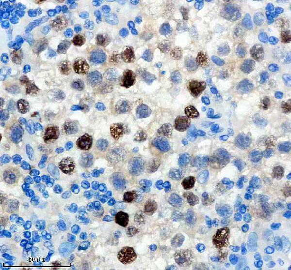
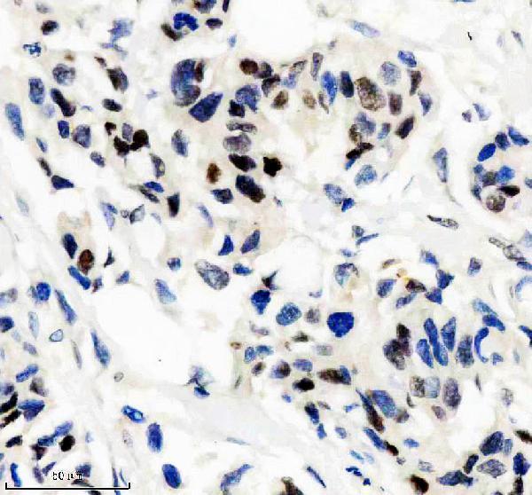
The catalog antibody has an IHC image from a paraffin-embedded human breast cancer section (M00856 image caption); its listed reactivity is human (catalog reactivity: M00856).

Real IHC data IHC analysis of NCOA1 using anti-NCOA1 antibody (M00856). NCOA1 was detected in a paraffin-embedded section of human breast cancer tissue. Heat mediated antigen retrieval was performed in EDTA buffer (pH 8.0, epitope retrieval solution). The tissue section was blocked with 10% goat serum. The tissue section was then incubated with 1: 50 rabbit anti-NCOA1 Antibody (M00856) overnight at 4°C. Peroxidase Conjugated Goat Anti-rabbit IgG was used as secondary antibody and incubated for 30 minutes at 37°C. The tissue section was developed using HRP Conjugated Rabbit IgG Super Vision Assay Kit (Catalog # SV0002) with DAB as the chromogen.
Anti-KAT13A / SRC1 Monoclonal Antibody
Cat # M00856

M00856 is listed for IHC and human reactivity (catalog applications and reactivity: M00856). Its IHC image shows a paraffin-embedded human breast cancer section (M00856 image caption).

Which to pick: Choose M00856 for human tissue IHC: it is a rabbit monoclonal antibody listed for IHC, with a paraffin-section image and a 1:50 incubation in that caption (catalog: M00856; M00856 image caption). The caption does not report the fixative (M00856 image caption). IF/ICC is absent from its application list, and no listed SKU has reactivity beyond human (catalog applications and reactivity: M00856).

Each figure is that product's own IHC / IF validation image from its datasheet.

References

  1. UniProt Consortium. UniProt entry Q15788 (NCOA1_HUMAN, Nuclear receptor coactivator 1).
  2. Human Protein Atlas. NCOA1 tissue IHC expression (reliability: Supported).
  3. Human Protein Atlas. NCOA1 subcellular location (ICC-IF): Mainly localized to the nucleoplasm and cytosol. In addition localized to the plasma membrane and vesicles..
  4. Human Protein Atlas. NCOA1 antibody validation summary (3 antibodies).
  5. The oncogenic roles of nuclear receptor coactivator 1 in human esophageal carcinoma. Cancer medicine 2018 — PMC6198200.
  6. NCOA1 promotes angiogenesis in breast tumors by simultaneously enhancing both HIF1α- and AP-1-mediated VEGFa transcription. Oncotarget 2015 — PMC4695159.
  7. High expression of miR-105-1 positively correlates with clinical prognosis of hepatocellular carcinoma by targeting oncogene NCOA1. Oncotarget 2017 — PMC5355313.
  8. Expression and significance of androgen receptor coactivators in urothelial carcinoma of the bladder. Endocrine-related cancer 2009 — PMC2674368.
  9. PubMed PMID:8754792 — UniProt-cited evidence.
  10. PubMed PMID:9427757 — UniProt-cited evidence.
  11. PubMed PMID:9575154 — UniProt-cited evidence.