NCOA3 / Nuclear receptor coactivator 3 · IHC design guide

Design Immunohistochemistry for NCOA3

Plan NCOA3 chromogenic IHC on paraffin sections using the catalog antibody at 1:100–1:300 (datasheet: A01337-2). Assess cytoplasmic and nuclear staining across tissues (HPA tissue IHC), allowing for TNF-dependent nuclear translocation (UniProt).

Evidence assembled Oct 2026 · For research use; verify linked source records and product datasheet before use
Immunohistochemistry protocol sheet for NCOA3 (IHC for NCOA3): expected localisation Cytoplasmic and nuclear tissue staining (HPA tissue IHC), antibody A01337-2, validated IHC image, and IHC protocol steps
Printable NCOA3 IHC protocol sheet — expected localisation Cytoplasmic and nuclear tissue staining (HPA tissue IHC), antibody A01337-2, controls and protocol steps. Open the full NCOA3 IHC guide →

NCOA3 Immunohistochemistry Experimental Design Guide

Expected localisation, validated protocols, controls and antibodies — the at-a-glance facts below, then the full design guide.

Must know before staining
Expected localisation Cytoplasmic and nuclear tissue staining (HPA tissue IHC)
Staining pattern Widespread cytoplasmic and nuclear staining (HPA tissue IHC)
Antigen retrieval Tris-EDTA pH 9.0 HIER, 95–98 °C, 20 min (rule: nuclear antigen)
Positive control ⓘ Testis+4 more · see all
Negative control ⓘ Caudate
Important caveats
Reasons your staining may differ from the expected pattern.
Fixation Keep paraffin-section fixation consistent (standard IHC practice; not target-specific)
Caveat Caudate glia may lack detectable staining (HPA tissue IHC)
Regulation TNF activation drives nuclear entry (UniProt)
Isoform / epitope Five isoforms; epitope coverage is undetermined (UniProt)
Section 1

Recommended NCOA3 IHC & IF Protocols

The catalog antibody’s IHC-P protocol is accompanied by four published NCOA3 IHC protocols (PMC2784204; PMC5690823; PMC4257137; PMC4476953).

Recommended immunohistochemistry (IHC-P) protocol parameters
SampleParaffin-embedded Mouse Brain tissue; fixative not specified (datasheet A01337-2)
FixationImage fixative and duration unreported (datasheet A01337-2); verify before use.
Sectioning4–5 µm sections on charged slides (standard)
DeparaffinisationXylene, graded ethanol series to water (standard)
Antigen retrievalHeat-induced epitope retrieval in Tris-EDTA buffer, pH 9.0, 20 min at 95–98 °C (standard rule: nuclear antigen)
Peroxidase block3% H2O2, 10 min, room temperature (standard)
Blocking10% normal serum of the secondary host, 30 min, room temperature (standard)
Primary antibodyRabbit anti-NCOA3, 1:100-1:300 (datasheet A01337-2)
Primary incubationOvernight at 4 °C (standard)
DetectionHRP-polymer secondary, DAB chromogen 5–10 min (standard)
CounterstainHematoxylin, blue, dehydrate and mount (standard)
Expected resultNCOA3-positive staining in spermatogonia cells of testis (HPA tissue IHC: High). HPA tissue profile: Ubiquitous cytoplasmic and nuclear expression in all tissues. No signal in the no-primary control.
💡Decision noteStart with Tris-EDTA pH 9.0 retrieval at 95–98 °C for 20 min (page retrieval); compare the published retrieval conditions where specified (PMC4257137; PMC4476953).
Section 2

What Is the Expected NCOA3 Staining Pattern?

In paraffin-section IHC, expect NCOA3 in cytoplasm and nuclei across many cell types; HPA describes ubiquitous staining with Supported reliability and medium consistency with RNA data (HPA tissue IHC). Spermatogonia show a high signal, while several other listed cell types show medium signal (HPA tissue IHC). NCOA3 has no transmembrane segment, so a membrane-restricted pattern lacks support (UniProt Q9Y6Q9 topology).

What am I looking at on my slide?
Cytoplasmic and nuclear staining in spermatogonia, with a comparatively strong signal.This fits the high staining reported for spermatogonia and the broad two-compartment tissue profile (HPA tissue IHC). Compare cells within the same section before judging intensity: HPA levels describe observed patterns, not a fixed staining threshold for every preparation (HPA tissue IHC; general IHC practice).
Cytoplasmic and nuclear staining in adipocytes or glandular cells at moderate intensity.Medium staining is reported in adipocytes of adipose tissue and breast, and in glandular cells of adrenal gland and appendix (HPA tissue IHC). Either compartment can be credible: tissue IHC reports both, while the relative balance may depend on biological context (HPA tissue IHC; UniProt Q9Y6Q9 subcellular location).
A crisp signal confined to cell membranes, with little cellular staining elsewhere.Treat this as a localization mismatch worth checking. NCOA3 has no transmembrane segment, and the supplied localization evidence places it in cytoplasm and nucleus (UniProt Q9Y6Q9 topology and subcellular location; HPA tissue IHC). Recheck morphology, controls and detection before calling it NCOA3 (general IHC practice).
Strong staining in caudate glial cells, especially if the same color persists in a no-primary control.HPA reports NCOA3 as not detected in caudate glial cells, although that observation is not an absolute exclusion in every specimen (HPA tissue IHC). Persistence without primary antibody points toward endogenous detection activity or nonspecific detection; investigate rather than assigning the signal to NCOA3 (general IHC practice).
Widespread, hazy color that obscures nuclei and cell boundaries.This is difficult to score against HPA's cellular cytoplasmic and nuclear pattern (HPA tissue IHC). Background from detection reagents, insufficient blocking or excessive antibody concentration are general IHC possibilities, not documented NCOA3-specific effects (general IHC practice). Interpret only distinct cellular staining after the background is resolved.
💡Expected NCOA3 appearanceA convincing positive shows distinguishable cytoplasmic and nuclear staining, strongest among the listed examples in spermatogonia; membrane-only or structureless diffuse color is suspect (HPA tissue IHC; UniProt Q9Y6Q9 topology; general IHC practice).
How each factor affects the staining
Tissue and cell choiceSpermatogonia are a high-staining reference; adipocytes and several epithelial or glandular populations are medium, while caudate glial cells are not detected (HPA tissue IHC). These are observed levels, so assess the named cell population rather than calling an entire section positive or negative (HPA tissue IHC; general IHC practice).
Compartment and biological stateTissue IHC reports both cytoplasmic and nuclear staining; UniProt describes NCOA3 as mainly cytoplasmic and weakly nuclear, with nuclear translocation after TNF activation and phosphorylation (HPA tissue IHC; UniProt Q9Y6Q9 subcellular location). A shift in compartment balance can be biologically plausible, but staining alone does not establish TNF activation (UniProt Q9Y6Q9; general IHC practice).
Antibody evidenceHPA rates the tissue profile Supported, with medium consistency between antibody staining and RNA expression (HPA tissue IHC). HPA lists IHC support for HPA024210 and CAB009800; neither entry here establishes the performance of an unspecified catalog antibody (HPA antibody validation). Use each antibody's own controls when interpreting a new stain (general IHC practice).
Retrieval and detectionTarget-specific fixation effects are not established by the supplied assay evidence. Verify with a matched IHC source before attributing a result to fixation.
IF/ICC: should the signal be nuclear?Yes. HPA ICC-IF places NCOA3 mainly in nucleoplasm, with additional cytosol localization, in its subcellular summary (HPA subcellular ICC-IF). That IF observation can inform localization checks, but it does not set a paraffin IHC protocol or require every IHC-positive cell to be predominantly nuclear (HPA subcellular ICC-IF; HPA tissue IHC).
Why is my staining missing, weak or wrong?
SituationLikely causeNext action
No stain in spermatogonia expected to be positive.The high HPA signal makes a wholly blank result worth investigating, but does not identify the failed step (HPA tissue IHC).Check that the named cells are present, then inspect the primary-antibody step, retrieval and chromogen controls using standard IHC checks (general IHC practice). Avoid assigning NCOA3-specific fixation sensitivity; none is supplied (HPA tissue IHC; UniProt Q9Y6Q9).
Only a membrane rim is visible.A membrane-restricted pattern conflicts with the supplied cytoplasmic and nuclear localization and lack of a transmembrane segment (HPA tissue IHC; UniProt Q9Y6Q9 topology).Confirm the rim follows cells rather than section edges or deposits; compare no-primary and appropriate control sections before scoring it as specific (general IHC practice).
Strong color appears in caudate glial cells.HPA reports this cell population as not detected; strong color there raises a specificity or detection question, without proving cross-reactivity (HPA tissue IHC).Review cell identity and compare a no-primary control. If color remains, address endogenous detection activity; if it disappears, reassess primary-antibody specificity (general IHC practice).
Diffuse background hides the cellular pattern.Excess chromogen development, inadequate blocking or excessive primary concentration can cause background in IHC (general IHC practice).Inspect no-primary and reagent controls, then adjust blocking, antibody concentration or development one variable at a time (general IHC practice). Score NCOA3 only where distinct cellular staining remains (HPA tissue IHC; general IHC practice).
Nuclear signal dominates a section with little cytoplasmic color.HPA ICC-IF emphasizes nucleoplasm, while UniProt describes mainly cytoplasmic localization and stimulus-linked nuclear translocation; the sources support context-dependent localization (HPA subcellular ICC-IF; UniProt Q9Y6Q9).Check that nuclear staining is cellular and reproducible across relevant controls; report the observed compartment without inferring TNF activation from the image alone (general IHC practice; UniProt Q9Y6Q9).
A listed medium-staining tissue appears weaker than testis.This can match HPA's different reported levels: high in spermatogonia and medium in several other listed populations (HPA tissue IHC).Compare the correct cell populations under the same staining run, document relative intensity, and avoid calling weak-but-distinct cellular staining negative solely by comparison with spermatogonia (HPA tissue IHC; general IHC practice).

Sample controls for NCOA3 IHC & IF

🧪Run testis first; spermatogonia should stain strongly (HPA: High in spermatogonia). Use caudate glial cells as the negative tissue (HPA: Not detected in caudate glial cells); compare other cells on the testis slide with spermatogonia, but do not require them to be unstained because NCOA3 is widely expressed (UniProt Q9Y6Q9: tissue specificity).
Positive control tissue: Testis (Spermatogonia cells, HPA High)
Negative control tissue: Caudate (HPA Not detected)
ICC-IF cell lines (HPA subcellular resource): HPA ICC-IF images show NCOA3 in A-431, U-251MG, U2OS, with annotated localisation: Nucleoplasm (supported) (HPA subcellular).
Technical controls: Include a no-primary (secondary-only) slide, host- and isotype-matched control IgG for a monoclonal primary or host-matched nonimmune IgG for a polyclonal primary, and an NCOA3 knockout specimen if available (standard IHC practice). Check endogenous peroxidase background in the testis section before interpreting chromogenic signal (standard IHC practice).
⚠️Feasibility: A target-specific fixation window and retrieval dependency are unreported in the supplied evidence; the selected A01337-2 mouse-brain paraffin-section caption also leaves the fixative unreported (A01337-2 tissue-IHC caption). The supplied evidence does not establish whether frozen sections or IF are easier than paraffin IHC; ICC-IF images support nucleoplasmic and cytosolic localisation in A-431, U-251MG and U2OS cells (HPA: subcellular localisation). No testis-specific artefact is reported in the supplied evidence; assess background with the technical controls (standard IHC practice).

HPA tissue IHC evidence for NCOA3

Comprehensive Human Protein Atlas IHC scoring per tissue (reliability: Supported — Medium consistency between antibody staining and RNA expression data.). Rows are taken directly from the HPA tissue chart — click any row's HPA link to view the source.

Positive expression · recommended positive controls

TissueCell typeLevelEvidenceSource
Testis Spermatogonia cells High Protein (IHC) HPA →
Adipose tissue Adipocytes Medium Protein (IHC) HPA →
Adrenal gland Glandular cells Medium Protein (IHC) HPA →
Appendix Glandular cells Medium Protein (IHC) HPA →
Bone marrow Hematopoietic cells Medium Protein (IHC) HPA →

Undetected expression · recommended negative controls

TissueCell typeLevelEvidenceSource
Caudate Glial cells Not detected Protein (IHC) HPA →
Section 3

Advanced NCOA3 IHC Tips

Troubleshoot NCOA3 staining in paraffin sections by assessing retrieval, cell compartment, controls, and scoring before interpreting chromogenic signal.

What should I change if NCOA3 staining is weak after antigen retrieval?
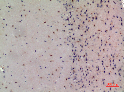
Start with Tris-EDTA pH 9.0 heat-induced epitope retrieval at 95–98 °C for 20 min (page retrieval setting). Keep section thickness, heating, cooling, and antibody incubation consistent while comparing a weak section with a control section in the same run (standard IHC practice). The catalog image shows staining in paraffin-embedded mouse brain, but its caption does not report the fixative or retrieval method (catalog caption: A01337-2). If staining remains weak, trial another validated retrieval condition on adjacent sections and compare signal with tissue morphology and background (standard IHC practice). Do not treat the page setting as a demonstrated optimum for every specimen (page retrieval setting).
Could fixation explain inconsistent NCOA3 staining between paraffin blocks?
Target-specific fixation sensitivity is unknown from the supplied evidence, so record each block’s fixative and fixation duration before attributing differences to NCOA3 (catalog caption: A01337-2; standard IHC practice). The catalog caption identifies paraffin-embedded mouse brain but does not state its fixative (catalog caption: A01337-2). Compare similarly processed sections in the same staining run, using the page’s Tris-EDTA pH 9.0 retrieval at 95–98 °C for 20 min (page retrieval setting; standard IHC practice). Inspect nuclear detail and section integrity while comparing signal, since damaged morphology makes compartment scoring unreliable (standard IHC practice). Document any fixation difference as a possible processing variable, not a proven NCOA3-specific effect (standard IHC practice).
How should I interpret nuclear versus cytoplasmic NCOA3 staining?
Score nuclear and cytoplasmic chromogenic staining separately because both compartments are supported for NCOA3 (HPA tissue IHC: ubiquitous cytoplasmic and nuclear expression). HPA subcellular data place it mainly in the nucleoplasm with additional cytosolic localisation, whereas UniProt describes mainly cytoplasmic and weakly nuclear localisation (HPA subcellular: supported locations; UniProt Q9Y6Q9: subcellular location). UniProt also reports nuclear translocation after TNF activation and phosphorylation, so a different nuclear fraction may reflect biological context (UniProt Q9Y6Q9: subcellular location). Compare adjacent cell types and matched processing conditions before making that inference (standard IHC practice). Treat isolated cell-border staining without convincing intracellular signal as suspect, since NCOA3 has no transmembrane segment (UniProt Q9Y6Q9: topology).
Can an NCOA3 epitope explain different staining patterns across specimens?
NCOA3 has 5 listed isoforms, and the supplied antibody caption does not identify the epitope recognised by A01337-2 (UniProt Q9Y6Q9: isoforms; catalog caption: A01337-2). Its bHLH domain spans residues 25–82, and its PAS domain spans 110–180 (UniProt Q9Y6Q9: domains). UniProt also lists multiple modified residues, including phosphoserines at 214, 551, and 569 (UniProt Q9Y6Q9: modified residues). Ask for the antibody’s immunogen or epitope information before claiming that an isoform or modification accounts for a staining difference (standard IHC practice). Until then, compare nuclear and cytoplasmic patterns using the same retrieval and detection conditions (page retrieval setting; standard IHC practice).
How can I check an NCOA3 IHC pattern with multiplex IF?
Use multiplex IF as a separate check of the cell and compartment assignments made on chromogenic sections (standard IHC/IF practice). In testis, pair NCOA3 with a validated marker for spermatogonia, since HPA reports high staining in those cells (HPA tissue IHC: testis, spermatogonia, High). Choose a far-red channel when tissue autofluorescence obscures shorter wavelengths, and include single-stain controls to assess bleed-through (standard IF practice). Because NCOA3 is intracellular and has no transmembrane segment, permeabilise for access to its cytoplasmic and nuclear epitopes, for example with 0.1% Triton X-100 for 5–10 min, then assess morphology (UniProt Q9Y6Q9: topology and location; standard IF practice). HPA reports nucleoplasmic and additional cytosolic IF localisation, which provides a compartment comparison (HPA subcellular: supported locations).
How can I distinguish weak NCOA3 signal from chromogenic background?
Run a no-primary control through the same chromogenic detection steps and compare it with the stained section (standard IHC practice). For a peroxidase and DAB workflow, block endogenous peroxidase and check whether residual colour persists without primary antibody; these are general assay steps, not NCOA3-specific validation (standard IHC practice). Examine folds, edges, and damaged regions before adjusting antibody concentration, because uneven reagent access can produce local artefacts (standard IHC practice). Compare intracellular staining in candidate cells with the control, using nuclear and cytoplasmic compartments as separate observations (HPA tissue IHC: cytoplasmic and nuclear expression; standard IHC practice). If diffuse background dominates, retitrate the antibody and review blocking and wash conditions while keeping retrieval fixed (standard IHC practice).
How should I quantify NCOA3 across chromogenic IHC sections? ⚠ ANSWER MARKED FOR VERIFICATION
Define the cell population and tissue region before scoring, and analyse comparable regions across sections (standard IHC practice). Report nuclear and cytoplasmic staining separately, since both are documented for NCOA3 (HPA tissue IHC: cytoplasmic and nuclear expression). An H-score sums the percentage of cells at each intensity level multiplied by that level, giving a range of 0–300 for intensities 0–3 (standard IHC practice). Alternatively, report percent positive cells or positive-cell density per mm², alongside the compartment and positivity threshold used (standard IHC practice). Normalise counts to eligible cells or measured tissue area, and exclude folds and nonviable regions consistently (standard IHC practice).
When is an apparent NCOA3-positive cell likely to be an artefact?
Accept a signal only when it is intracellular, follows intact cell morphology, and exceeds the corresponding no-primary control (HPA tissue IHC: cytoplasmic and nuclear expression; standard IHC practice). Nucleus-only staining is not automatically suspect, because HPA supports nucleoplasmic localisation, while UniProt also describes cytoplasmic localisation (HPA subcellular: supported locations; UniProt Q9Y6Q9: subcellular location). Check whether the cell assignment is plausible: HPA reports high staining in testis spermatogonia but no detection in caudate glial cells (HPA tissue IHC: positive and negative cell annotations). Treat colour confined to section edges, folds, or necrotic areas as suspect (standard IHC practice). If staining survives the no-primary control in a peroxidase assay, investigate endogenous enzyme signal before calling it NCOA3 (standard IHC practice).
Boster reagents

Best NCOA3 / Nuclear receptor coactivator 3 IHC Antibodies

Two anti-NCOA3 antibodies have paraffin-section IHC images from mouse brain and human brain (catalog image captions); both list Human, Mouse and Rat reactivity (catalog applications and reactivity).

Real IHC data Immunohistochemistry validation of NCOA3 using Anti-NCoA-3 Antibody (A01337-2). Immunohistochemistry (IHC) analysis of paraffin-embedded Mouse Brain
Anti-NCoA-3 Antibody
Cat # A01337-2
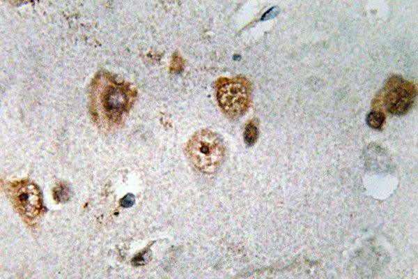
Real IHC data Immunohistochemistry (IHC) analyzes of NCoA-3 (S587) pAb in paraffin-embedded human brain tissue.
Anti-NCoA-3 (S587) Antibody
Cat # A01337S587

A01337-2 has a paraffin-embedded mouse brain IHC image and lists IHC and IF applications (catalog image caption and applications). A01337S587 has a paraffin-embedded human brain IHC image and lists IHC, but not IF, as an application (catalog image caption and applications).

Which to pick: For tissue IHC, choose A01337-2 when the mouse brain image is most relevant, or A01337S587 when the human brain image is most relevant; both captions identify paraffin-embedded tissue, but neither reports the fixative (catalog image captions). For IF, choose A01337-2 because IF is listed for that SKU; neither SKU has an IF image or explicit ICC validation in the payload (catalog applications and image captions). Both are rabbit polyclonal antibodies listing Human, Mouse and Rat reactivity, so species coverage alone does not distinguish them (catalog host, dilution_raw and reactivity).

Each figure is that product's own IHC / IF validation image from its datasheet.

References

  1. UniProt Consortium. UniProt entry Q9Y6Q9 (NCOA3_HUMAN, Nuclear receptor coactivator 3).
  2. Human Protein Atlas. NCOA3 tissue IHC expression (reliability: Supported).
  3. Human Protein Atlas. NCOA3 subcellular location (ICC-IF): Mainly localized to the nucleoplasm. In addition localized to the cytosol..
  4. Human Protein Atlas. NCOA3 antibody validation summary (2 antibodies).
  5. A multimarker prognostic assay for primary cutaneous melanoma. Clinical cancer research : an official journal of the American Association for Cancer Research 2009 — PMC2784204.
  6. Prospective Validation of Molecular Prognostic Markers in Cutaneous Melanoma: A Correlative Analysis of E1690. Clinical cancer research : an official journal of the American Association for Cancer Research 2017 — PMC5690823.
  7. Prostate cancer. Ubiquitylome analysis identifies dysregulation of effector substrates in SPOP-mutant prostate cancer. Science (New York, N.Y.) 2014 — PMC4257137.
  8. NCOA3-mediated upregulation of mucin expression via transcriptional and post-translational changes during the development of pancreatic cancer. Oncogene 2015 — PMC4476953.
  9. PubMed PMID:9346901 — UniProt-cited evidence.
  10. PubMed PMID:9267036 — UniProt-cited evidence.
  11. PubMed PMID:9252329 — UniProt-cited evidence.