NCOA5 / Nuclear receptor coactivator 5 · IHC design guide

Design Immunohistochemistry for NCOA5

Plan NCOA5 paraffin-section IHC around a general nuclear staining pattern (HPA tissue IHC). The guide covers tissue controls and a catalog antibody IHC range of 2–5 μg/ml (HPA tissue IHC; datasheet A07810-2).

Evidence assembled Oct 2026 · For research use; verify linked source records and product datasheet before use
Immunohistochemistry protocol sheet for NCOA5 (IHC for NCOA5): expected localisation General nuclear staining in tissue (HPA tissue IHC), antibody A07810-2, validated IHC image, and IHC protocol steps
Printable NCOA5 IHC protocol sheet — expected localisation General nuclear staining in tissue (HPA tissue IHC), antibody A07810-2, controls and protocol steps. Open the full NCOA5 IHC guide →

NCOA5 Immunohistochemistry Experimental Design Guide

Expected localisation, validated protocols, controls and antibodies — the at-a-glance facts below, then the full design guide.

Must know before staining
Expected localisation General nuclear staining in tissue (HPA tissue IHC)
Staining pattern Nuclear staining across multiple cell types (HPA tissue IHC)
Antigen retrieval EDTA pH 8.0 HIER, heat-mediated (datasheet A07810-2)
Positive control ⓘ Adrenal gland+4 more · see all
Negative control ⓘ Adipose tissue
Important caveats
Reasons your staining may differ from the expected pattern.
Fixation Keep fixation consistent across sections (standard IHC practice; not target-specific)
Caveat Reference tissue pattern awaits verification (HPA tissue IHC)
Regulation Widely expressed (UniProt)
Isoform / epitope No isoforms or processing variants annotated (UniProt)
Section 1

Recommended NCOA5 IHC & IF Protocols

The catalog antibody has an IHC-P protocol (datasheet: A07810-2). Published NCOA5 protocols add examples from ovarian, colorectal, and liver tumors (PMC10183562; PMC5746116; PMC12052255; PMC12509451).

Recommended immunohistochemistry (IHC-P) protocol parameters
SampleParaffin-embedded human liver cancer tissue; fixative not specified (datasheet A07810-2)
FixationImage fixative and duration unreported (datasheet A07810-2); verify before use.
Sectioning4–5 µm sections on charged slides (standard)
DeparaffinisationXylene, graded ethanol series to water (standard)
Antigen retrievalHeat retrieval: EDTA pH 8.0 (datasheet A07810-2); 20 min, 95–100 °C (standard)
Peroxidase block3% H2O2, 10 min, room temperature (standard)
Blocking10% goat serum (datasheet A07810-2)
Primary antibodyRabbit anti-NCOA5, 2-5 μg/ml (datasheet A07810-2)
Primary incubationOvernight at 4 °C (datasheet A07810-2)
DetectionHRP-conjugated secondary, DAB chromogen (datasheet A07810-2)
CounterstainHematoxylin, blue, dehydrate and mount (standard)
Expected resultNCOA5-positive staining in glandular cells of adrenal gland (HPA tissue IHC: High). HPA tissue profile: General nuclear expression. No signal in the no-primary control.
💡Decision noteStart with heat-mediated EDTA at pH 8.0 for the catalog antibody (datasheet: A07810-2). Citrate at pH 6.0 is a published alternative (PMC10183562; PMC5746116; PMC12509451).
Section 2

What Is the Expected NCOA5 Staining Pattern?

NCOA5 is a nuclear protein with no transmembrane segment (UniProt Q9HCD5). In paraffin sections, expect nuclear staining in cells with reported expression, including adrenal glandular cells and bronchial respiratory epithelial cells (HPA: High in both). Expression is widespread, so assess cell identity and compartment together (UniProt: widely expressed; HPA: general nuclear expression). The HPA tissue IHC pattern remains uncertain pending external verification (HPA: reliability Uncertain).

What am I looking at on my slide?
Nuclear chromogen in adrenal glandular or bronchial respiratory epithelial cells.This fits the reported compartment and high-staining cell populations (UniProt Q9HCD5: nucleus; HPA: High in both). Compare nuclear signal with the section's negative control before scoring. A fitting pattern supports interpretation, but the HPA tissue IHC result is pending external verification (HPA: reliability Uncertain).
Predominantly cytoplasmic or surface staining, with little nuclear signal.Treat this as a suspect IHC pattern because the expected tissue pattern is nuclear and the protein has no transmembrane segment (HPA: general nuclear expression; UniProt Q9HCD5: nucleus, no transmembrane segment). Review morphology, control staining, and chromogen deposition before calling the cells positive.
Strong staining appears mainly in adipocytes while expected positive cells are weak.HPA reports NCOA5 as not detected in adipocytes, while several glandular and epithelial populations stain strongly (HPA: adipocytes Not detected; adrenal glandular and bronchial epithelial cells High). Consider antibody cross-reactivity or endogenous detection activity; the discrepancy alone does not identify which caused it (general IHC practice).
Diffuse color covers nuclei, cytoplasm, and tissue spaces.A diffuse deposit makes nuclear localization hard to judge against the reported pattern (HPA: general nuclear expression). Check whether color also appears in a control lacking primary antibody; background there points toward the detection system or endogenous activity rather than specific NCOA5 staining (general IHC practice).
No detectable nuclear signal in an expected positive section.Absence in adrenal glandular or bronchial epithelial cells conflicts with their reported high staining (HPA: High in both). First check tissue preservation, assay controls, and whether nuclei are identifiable (general IHC practice). A single negative section cannot establish absent expression, especially with uncertain tissue IHC reliability (HPA: reliability Uncertain).
💡Expected NCOA5 appearanceA convincing positive is discernible nuclear staining in reported high-staining cells, such as adrenal glandular cells; diffuse tissue-wide color or predominant surface staining is suspect (HPA: High in adrenal glandular cells; HPA: general nuclear expression; UniProt Q9HCD5: no transmembrane segment).
How each factor affects the staining
Cellular compartmentUse identifiable nuclei as the primary scoring compartment in IHC (UniProt Q9HCD5: nucleus; HPA: general nuclear expression). Cytoplasmic color alone does not reproduce the reported tissue pattern (HPA: general nuclear expression).
Tissue and cell populationExpression is widespread rather than restricted to one tissue (UniProt: widely expressed; HPA: low RNA tissue specificity). HPA reports High staining in selected glandular, epithelial, and neuronal populations, but Not detected in adipocytes (HPA: tissue IHC).
Strength of supporting evidenceTreat matching IHC staining as provisional: the tissue profile is pending external verification, and antibody HPA050231 has Uncertain IHC validation (HPA: tissue reliability Uncertain; HPA: HPA050231 IHC Uncertain).
Protein topology and processingThe annotated protein has no transmembrane segment or signal peptide, and its recorded chain spans residues 1–579 (UniProt Q9HCD5). These annotations support a nuclear interpretation; they do not establish an antigen retrieval requirement or fixation sensitivity.
Why is my staining missing, weak or wrong?
SituationLikely causeNext action
Expected positive cells show no nuclear staining.The assay may have failed, or this section may differ from the reported high-staining examples (HPA: adrenal glandular and bronchial epithelial cells High; HPA: reliability Uncertain).Confirm that nuclei and morphology are preserved, then review the IHC-validated antibody's supplied conditions and a concurrently processed positive control (general IHC practice).
Color is diffuse across the section.Nonspecific deposition or endogenous detection activity can obscure the expected nuclear pattern (general IHC practice; HPA: general nuclear expression).Inspect a section processed without primary antibody; if color persists, check the detection reagents and relevant endogenous-activity blocking steps (general IHC practice).
Signal is mainly cytoplasmic or at cell borders.The distribution conflicts with the reported nuclear tissue pattern and lacks support from NCOA5 topology (HPA: general nuclear expression; UniProt Q9HCD5: no transmembrane segment).Check nuclear counterstain and morphology, compare controls, and avoid scoring compartment-mismatched color as a confirmed positive (general IHC practice).
Adipocytes stain strongly.That result conflicts with the HPA adipocyte observation; cross-reactivity or endogenous detection activity is possible (HPA: adipocytes Not detected; general IHC practice).Compare with a no-primary control and with reported positive cell populations on a suitable section before assigning specificity (HPA: tissue IHC; general IHC practice).
Reported positive cell populations vary in intensity.HPA's High calls identify reported populations, not a guaranteed intensity for every cell or section (HPA: tissue IHC; HPA: reliability Uncertain).Score cell type, compartment, and intensity separately, using the same controls and scoring criteria across sections (general IHC practice).
Q: What if IF/ICC shows filament staining as well as nucleoplasmic signal?A: HPA reports both nucleoplasm and actin filaments as supported ICC-IF locations (HPA: subcellular ICC-IF).Interpret that image using the separate IF/ICC guide; do not transfer its filament pattern into the expected chromogenic tissue IHC score (HPA: general nuclear tissue expression; HPA: subcellular ICC-IF).

Sample controls for NCOA5 IHC & IF

🧪Run adrenal gland first and look for staining in glandular cells (HPA: High in adrenal gland glandular cells). Use adipose tissue as the negative tissue (HPA: Not detected in adipocytes); on the adrenal slide, use cells outside the glandular population as internal background references only after confirming that they lack staining.
Positive control tissue: Adrenal gland (Glandular cells, HPA High)
Negative control tissue: Adipose tissue (HPA Not detected)
ICC-IF cell lines (HPA subcellular resource): HPA ICC-IF images show NCOA5 in A-431, MCF-7, U2OS, NIH 3T3, with annotated localisation: Nucleoplasm (supported), Actin filaments (supported) (HPA subcellular).
Technical controls: Include a no-primary (secondary-only) control, a concentration-matched rabbit IgG isotype control (selected-SKU tissue-IHC caption: rabbit primary antibody), and NCOA5-knockout tissue as a biological negative. For adrenal DAB staining, quench endogenous peroxidase and inspect intrinsic pigment or vascular background before scoring (standard IHC practice; HPA: adrenal gland positive tissue).
⚠️Feasibility: A target-specific fixation window or fixation effect is unreported, and the exact selected-SKU paraffin-section caption does not state a fixative. Heat retrieval in EDTA at pH 8.0 is reported for that IHC example, but whether retrieval is required has not been established (selected-SKU tissue-IHC caption). Frozen-section or tissue-IF ease cannot be ranked from the supplied evidence; inspect adrenal pigment and peroxidase background during chromogenic scoring (standard IHC practice).

HPA tissue IHC evidence for NCOA5

Comprehensive Human Protein Atlas IHC scoring per tissue (reliability: Uncertain — Pending external verification.). Rows are taken directly from the HPA tissue chart — click any row's HPA link to view the source.

Positive expression · recommended positive controls

TissueCell typeLevelEvidenceSource
Adrenal gland Glandular cells High Protein (IHC) HPA →
Bronchus Respiratory epithelial cells High Protein (IHC) HPA →
Caudate Neuronal cells High Protein (IHC) HPA →
Cerebellum Cells in granular layer High Protein (IHC) HPA →
Cerebral cortex Neuronal cells High Protein (IHC) HPA →

Undetected expression · recommended negative controls

TissueCell typeLevelEvidenceSource
Adipose tissue Adipocytes Not detected Protein (IHC) HPA →
Section 3

Advanced NCOA5 IHC Tips

Use compartment-aware controls and the catalog antibody’s documented paraffin-section workflow to troubleshoot NCOA5 staining; interpret tissue patterns cautiously.

How should I adjust retrieval when nuclear NCOA5 staining is weak?
Use heat-mediated antigen retrieval in EDTA at pH 8.0 for paraffin-section NCOA5 IHC (datasheet A07810-2). The documented section was incubated with 2 μg/ml primary antibody overnight at 4°C, so check that exposure and the retrieval step before changing both variables together (datasheet A07810-2). Compare retrieved and unretrieved serial sections while keeping detection and DAB development consistent, and look for nuclear signal rather than diffuse tissue color (UniProt Q9HCD5; standard IHC practice). If staining remains weak, vary heating duration in small steps and inspect tissue integrity; the caption reports neither a heating time nor a validated alternative buffer (datasheet A07810-2).
Can fixation explain weak NCOA5 staining in paraffin sections?
The catalog image documents paraffin-embedded human liver cancer tissue but does not state its fixative, so NCOA5-specific fixation sensitivity is unknown (datasheet A07810-2). Record fixative, fixation duration, processing history, and section age for each specimen before comparing staining intensity across batches (standard IHC practice). Keep those variables consistent while applying the documented EDTA retrieval at pH 8.0 and 2 μg/ml primary antibody overnight at 4°C (datasheet A07810-2). If a batch stains poorly, compare a known staining control section processed alongside it and inspect morphology; neither tissue-expression patterns nor protein modifications establish a fixation effect (HPA: uncertain tissue-IHC reliability; UniProt Q9HCD5; standard IHC practice).
What should I make of cytoplasmic NCOA5 staining?
Nuclear staining is the principal expectation for NCOA5 in chromogenic sections (UniProt Q9HCD5: nucleus; HPA tissue IHC: general nuclear expression). HPA subcellular imaging also supports nucleoplasmic and actin-filament localisation, so a cytoplasmic pattern needs compartment-specific review rather than automatic acceptance or rejection (HPA subcellular). Compare the same cell populations across serial sections, check whether nuclear signal accompanies cytoplasmic color, and inspect pigment or diffuse DAB deposition in a no-primary control (standard IHC practice). The protein has no annotated transmembrane segment, making a crisp cell-surface rim an unexpected pattern that warrants antibody and detection controls (UniProt Q9HCD5 topology; standard IHC practice).
Could epitope accessibility explain inconsistent nuclear staining?
No isoforms or domains are annotated for NCOA5 in the supplied record, and the catalog caption does not identify the antibody epitope (UniProt Q9HCD5; datasheet A07810-2). The record lists 16 modified residues, including several near the amino terminus, but it does not establish that any modification changes this antibody’s staining (UniProt Q9HCD5). Compare EDTA retrieval at pH 8.0 under matched processing conditions, then assess whether nuclear staining changes while tissue morphology remains intact (datasheet A07810-2; standard IHC practice). If inconsistent signal persists, use an independently validated antibody with a documented, distinct epitope when available; do not assign an isoform or modification from DAB appearance alone (standard IHC practice).
How can IF help resolve an ambiguous IHC localisation pattern?
Use IF/ICC as a separate localisation check, since HPA subcellular imaging supports both nucleoplasmic and actin-filament NCOA5 signal (HPA subcellular). For multiplexing, pair NCOA5 with a marker identifying the expected cell population, such as respiratory epithelial cells in bronchus, and include a nuclear counterstain (HPA tissue IHC: high in bronchial respiratory epithelial cells; standard IF practice). Choose spectrally separated fluorophores, favoring a far-red channel when tissue autofluorescence obscures shorter wavelengths, and inspect single-channel controls (standard IF practice). Because NCOA5 is nuclear and lacks a transmembrane segment, permeabilisation must allow antibody access to intracellular epitopes; optimise it against cell morphology and background (UniProt Q9HCD5; standard IF practice).
How do I separate NCOA5 signal from DAB background?
The documented paraffin-section example used 10% goat serum blocking, 2 μg/ml primary antibody overnight at 4°C, and a peroxidase-linked secondary for 30 minutes at 37°C (datasheet A07810-2). Start by matching those documented conditions, then compare a no-primary section to identify secondary or detection-system background (datasheet A07810-2; standard IHC practice). An endogenous-peroxidase block and controlled DAB development are general chromogenic-IHC steps; their use is not established by the supplied caption (standard IHC practice; datasheet A07810-2). If diffuse color persists, inspect washing, tissue folds, and section edges before interpreting intensity as NCOA5, whose principal expected compartment is nuclear (standard IHC practice; UniProt Q9HCD5).
What is a defensible way to quantify NCOA5 IHC? ⚠ ANSWER MARKED FOR VERIFICATION
Define the scored cell population and assess nuclear DAB separately from cytoplasmic color, because the reported tissue pattern is generally nuclear (HPA tissue IHC; standard IHC practice). An H-score combines the percentage of cells at intensity grades 0–3 into a 0–300 score; alternatively, report percentage of nuclear-positive cells or positive-cell density per mm² (standard IHC practice). Normalise percentages to all evaluable cells of the same type and densities to the measured viable tissue area, excluding folds and necrosis (standard IHC practice). Apply the same thresholds, exposure, and sampling plan across sections, and avoid treating HPA’s uncertain tissue-IHC profile as a validated clinical cutoff (HPA: uncertain reliability; standard IHC practice).
Which staining patterns support a true NCOA5-positive result?
Prioritise reproducible nuclear staining in intact cells, consistent with NCOA5’s nuclear annotation and HPA’s general nuclear tissue pattern (UniProt Q9HCD5; HPA tissue IHC). HPA reports high staining in several glandular and neuronal populations and no detection in adipocytes, but its tissue-IHC reliability is uncertain, so these are comparison points rather than definitive controls (HPA tissue IHC). Question isolated cell-surface rims, strong section-edge gradients, necrotic deposits, or color that persists without primary antibody; inspect endogenous-peroxidase background in the chromogenic workflow (UniProt Q9HCD5 topology; standard IHC practice). Confirm a disputed pattern with matched serial sections and an independently validated reagent where available before attributing it to NCOA5 (standard IHC practice).
Boster reagents

Best NCOA5 / Nuclear receptor coactivator 5 IHC Antibodies

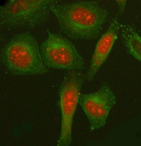
The catalog antibodies have paraffin-section IHC images from human tissue (A07810 and A07810-2 captions) and mouse brain (A07810-2 caption); A07810-2 also has an IF image from HeLa cells (A07810-2 IF caption).

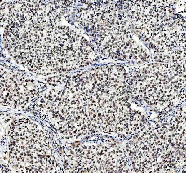
Real IHC data IHC analysis of NCOA5 using anti-NCOA5 antibody (A07810-2). NCOA5 was detected in a paraffin-embedded section of human liver cancer tissue. Heat mediated antigen retrieval was performed in EDTA buffer (pH 8.0, epitope retrieval solution). The tissue section was blocked with 10% goat serum. The tissue section was then incubated with 2 μg/ml rabbit anti-NCOA5 Antibody (A07810-2) overnight at 4°C. Peroxidase Conjugated Goat Anti-rabbit IgG was used as secondary antibody and incubated for 30 minutes at 37°C. The tissue section was developed using HRP Conjugated Rabbit IgG Super Vision Assay Kit (Catalog # SV0002) with DAB as the chromogen.
Anti-NCOA5 Antibody ®
Cat # A07810-2
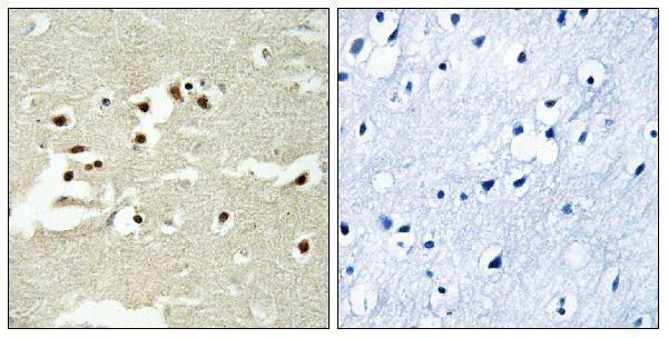
Real IHC data Immunohistochemistry analysis of paraffin-embedded human brain tissue, using NCOA5 Antibody. The picture on the right is blocked with the synthesized peptide.
Anti-Nuclear receptor coactivator 5 NCOA5 Antibody
Cat # A07810

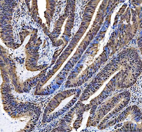
A07810 will render with paraffin-section human brain IHC and a peptide-blocked comparison (A07810 IHC caption). A07810-2 will render with paraffin-section human liver cancer IHC; its additional captions document human colorectal cancer, placenta and mouse brain IHC, plus HeLa cell IF (A07810-2 image captions).

Which to pick: Choose A07810-2 for tissue IHC when its documented paraffin-section examples and retrieval conditions are useful (A07810-2 IHC captions); A07810 offers a human brain paraffin-section example with a peptide-blocked comparison (A07810 IHC caption). For IF/ICC, A07810-2 has a HeLa cell IF image, while A07810 lists IF/ICC applications without an IF image in this payload (A07810-2 IF caption; A07810 catalog). For cross-species planning, A07810-2 lists human, mouse and rat reactivity, though its IHC images show human and mouse samples only; the fixative is unreported in both products’ IHC captions (A07810-2 catalog and IHC captions; A07810 IHC caption).

Each figure is that product's own IHC / IF validation image from its datasheet.

References

  1. UniProt Consortium. UniProt entry Q9HCD5 (NCOA5_HUMAN, Nuclear receptor coactivator 5).
  2. Human Protein Atlas. NCOA5 tissue IHC expression (reliability: Uncertain).
  3. Human Protein Atlas. NCOA5 subcellular location (ICC-IF): Localized to the nucleoplasm and actin filaments..
  4. Human Protein Atlas. NCOA5 antibody validation summary (1 antibodies).
  5. Expression and clinical significance of NCOA5 in epithelial ovarian cancer. Frontiers in oncology 2023 — PMC10183562.
  6. NCOA5 promotes proliferation, migration and invasion of colorectal cancer cells via activation of PI3K/AKT pathway. Oncotarget 2017 — PMC5746116.
  7. NCOA5 induces sorafenib resistance in hepatocellular carcinoma by inhibiting ferroptosis. Cell death discovery 2025 — PMC12052255.
  8. Inhibition of nuclear receptor coactivator 5 overcomes acquired lenvatinib resistance driven by protein kinase B-mammalian target of rapamycin signaling in hepatocellular carcinoma. Anti-cancer drugs 2025 — PMC12509451.
  9. PubMed PMID:11113208 — UniProt-cited evidence.
  10. PubMed PMID:15073177 — UniProt-cited evidence.
  11. PubMed PMID:11780052 — UniProt-cited evidence.