NCOR2 / Nuclear receptor corepressor 2 · IHC design guide

Design Immunohistochemistry for NCOR2

Plan NCOR2 chromogenic IHC around its general nuclear tissue pattern (HPA tissue IHC). Cerebellar molecular-layer cells and placental decidual cells show high staining and can serve as reference tissues (HPA tissue IHC).

Evidence assembled Oct 2026 · For research use; verify linked source records and product datasheet before use
Immunohistochemistry protocol sheet for NCOR2 (IHC for NCOR2): expected localisation Predominantly nuclear in tissue (HPA tissue IHC), antibody A01412-3, validated IHC image, and IHC protocol steps
Printable NCOR2 IHC protocol sheet — expected localisation Predominantly nuclear in tissue (HPA tissue IHC), antibody A01412-3, controls and protocol steps. Open the full NCOR2 IHC guide →

NCOR2 Immunohistochemistry Experimental Design Guide

Expected localisation, validated protocols, controls and antibodies — the at-a-glance facts below, then the full design guide.

Must know before staining
Expected localisation Predominantly nuclear in tissue (HPA tissue IHC)
Staining pattern General nuclear staining across tissue cell types (HPA tissue IHC)
Antigen retrieval EDTA pH 8.0 HIER, heat-mediated (datasheet A01412-3)
Positive control ⓘ Cerebellum+4 more · see all
Negative control ⓘ Liver+2 more · see all
Important caveats
Reasons your staining may differ from the expected pattern.
Fixation Keep fixation consistent across sections. (standard IHC practice; not target-specific)
Caveat Staining and RNA have medium consistency (HPA tissue IHC)
Regulation Stimulus-dependent changes unreported (UniProt)
Isoform / epitope 4 isoforms; epitope coverage is unknown (UniProt)
Section 1

Recommended NCOR2 IHC & IF Protocols

The catalog antibody’s IHC-P protocol (datasheet: A01412-3) is followed by four published NCOR2 IHC protocols (PMC10374731; PMC9133634; PMC3828259; PMC11952456).

Recommended immunohistochemistry (IHC-P) protocol parameters
SampleParaffin-embedded human breast cancer tissue; fixative not specified (datasheet A01412-3)
FixationImage fixative and duration unreported (datasheet A01412-3); verify before use.
Sectioning4–5 µm sections on charged slides (standard)
DeparaffinisationXylene, graded ethanol series to water (standard)
Antigen retrievalHeat retrieval: EDTA pH 8.0 (datasheet A01412-3); 20 min, 95–100 °C (standard)
Peroxidase block3% H2O2, 10 min, room temperature (standard)
Blocking10% goat serum (datasheet A01412-3)
Primary antibodyRabbit anti-NCOR2, 2-5μg/ml (datasheet A01412-3)
Primary incubationOvernight at 4 °C (datasheet A01412-3)
DetectionHRP-conjugated secondary, DAB chromogen (datasheet A01412-3)
CounterstainHematoxylin, blue, dehydrate and mount (standard)
Expected resultNCOR2-positive staining in cells in molecular layer of cerebellum (HPA tissue IHC: High). HPA tissue profile: General nuclear expression. No signal in the no-primary control.
💡Decision noteStart with heat-mediated EDTA retrieval at pH 8.0 (datasheet: A01412-3); compare citrate retrieval if needed (PMC9133634: microwave, 5 min).
Section 2

What Is the Expected NCOR2 Staining Pattern?

NCOR2 should appear predominantly in nuclei across many cell types: UniProt places it in the nucleus and reports no transmembrane segment (UniProt Q9Y618), while HPA describes general nuclear tissue staining (HPA tissue IHC). Strong examples are cells in the cerebellar molecular layer and placental decidual cells (HPA: High). HPA rates its tissue IHC evidence Approved, with medium consistency between antibody staining and RNA expression (HPA tissue IHC).

What am I looking at on my slide?
Nuclear staining in cerebellar molecular-layer cells or placental decidual cells.This matches the expected compartment and two reported high-staining cell populations (UniProt Q9Y618: nucleus; HPA: High in these cells). Judge the named cells rather than assigning one score to the whole section; HPA reports staining by cell population (HPA tissue IHC).
Predominantly cytoplasmic or membrane-like staining, with little nuclear signal.Treat this as a localization mismatch requiring investigation: NCOR2 is nuclear and has no transmembrane segment (UniProt Q9Y618). Check whether the apparent signal follows tissue edges, pigment, or background before interpreting it as NCOR2 (general IHC practice).
Strong staining in liver cholangiocytes, oral squamous epithelium, or skeletal myocytes.HPA reports NCOR2 as not detected in those specific populations (HPA tissue IHC). Confirm cell identity and compare a known-positive population on the same run; persistent staining could reflect antibody cross-reactivity or chromogenic detection activity (general IHC interpretation).
Haze across nuclei, cytoplasm, and extracellular space.A widespread, poorly bounded deposit does not resemble the reported general nuclear pattern (HPA tissue IHC). Assess the no-primary control, wash quality, and detection background before scoring cell-specific nuclear staining (general IHC practice).
No nuclear signal in the expected high-staining populations.Absence in cerebellar molecular-layer cells or placental decidual cells conflicts with the reported high staining (HPA tissue IHC). First verify tissue identity, section quality, and positive-control performance; a failed run cannot establish NCOR2 absence (general IHC practice).
💡Expected NCOR2 appearanceCall a positive result when staining is predominantly nuclear in the relevant cells, especially strong in cerebellar molecular-layer cells or placental decidual cells (UniProt Q9Y618: nucleus; HPA: High); dominant cytoplasmic or diffuse extracellular color is suspect (HPA: general nuclear expression; general IHC interpretation).
How each factor affects the staining
Cell population and reference intensityUse HPA's cell-specific levels: High in cerebellar molecular-layer and placental decidual cells; Medium in adipocytes, adrenal glandular cells, and cortical neurons; Low in caudate neurons and splenic red-pulp cells (HPA tissue IHC). A low population is a weak benchmark for troubleshooting an apparently negative run (general IHC practice).
Antibody evidenceThe listed rabbit polyclonal antibody HPA001928 is Approved for IHC, while the overall tissue IHC record has medium staining–RNA consistency (HPA antibodies; HPA tissue IHC). These ratings support use as a reference pattern but do not make every unexpected positive or negative conclusive.
Isoforms and molecular featuresUniProt lists four NCOR2 isoforms and a full-length chain of residues 1–2514, without a signal peptide, propeptide, or transmembrane segment (UniProt Q9Y618). No epitope position is supplied, so isoform-specific staining and epitope-dependent retrieval behavior cannot be predicted from this record.
IF/ICC Q: What localization should agree with tissue IHC?A: Predominantly nucleoplasmic signal is expected in IF/ICC (HPA subcellular: supported nucleoplasm), consistent with nuclear IHC staining (HPA tissue IHC). HPA lists images from A-431, U-251MG, and U2OS (HPA subcellular); IF/ICC method choices belong in its separate guide.
Why is my staining missing, weak or wrong?
SituationLikely causeNext action
The expected positive tissue shows no staining.The high-staining reference population may be absent from the field, or the IHC run may have failed (HPA: High in cerebellar molecular-layer cells and placental decidual cells; general IHC practice).Confirm the named population on the section, inspect morphology, and compare a working positive control processed in the same run before interpreting absence (general IHC practice).
Nuclear staining is weaker than expected.The field may contain a naturally lower-staining population: HPA records Low in caudate neurons and splenic red-pulp cells, versus High in the two reference populations (HPA tissue IHC).Compare like cell populations and score nuclear staining within each population; check the run control before adjusting IHC conditions (HPA tissue IHC; general IHC practice).
Signal is mainly cytoplasmic or outlines membranes.The distribution conflicts with nuclear NCOR2 and its lack of a transmembrane segment (UniProt Q9Y618); nonspecific staining or misread tissue features are possible (general IHC interpretation).Recheck the matched no-primary control and tissue morphology, then prioritize bounded nuclear signal for interpretation (general IHC practice).
Color is diffuse across the section.Widespread background can obscure the reported general nuclear pattern (HPA tissue IHC; general IHC practice).Inspect no-primary background and review blocking, washes, detection exposure, and counterstain balance using the laboratory's established IHC procedure (general IHC practice).
A reported not-detected cell population stains strongly.HPA reports no detection in liver cholangiocytes, oral squamous epithelial cells, and skeletal myocytes (HPA tissue IHC); cross-reactivity or endogenous detection activity is possible (general IHC interpretation).Verify cell identity, compare a high-staining reference and a no-primary control, and investigate endogenous enzyme activity if using an enzyme-based chromogen (HPA tissue IHC; general IHC practice).
Nuclear staining varies between tissue regions.HPA reports different levels by cell population, from High to Low or not detected (HPA tissue IHC). Variation may therefore reflect cellular composition rather than a uniform technical failure.Score identified cell populations separately and compare equivalent regions across sections; use run controls to investigate variation that does not track cell identity (general IHC practice).

Sample controls for NCOR2 IHC & IF

🧪Run cerebellum first: cells in the molecular layer should show NCOR2 staining (HPA: High in cells in the molecular layer). Use skeletal muscle myocytes as the negative tissue (HPA: Not detected in myocytes); cells without nuclear staining on the cerebellum slide provide an internal comparison, but are not a validated negative population (HPA: cerebellum row; UniProt Q9Y618: nucleus).
Positive control tissue: Cerebellum (Cells in molecular layer, HPA High)
Negative control tissue: Liver (HPA Not detected)
ICC-IF cell lines (HPA subcellular resource): HPA ICC-IF images show NCOR2 in A-431, U-251MG, U2OS, with annotated localisation: Nucleoplasm (supported) (HPA subcellular).
Technical controls: Include a no-primary, secondary-only control, a rabbit IgG isotype control matched to the primary antibody’s clonality, and an NCOR2-knockout biological negative (selected IHC caption: rabbit primary; standard IHC practice). Block endogenous peroxidase for chromogenic detection and assess pigment-related background in cerebellum (selected IHC caption: HRP/DAB; standard IHC practice).
⚠️Feasibility: The selected A01412-3 paraffin-section caption reports heat-mediated retrieval in EDTA at pH 8.0, but does not report the fixative or establish whether retrieval is required (selected IHC caption). No target-specific fixation window or fixation effect is reported in the supplied evidence, and the HPA staining rows do not establish specimen processing (supplied HPA rows; selected IHC caption). IHC-P has a supplied tissue example; the evidence does not establish that frozen sections or IF are easier, while cerebellar pigment can complicate signal assessment (selected IHC caption; standard IHC/IF practice).

HPA tissue IHC evidence for NCOR2

Comprehensive Human Protein Atlas IHC scoring per tissue (reliability: Approved — Medium consistency between antibody staining and RNA expression data.). Rows are taken directly from the HPA tissue chart — click any row's HPA link to view the source.

Positive expression · recommended positive controls

TissueCell typeLevelEvidenceSource
Cerebellum Cells in molecular layer High Protein (IHC) HPA →
Placenta Decidual cells High Protein (IHC) HPA →
Adipose tissue Adipocytes Medium Protein (IHC) HPA →
Adrenal gland Glandular cells Medium Protein (IHC) HPA →
Appendix Glandular cells Medium Protein (IHC) HPA →

Undetected expression · recommended negative controls

TissueCell typeLevelEvidenceSource
Liver Cholangiocytes Not detected Protein (IHC) HPA →
Oral mucosa Squamous epithelial cells Not detected Protein (IHC) HPA →
Skeletal muscle Myocytes Not detected Protein (IHC) HPA →
Section 3

Advanced NCOR2 IHC Tips

Use nuclear staining, matched controls and consistent processing to troubleshoot NCOR2 in paraffin section IHC; interpret IF findings separately.

How should I retrieve NCOR2 in paraffin sections when nuclear staining is weak?
Start with heat mediated antigen retrieval in EDTA at pH 8.0 (datasheet A01412-3). The selected paraffin section image used that retrieval before staining with 2 μg/ml primary antibody overnight at 4°C (caption A01412-3). If nuclei remain weak, compare a controlled range of heating durations on adjacent sections while holding antibody concentration and detection conditions constant (standard IHC practice). Check whether increased heating improves nuclear signal without lifting tissue or increasing diffuse staining (standard IHC practice). If needed, test another retrieval buffer only as a documented fallback and compare it directly with EDTA pH 8.0 (standard IHC practice).
Could fixation explain weak or uneven NCOR2 staining?
Target specific sensitivity to fixation is unknown because the selected paraffin section caption does not state a fixative (caption A01412-3). Compare sections with documented, consistent fixation and processing histories before changing retrieval or primary antibody concentration (standard IHC practice). Patchy staining can reflect uneven fixation or processing, so inspect whether weak regions track section thickness, tissue folds or damaged areas (standard IHC practice). Include a reference section in the same staining run and judge nuclear signal under matched retrieval and detection conditions (standard IHC practice). Neither the reported nuclear location nor the listed modifications establish a fixation response (UniProt Q9Y618).
Where should NCOR2 staining appear, and how should I assess cytoplasmic signal?
Prioritise staining within nuclei: NCOR2 is annotated as nuclear (UniProt Q9Y618), and tissue IHC shows general nuclear expression (HPA tissue IHC). The supported subcellular location is nucleoplasm (HPA subcellular), so score chromogen against a counterstain that lets you identify individual nuclei (standard IHC practice). If diffuse cytoplasmic colour dominates, review the no primary control, section edges and detection background before assigning it to NCOR2 (standard IHC practice). Compare nuclear staining in the same run across morphologically intact cells; HPA reports high staining in cerebellar molecular layer cells and placental decidual cells (HPA tissue IHC).
How can isoforms and epitope accessibility affect an NCOR2 IHC result?
NCOR2 has 4 annotated isoforms and SANT domains at residues 427–478 and 610–661 (UniProt Q9Y618). The supplied caption identifies the catalog antibody and its working concentration, but does not identify its epitope (caption A01412-3). Check the antibody's mapped immunogen or epitope before claiming that staining represents every isoform, and compare serial sections under matched retrieval conditions (standard IHC practice). NCOR2 also has listed modified residues, including phosphorylation and asymmetric dimethylarginine (UniProt Q9Y618); their presence alone does not establish that this antibody detects a particular modification state (standard IHC interpretation).
How should I investigate NCOR2 localisation by IF alongside cell type markers?
For IF, pair NCOR2 with a marker identifying the expected cell type, such as decidual cells in placenta, where high staining is reported (HPA tissue IHC). Select a spectrally separated, preferably far red fluorophore after checking tissue autofluorescence in an unstained control, and use a nuclear counterstain to assess overlap (standard IF practice). NCOR2 is nuclear and has no transmembrane segment (UniProt Q9Y618), so permeabilise sufficiently for antibody access to nuclear epitopes; the antibody's precise epitope is unspecified (caption A01412-3). Evaluate each channel separately with single stain controls, since spectral bleed through can make apparent nuclear colocalisation misleading (standard IF practice).
What should I adjust when NCOR2 DAB staining appears diffuse?
The selected paraffin section workflow used 10% goat serum blocking, 2 μg/ml rabbit primary antibody overnight at 4°C, and DAB development (caption A01412-3). Run a no primary control, then inspect nonspecific secondary binding, endogenous peroxidase activity and overdevelopment before changing the antibody concentration (standard IHC practice). Apply a peroxidase block and keep DAB development time consistent across comparison sections; these are general chromogenic IHC steps, not NCOR2 specific evidence (standard IHC practice). If background persists, compare shorter development or greater primary dilution against preserved nuclear signal in an intact reference region (standard IHC practice).
How should I quantify NCOR2 staining across tissue sections? ⚠ ANSWER MARKED FOR VERIFICATION
Define morphologically comparable regions and score the percentage of positive nuclei together with nuclear intensity, using an H-score if intensity categories are reproducible (standard IHC practice). Normalise positive nuclei to the total count of evaluable nuclei in the same cell population, and report region area if using positive cells per mm² (standard IHC practice). Keep retrieval, DAB development, illumination and scoring thresholds consistent, and exclude folds, necrosis and poorly preserved tissue (standard IHC practice). Report cell type and compartment because NCOR2 shows general nuclear expression, while HPA rates antibody staining and RNA agreement as medium consistency (HPA tissue IHC).
How can I distinguish genuine NCOR2 staining from tissue artefact?
A credible result shows nuclear staining in intact cells, consistent with NCOR2's nuclear annotation and supported nucleoplasmic localisation (UniProt Q9Y618; HPA subcellular). Check cell identity: HPA reports high staining in placental decidual cells, whereas skeletal muscle myocytes were not detected in its tissue profile (HPA tissue IHC). Treat isolated edge staining, necrotic deposits and signal present in a no primary control as potential artefacts; assess endogenous peroxidase when diffuse DAB persists (standard IHC practice). Replicate the pattern on adjacent sections with matched retrieval and detection, and interpret intensity cautiously because HPA reports medium staining to RNA consistency (HPA tissue IHC).
Boster reagents

Best NCOR2 / Nuclear receptor corepressor 2 IHC Antibodies

Anti-NCOR2 antibodies have human paraffin-section IHC data and U2OS-cell IF/ICC data (catalog image captions: A01412-3 IHC; A01412-2 and A01412-3 IF).

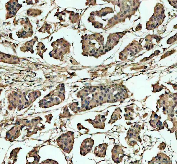
Real IHC data IHC analysis of NCOR2 using anti-NCOR2 antibody (A01412-3). NCOR2 was detected in a paraffin-embedded section of human breast cancer tissue. Heat mediated antigen retrieval was performed in EDTA buffer (pH 8.0, epitope retrieval solution). The tissue section was blocked with 10% goat serum. The tissue section was then incubated with 2 μg/ml rabbit anti-NCOR2 Antibody (A01412-3) overnight at 4°C. Peroxidase Conjugated Goat Anti-rabbit IgG was used as secondary antibody and incubated for 30 minutes at 37°C. The tissue section was developed using HRP Conjugated Rabbit IgG Super Vision Assay Kit (Catalog # SV0002) with DAB as the chromogen.
Anti-NCOR2 Antibody ®
Cat # A01412-3
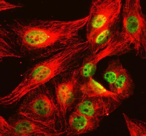
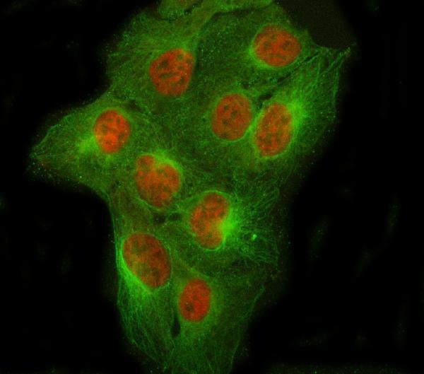
Real IF data IF analysis of NCOR2 using anti-NCOR2 antibody (A01412-2) and anti-Beta Tubulin antibody (M01857-3). NCOR2 was detected in immunocytochemical section of U2OS cell. Enzyme antigen retrieval was performed using IHC enzyme antigen retrieval reagent (AR0022) for 15 mins. The cells were blocked with 10% goat serum. And then incubated with 5 μg/mL rabbit anti-NCOR2 Antibody (A01412-2) and mouse anti-Beta Tubulin antibody (M01857-3) overnight at 4°C. Cy3 Conjugated Goat Anti-Rabbit IgG (BA1032) and DyLight®488 Conjugated Goat Anti-Mouse IgG (BA1126) were used as secondary antibody at 1:500 dilution and incubated for 30 minutes at 37°C. Visualize using a fluorescence microscope and filter sets appropriate for the label used.
Anti-NCOR2 Antibody ®
Cat # A01412-2

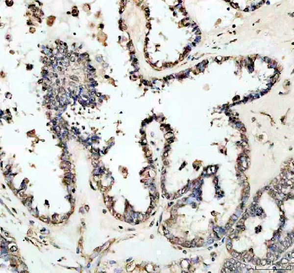
A01412-3 is listed for human IHC and IF/ICC, with IHC images from paraffin-embedded human breast and ovarian cancer sections and an IF image from U2OS cells (catalog applications and image captions). A01412-2 is listed for human IF/ICC, with an IF image from U2OS cells; IHC is absent from its application list (catalog applications and image caption).

Which to pick: Choose A01412-3 for paraffin-section tissue IHC: its breast cancer image used EDTA retrieval at pH 8.0 and 2 μg/ml primary antibody; the fixative is unreported (A01412-3 IHC image caption). For IF/ICC, choose A01412-2 or A01412-3 based on their respective U2OS images; clonality is unreported for both (catalog IF image captions; clone fields). Neither SKU has documented cross-species reactivity beyond human (catalog reactivity fields).

Each figure is that product's own IHC / IF validation image from its datasheet.

References

  1. UniProt Consortium. UniProt entry Q9Y618 (NCOR2_HUMAN, Nuclear receptor corepressor 2).
  2. Human Protein Atlas. NCOR2 tissue IHC expression (reliability: Approved).
  3. Human Protein Atlas. NCOR2 subcellular location (ICC-IF): Localized to the nucleoplasm..
  4. Human Protein Atlas. NCOR2 antibody validation summary (1 antibodies).
  5. Nuclear receptor co-repressor NCOR2 and its relation to GPER with prognostic impact in ovarian cancer. Journal of cancer research and clinical oncology 2023 — PMC10374731.
  6. Genetic Alterations in Papillary Thyroid Carcinoma With Hashimoto(')s Thyroiditis: ANK3, an Indolent Maintainer of Papillary Thyroid Carcinoma. Frontiers in oncology 2022 — PMC9133634.
  7. microRNA-100 targets SMRT/NCOR2, reduces proliferation, and improves survival in glioblastoma animal models. PloS one 2013 — PMC3828259.
  8. NCOR2 represses MHC class I molecule expression to drive metastatic progression of breast cancer. bioRxiv : the preprint server for biology 2025 — PMC11952456.
  9. PubMed PMID:8813722 — UniProt-cited evidence.
  10. PubMed PMID:10077563 — UniProt-cited evidence.
  11. PubMed PMID:10097068 — UniProt-cited evidence.