This website uses cookies to ensure you get the best experience on our website.


- Table of Contents
Plan chromogenic NCSTN IHC in paraffin sections around the cytoplasmic staining reported in most tissues (HPA tissue IHC). Use lung macrophages or pancreatic exocrine cells as high-staining reference populations (HPA tissue IHC), and keep fixation consistent when comparing sections.

Expected localisation, validated protocols, controls and antibodies — the at-a-glance facts below, then the full design guide.
| Expected localisation | Cytoplasmic staining in most tissues (HPA tissue IHC) | |
| Staining pattern | Cytoplasmic staining across most tissues (HPA tissue IHC) | |
| Antigen retrieval | Citrate pH 6.0 HIER, 95–98 °C, 20 min (rule: cytoplasmic / membrane antigen) | |
| Positive control | Lung+4 more · see all | |
| Negative control | Adipose tissue+4 more · see all |
| Fixation | Keep fixation conditions consistent across sections. (standard IHC practice; not target-specific) | |
| Caveat | Staining shows low consistency with RNA expression (HPA tissue IHC) | |
| Regulation | Specific expression regulation not reported (UniProt) | |
| Isoform / epitope | 2 isoforms; check epitope against extracellular and cytoplasmic regions (UniProt) |
The catalog antibody’s IHC-P protocol is paired with published NCSTN IHC methods from two articles (PMC7092570; PMC11668073).
| Sample | Tissue sections; selected-image fixative not specified (standard IHC workflow) |
| Fixation | Image fixative and duration unreported (datasheet A03144); verify before use. |
| Sectioning | 4–5 µm sections on charged slides (standard) |
| Deparaffinisation | Xylene, graded ethanol series to water (standard) |
| Antigen retrieval | Heat-induced epitope retrieval in citrate buffer, pH 6.0, 20 min at 95–98 °C (standard rule: cytoplasmic / membrane antigen) |
| Peroxidase block | 3% H2O2, 10 min, room temperature (standard) |
| Blocking | 10% normal serum of the secondary host, 30 min, room temperature (standard) |
| Primary antibody | Rabbit anti-NCSTN, 5 μg/mL (datasheet A03144) |
| Primary incubation | Overnight at 4 °C (standard) |
| Detection | HRP-polymer secondary, DAB chromogen 5–10 min (standard) |
| Counterstain | Hematoxylin, blue, dehydrate and mount (standard) |
| Expected result | NCSTN-positive staining in macrophages of lung (HPA tissue IHC: High). HPA tissue profile: Cytoplasmic expression in most tissues. No signal in the no-primary control. |
In paraffin sections, expect predominantly cytoplasmic NCSTN staining across many tissues, with strong staining reported in lung macrophages, pancreatic exocrine glandular cells and cells in testicular seminiferous ducts (HPA: tissue IHC). NCSTN has a large extracellular region, one transmembrane segment and a short cytoplasmic tail, so membrane-associated staining may also be plausible (UniProt Q92542 topology). Interpret intensity cautiously: HPA rates its tissue IHC profile Approved but reports low consistency with RNA expression (HPA: tissue IHC reliability).
| Cytoplasmic staining in lung macrophages or pancreatic exocrine glandular cells, with stronger signal than nearby unstained cells (HPA: High in these cell types). | This matches reported tissue IHC. Score the named cell population and its compartment, rather than treating every cell in the section as an equivalent positive control (HPA: tissue IHC; general IHC practice). |
| Signal confined to nuclei, with no convincing cytoplasmic or membrane-associated pattern. | An exclusively nuclear pattern conflicts with HPA's cytoplasmic tissue profile and UniProt's membrane and vesicle annotations; investigate staining artefact before assigning it to NCSTN (HPA: tissue IHC; UniProt Q92542 subcellular location). |
| Strong staining in adipocytes, cardiomyocytes or skeletal myocytes instead of the expected positive cell populations (HPA: Not detected in these cells). | Consider cross-reactivity or endogenous detection activity, especially if the signal also appears in a no-primary control. HPA's low antibody–RNA consistency makes an unexpected result a finding to verify, rather than proof of either cause (HPA: tissue IHC reliability; general IHC practice). |
| Color spread broadly across tissue, including spaces or cell types without a coherent cellular pattern. | Treat diffuse signal as background until a no-primary control and inspection of blocking, washes and chromogen development support specific staining (general IHC practice). It does not match HPA's cell-associated cytoplasmic profile (HPA: tissue IHC). |
| No staining in lung macrophages or pancreatic exocrine glandular cells (HPA: High in these cell types). | A blank known-positive population raises a sensitivity or workflow concern; confirm that the cells are present, then assess antibody dilution, retrieval and detection with appropriate controls (HPA: tissue IHC; general IHC practice). |
| Cell population and comparator | HPA reports High staining in three named populations, Medium staining in selected glandular, respiratory epithelial and neuronal cells, and no detection in specified others. Choose and score the actual cell type within each section; a tissue name alone does not define every cell's expected result (HPA: tissue IHC). |
| Topology and compartment | NCSTN spans the membrane at residues 670–690, with residues 34–669 extracellular and 691–709 cytoplasmic (UniProt Q92542 topology). This supports a membrane-associated protein, while HPA describes the observed tissue IHC pattern as cytoplasmic; topology alone does not predict an antibody's visible outline (HPA: tissue IHC). |
| Evidence and antibody validation | HPA marks tissue IHC reliability Approved despite low staining–RNA consistency. It lists IHC approval for HPA054846 and CAB021982, without an IHC Enhanced designation in the supplied record. Treat unusual tissue results as requiring controls and independent confirmation (HPA: tissue IHC reliability; HPA: antibody validation). |
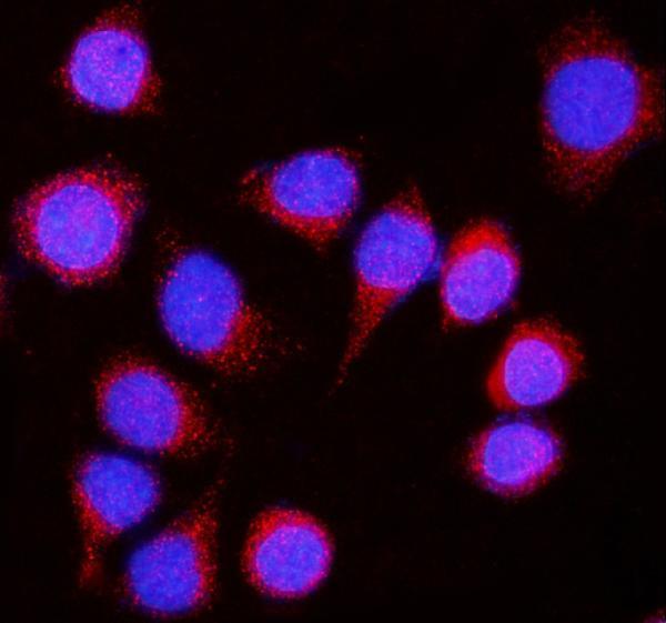
| IF/ICC Q: Should its pattern match IHC? | A: HPA reports approved cytosolic ICC-IF localization and images in HaCaT, SiHa and U2OS; HPA070642 has ICC approval, while the supplied IHC-approved antibodies are separately listed. Use that evidence to interpret IF/ICC images, without assuming identical antibody performance or prescribing an IF protocol here (HPA: subcellular ICC-IF; HPA: antibody validation). |
| Situation | Likely cause | Next action |
|---|---|---|
| Known-positive lung macrophages are blank (HPA: High in macrophages). | The target cells may be absent from the sampled field, or the IHC workflow may lack sensitivity; the image alone cannot distinguish these possibilities (HPA: tissue IHC; general IHC practice). | Verify macrophages morphologically, compare a known-positive section, and check retrieval, antibody dilution and detection controls using the chosen antibody's validated IHC conditions (general IHC practice). |
| Pancreatic exocrine glandular cells stain weakly despite an expected High HPA level (HPA: tissue IHC). | Section or assay variation may reduce contrast; HPA's tissue profile is an observed reference, not a guaranteed intensity for every preparation (HPA: tissue IHC reliability; general IHC practice). | Compare matched positive-control sections and review retrieval, dilution and development consistently before changing the biological interpretation (general IHC practice). |
| Signal appears mainly in nuclei. | Exclusive nuclear staining does not fit HPA's cytoplasmic IHC profile or UniProt's membrane and vesicle localization (HPA: tissue IHC; UniProt Q92542 subcellular location). | Inspect a no-primary control, confirm morphology and evaluate whether an independently validated IHC antibody reproduces the cellular pattern (general IHC practice; HPA: antibody validation). |
| Adipocytes or cardiomyocytes show strong signal (HPA: Not detected in these cells). | Cross-reactivity or endogenous detection activity is possible; HPA's low staining–RNA consistency prevents a definitive assignment from this discrepancy alone (HPA: tissue IHC reliability; general IHC practice). | Run a no-primary control, inspect signal location within the named cells and seek confirmation with another IHC-approved antibody when the result matters (general IHC practice; HPA: antibody validation). |
| Brown signal coats multiple structures without distinct stained cells. | Diffuse background can result from incomplete blocking or washes, or excessive chromogen development (general IHC practice). | Compare the no-primary slide, review blocking and wash steps, and shorten development if control background rises; then reassess whether cell-associated cytoplasmic signal remains (general IHC practice; HPA: tissue IHC). |
| A tissue reported as ‘Not detected’ contains a few stained cells. | HPA's negative entries name particular cell populations; for example, the adipose entry specifies adipocytes, so another cell type in that section cannot be classified from that entry alone (HPA: tissue IHC). | Identify the stained cells first, score them separately, and compare their pattern with a reported positive population and assay controls before calling the finding specific (HPA: tissue IHC; general IHC practice). |


Comprehensive Human Protein Atlas IHC scoring per tissue (reliability: Approved — Low consistency between antibody staining and RNA expression data.). Rows are taken directly from the HPA tissue chart — click any row's HPA link to view the source.
| Tissue | Cell type | Level | Evidence | Source |
|---|---|---|---|---|
| Lung | Macrophages | High | Protein (IHC) | HPA → |
| Pancreas | Exocrine glandular cells | High | Protein (IHC) | HPA → |
| Testis | Cells in seminiferous ducts | High | Protein (IHC) | HPA → |
| Adrenal gland | Glandular cells | Medium | Protein (IHC) | HPA → |
| Appendix | Glandular cells | Medium | Protein (IHC) | HPA → |
| Tissue | Cell type | Level | Evidence | Source |
|---|---|---|---|---|
| Adipose tissue | Adipocytes | Not detected | Protein (IHC) | HPA → |
| Bone marrow | Hematopoietic cells | Not detected | Protein (IHC) | HPA → |
| Heart muscle | Cardiomyocytes | Not detected | Protein (IHC) | HPA → |
| Lymph node | Germinal center cells | Not detected | Protein (IHC) | HPA → |
| Oral mucosa | Squamous epithelial cells | Not detected | Protein (IHC) | HPA → |
Troubleshoot NCSTN staining in paraffin section chromogenic IHC, with one entry for IF/ICC experimental design.
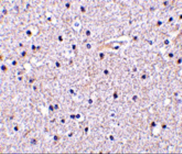
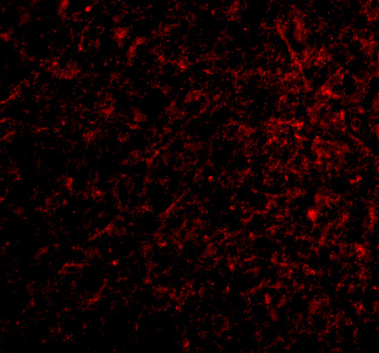
The rendered NCSTN antibodies have IHC data from human brain tissue and IF data from SiHa cells (catalog image captions); both list Human, Mouse, and Rat reactivity (catalog reactivity).
A03144 is listed for IHC-P and IF, with an IHC image of human brain tissue at 5 μg/mL (catalog applications; A03144 IHC image caption). A03144-1 is listed for ICC and IF, with an IF image of SiHa cells at 5 μg/mL (catalog applications; A03144-1 IF image caption).
Which to pick: Choose A03144 for paraffin-section IHC: its IHC-P application and human brain tissue image support that choice, while the fixative is unreported (catalog applications; A03144 IHC image caption). Choose A03144-1 for IF/ICC when the SiHa cell example is relevant (catalog applications; A03144-1 IF image caption). Both list Human, Mouse, and Rat reactivity for cross-species planning; clonality is unspecified for both rendered SKUs, and their pictured results are limited to the samples named above (catalog reactivity and clone fields; image captions).