This website uses cookies to ensure you get the best experience on our website.


- Table of Contents
Plan chromogenic NDE1 IHC in paraffin sections with the catalog antibody at 2–5 μg/ml (datasheet A03656-2). Use the reported cytoplasmic tissue pattern to guide scoring, while accounting for low consistency between antibody staining and RNA expression (HPA tissue IHC).

Expected localisation, validated protocols, controls and antibodies — the at-a-glance facts below, then the full design guide.
| Expected localisation | Cytoplasmic tissue staining (HPA tissue IHC) | |
| Staining pattern | Cytoplasmic staining in several tissues, strongest in lymphoid tissues (HPA tissue IHC) | |
| Antigen retrieval | EDTA pH 8.0 HIER, heat-mediated (datasheet A03656-2) | |
| Positive control | Caudate+4 more · see all | |
| Negative control | Adipose tissue+4 more · see all |
| Fixation | Keep paraffin-section fixation consistent (standard IHC practice; not target-specific) | |
| Caveat | Antibody staining has low consistency with RNA expression (HPA tissue IHC) | |
| Regulation | Expressed in developing neuroepithelium (UniProt) | |
| Isoform / epitope | 2 isoforms; epitope coverage needs checking (UniProt) |
The catalog antibody has an IHC-P protocol (datasheet: A03656-2). One published mouse brain IHC protocol provides a comparison (PMC4048543).
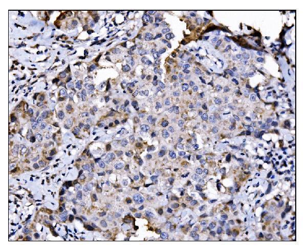
| Sample | Paraffin-embedded human breast cancer tissue; fixative not specified (datasheet A03656-2) |
| Fixation | Image fixative and duration unreported (datasheet A03656-2); verify before use. |
| Sectioning | 4–5 µm sections on charged slides (standard) |
| Deparaffinisation | Xylene, graded ethanol series to water (standard) |
| Antigen retrieval | Heat retrieval: EDTA pH 8.0 (datasheet A03656-2); 20 min, 95–100 °C (standard) |
| Peroxidase block | 3% H2O2, 10 min, room temperature (standard) |
| Blocking | 10% goat serum (datasheet A03656-2) |
| Primary antibody | Rabbit anti-NDE1, 2-5μg/ml (datasheet A03656-2) |
| Primary incubation | Overnight at 4 °C (datasheet A03656-2) |
| Detection | Streptavidin-biotin complex (SABC), DAB chromogen (datasheet A03656-2) |
| Counterstain | Hematoxylin, blue, dehydrate and mount (standard) |
| Expected result | NDE1-positive staining in neuronal cells of caudate (HPA tissue IHC: Medium). HPA tissue profile: Cytoplasmic expression in several tissues, most abundant in lymphoid tissues. No signal in the no-primary control. |
NDE1 is mainly cytoplasmic and cytoskeletal, with cell cycle dependent localization to centrosomes, spindle and cleavage furrow (UniProt Q9NXR1 localization). In paraffin tissue sections, expect cytoplasmic staining in selected cells, including neuronal cells, duodenal glandular cells and kidney tubule cells (HPA tissue IHC: Medium). Interpret cautiously: the HPA tissue assessment is Approved but reports low consistency between staining and RNA expression (HPA tissue IHC: reliability).
| Cytoplasmic chromogen in neuronal cells of cerebral cortex, hippocampus or caudate. | This fits the reported Medium staining in those cell populations (HPA tissue IHC). Score the named cells and their cytoplasm; staining elsewhere in the section does not establish the same pattern (HPA tissue IHC; standard IHC practice). |
| Cytoplasmic staining in duodenal glandular cells or kidney tubule cells. | Both are reported at Medium intensity (HPA tissue IHC). Compare the relevant cell population with adjacent tissue and controls when judging whether chromogen is cell associated (standard IHC practice). |
| Predominantly nuclear or continuous cell surface staining, without a cytoplasmic pattern. | Treat this as a localization mismatch requiring investigation, rather than confirmed NDE1: UniProt places NDE1 at cytoskeletal and cell division structures and reports no transmembrane segment (UniProt Q9NXR1 localization and topology). |
| Staining concentrated in adipocytes or bronchial respiratory epithelium. | Those cells are reported as Not detected (HPA tissue IHC). Reproducible staining there warrants checks for cross reactivity or endogenous detection activity (standard IHC practice); the HPA result alone does not prove its cause. |
| Diffuse chromogen across tissue, or no cell associated signal in a reported positive population. | Diffuse color is difficult to assign to NDE1 positive cells (standard IHC practice). Absence in cerebral cortical neurons, for example, conflicts with the reported Medium pattern, but HPA notes staining and RNA inconsistency (HPA tissue IHC). |
| Tissue and cell choice | HPA reports Medium staining in selected neuronal, duodenal glandular and kidney tubule cells, but Not detected in several other populations (HPA tissue IHC). Record the cell type, since a whole section can contain more than one staining pattern (standard IHC practice). |
| Strength of tissue evidence | The tissue assessment is Approved, with low consistency between antibody staining and RNA expression and a splice or transcript discrepancy caution (HPA tissue IHC: reliability). HPA024075 is IHC Approved; the supplied status does not say Enhanced (HPA antibodies). |
| Cell cycle and subcellular pattern | UniProt describes centrosome localization in interphase and S phase, partial spindle association during mitosis, kinetochore associated microtubule ends in metaphase and anaphase, and the cleavage furrow during cytokinesis (UniProt Q9NXR1 localization). These structures need not be obvious in every paraffin section (standard IHC practice). |
| Topology and processing | NDE1 has no transmembrane segment, signal sequence or propeptide, and its annotated chain spans residues 1–335 (UniProt Q9NXR1 topology and processing). Its reported association with cytoplasmic vesicles does not make a uniform cell surface rim the expected IHC result (UniProt Q9NXR1 localization). |
| Isoforms and epitope | Two isoforms are listed, but the supplied record gives no antibody epitope or isoform coverage (UniProt Q9NXR1 isoforms; HPA antibodies). If a result depends on isoform detection, check documented immunogen coverage before assigning a negative stain to lack of NDE1 (standard IHC practice). |
| IF/ICC Q&A: Is there an HPA image based location to compare? | No. HPA supplies no main ICC IF location and no cell lines with ICC IF images, while noting cell cycle correlated transcript variation (HPA subcellular). Use the separate IF/ICC guide for that application; the tissue IHC pattern does not validate an IF/ICC image (HPA tissue IHC; HPA subcellular). |
| Situation | Likely cause | Next action |
|---|---|---|
| No chromogen in reported positive neuronal, duodenal or kidney cells. | The result conflicts with HPA's Medium staining report, but the supplied evidence does not identify a target specific fixation or retrieval failure (HPA tissue IHC). | Check a known positive section, primary antibody coverage, detection reagents and the recorded retrieval conditions using routine IHC controls (standard IHC practice). Interpret persistent absence with HPA's low consistency caveat (HPA tissue IHC). |
| Color appears throughout the section without clear cell boundaries. | Diffuse color can arise from background detection or inadequately controlled staining steps (standard IHC practice); the supplied NDE1 sources do not assign a target specific cause. | Compare a no primary control, inspect washes and blocking, and judge whether signal remains localized to the reported cell populations (standard IHC practice; HPA tissue IHC). |
| Reported negative cells show strong staining. | HPA reports adipocytes and bronchial respiratory epithelial cells as Not detected (HPA tissue IHC); cross reactivity or endogenous detection activity are possible explanations (standard IHC practice). | Review no primary and detection controls, then compare a reported positive cell population on a separately processed section before calling the signal NDE1 (standard IHC practice; HPA tissue IHC). |
| Signal is mainly nuclear or forms a continuous membrane rim. | That distribution conflicts with the principal cytoskeletal and cytoplasmic localization and absent transmembrane segment (UniProt Q9NXR1 localization and topology). It is a warning sign, not proof of a particular artefact. | Check counterstain boundaries and a no primary control; assess whether any cell associated cytoplasmic pattern matches the HPA tissue observations (standard IHC practice; HPA tissue IHC). |
| Different positive tissues have unequal staining intensity. | HPA reports selected cell populations at Medium, others at Low or Not detected, and describes cytoplasmic expression in several tissues as most abundant in lymphoid tissues (HPA tissue IHC). | Score each named cell population against its own HPA observation; do not use a single intensity threshold for every tissue (HPA tissue IHC; standard IHC practice). |
| A negative result is attributed to fixation or a specific NDE1 isoform. | No target specific fixation effect or antibody epitope is supplied; UniProt lists 2 isoforms (UniProt Q9NXR1 isoforms; HPA antibodies). | Record processing and retrieval conditions, verify documented epitope coverage where available, and keep the explanation provisional until controlled comparison supports it (standard IHC practice). |


Comprehensive Human Protein Atlas IHC scoring per tissue (reliability: Approved — Low consistency between antibody staining and RNA expression data. Caution, Splice and/or transcript discrepancy exists.). Rows are taken directly from the HPA tissue chart — click any row's HPA link to view the source.
| Tissue | Cell type | Level | Evidence | Source |
|---|---|---|---|---|
| Caudate | Neuronal cells | Medium | Protein (IHC) | HPA → |
| Cerebral cortex | Neuronal cells | Medium | Protein (IHC) | HPA → |
| Duodenum | Glandular cells | Medium | Protein (IHC) | HPA → |
| Hippocampus | Neuronal cells | Medium | Protein (IHC) | HPA → |
| Kidney | Cells in tubules | Medium | Protein (IHC) | HPA → |
| Tissue | Cell type | Level | Evidence | Source |
|---|---|---|---|---|
| Adipose tissue | Adipocytes | Not detected | Protein (IHC) | HPA → |
| Adrenal gland | Glandular cells | Not detected | Protein (IHC) | HPA → |
| Bone marrow | Hematopoietic cells | Not detected | Protein (IHC) | HPA → |
| Breast | Adipocytes | Not detected | Protein (IHC) | HPA → |
| Bronchus | Respiratory epithelial cells | Not detected | Protein (IHC) | HPA → |
Troubleshoot NDE1 staining in paraffin sections by checking retrieval, cellular distribution, controls, and scoring against the available IHC evidence.
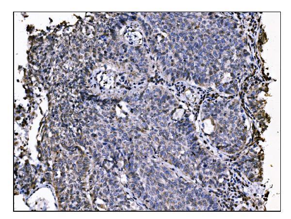
A03656-2 has real IHC data from paraffin-embedded human breast and lung cancer sections (catalog IHC captions). No IF data are supplied (catalog IF images).
A03656-2 is a rabbit anti-NDE1 antibody listed for human IHC (catalog host, reactivity, applications). Its IHC captions show staining of paraffin-embedded human breast and lung cancer sections (catalog IHC captions).
Which to pick: Choose A03656-2 for human tissue IHC: its captions document EDTA pH 8.0 retrieval and 2 μg/ml primary antibody on paraffin sections; the fixative is unreported (catalog IHC captions). No IF/ICC choice is supported because A03656-2 has no listed IF/ICC application or IF image (catalog applications, IF images). No cross-species choice is supported because A03656-2 lists human reactivity only (catalog reactivity).