This website uses cookies to ensure you get the best experience on our website.


- Table of Contents
Plan chromogenic NDFIP2 IHC in paraffin sections with the catalog antibody’s documented workflow (datasheet M08384). Compare cytoplasmic staining with the tissue profile while accounting for its uncertain reliability (HPA tissue IHC).

Expected localisation, validated protocols, controls and antibodies — the at-a-glance facts below, then the full design guide.
| Expected localisation | Cytoplasmic tissue staining (HPA tissue IHC); endosome and Golgi membranes (UniProt) | |
| Staining pattern | Cytoplasmic staining in most tissues (HPA tissue IHC) | |
| Antigen retrieval | EDTA pH 8.0 HIER, heat-mediated (datasheet M08384) | |
| Positive control | Appendix+4 more · see all | |
| Negative control | Adrenal gland+4 more · see all |
| Fixation | Keep fixation consistent across paraffin sections. (standard IHC practice; not target-specific) | |
| Caveat | Presumed off-target staining; splice/transcript discrepancy (HPA tissue IHC) | |
| Regulation | Low tissue specificity (HPA tissue RNA) | |
| Isoform / epitope | No annotated isoforms; cytoplasmic versus extracellular epitopes matter (UniProt) |
The catalog antibody's IHC-P protocol is paired with a published mouse kidney staining method (PMC5606929 Methods).
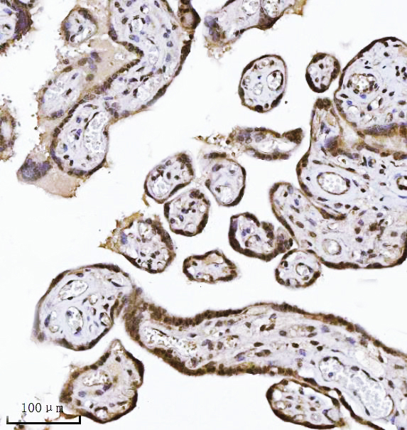
| Sample | Paraffin-embedded human placenta tissue; fixative not specified (datasheet M08384) |
| Fixation | Image fixative and duration unreported (datasheet M08384); verify before use. |
| Sectioning | 4–5 µm sections on charged slides (standard) |
| Deparaffinisation | Xylene, graded ethanol series to water (standard) |
| Antigen retrieval | Heat retrieval: EDTA pH 8.0 (datasheet M08384); 20 min, 95–100 °C (standard) |
| Peroxidase block | 3% H2O2, 10 min, room temperature (standard) |
| Blocking | 10% goat serum (datasheet M08384) |
| Primary antibody | Mouse monoclonal (clone 10D6D7) anti-NDFIP2, 2 μg/ml (datasheet M08384) |
| Primary incubation | Overnight at 4 °C (datasheet M08384) |
| Detection | HRP-conjugated secondary, DAB chromogen (datasheet M08384) |
| Counterstain | Hematoxylin, blue, dehydrate and mount (standard) |
| Expected result | NDFIP2-positive staining in lymphoid tissue of appendix (HPA tissue IHC: High). HPA tissue profile: Cytoplasmic expression in most tissues. No signal in the no-primary control. |
NDFIP2 is an endosome, multivesicular body and Golgi membrane protein with three transmembrane segments (UniProt Q9NV92 topology). In paraffin sections, expect chiefly cytoplasmic staining in the relevant cells, including colon glandular cells and esophageal squamous epithelial cells (HPA: High in both). Treat the pattern as provisional: HPA rates tissue IHC Uncertain, reports presumed off-target binding, and notes a splice or transcript discrepancy (HPA: tissue IHC reliability).
| Cytoplasmic staining in colon glandular cells or esophageal squamous epithelial cells, with visible tissue structure. | These are useful positive patterns because HPA reports High staining in those cells (HPA: colon glandular cells High; esophagus squamous epithelial cells High). A granular appearance can fit intracellular membranes (UniProt Q9NV92 subcellular location), but granularity alone does not establish antibody specificity (HPA: tissue IHC Uncertain). |
| Predominantly nuclear staining or a sharp outline confined to the cell surface. | That distribution is difficult to reconcile with endosome and Golgi membrane localization (UniProt Q9NV92 subcellular location). Check whether the pattern persists with appropriate controls before interpreting it as NDFIP2; HPA reports presumed off-target binding in tissue IHC (HPA: reliability description). |
| Strong staining in a cell population listed as Not detected, such as adrenal gland glandular cells. | This conflicts with that specific HPA observation (HPA: adrenal gland glandular cells Not detected). Consider cross-reactivity or endogenous detection activity, then check a no-primary control. A Not detected result for one cell population does not classify every cell in that organ (HPA: tissue IHC cell-specific entries). |
| Widespread, nearly uniform color over cells and surrounding tissue, obscuring cell boundaries. | This is hard to score as the cytoplasmic pattern HPA describes (HPA: cytoplasmic expression in most tissues). General IHC causes can include excess antibody or detection background; compare a no-primary control and inspect whether known positive cell populations remain distinguishable. |
| No staining in otherwise interpretable colon glandular cells or appendix lymphoid tissue. | Both are reported High by HPA (HPA: colon glandular cells High; appendix lymphoid tissue High). Review the IHC detection workflow and tissue preservation before calling the target absent. HPA rates its tissue IHC evidence Uncertain, so a single negative section cannot resolve expression or assay specificity (HPA: reliability). |
| Compartment and topology (UniProt Q9NV92 topology) | NDFIP2 has three transmembrane segments, a long cytoplasmic N-terminal region and endosome, multivesicular body and Golgi membrane locations (UniProt Q9NV92 topology and subcellular location). The expected tissue readout is cytoplasmic; the exact epitope of a selected antibody is not specified here. |
| Cell-specific tissue pattern (HPA: tissue IHC) | HPA reports High staining in appendix lymphoid, colon glandular, and esophagus and vagina squamous epithelial cells; it reports Medium staining in several neuronal populations (HPA: listed tissue cells). Choose comparisons by cell type, since HPA also lists Not detected populations within other organs. |
| Confidence in tissue interpretation (HPA: reliability) | HPA rates tissue IHC Uncertain and says presumed off-target binding was observed and disregarded; it also flags a splice or transcript discrepancy (HPA: reliability description). Use the reported pattern as a reference for comparison, not a standalone identity test. |
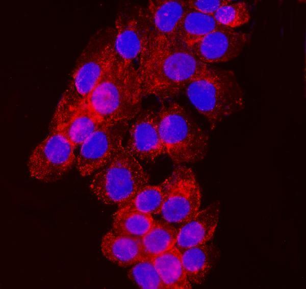
| IF/ICC pattern? (HPA: subcellular; HPA: HPA009160 ICC Supported) | For the separate IF/ICC guide, HPA reports supported localization to vesicles and lists A-431 and CACO-2 images (HPA: subcellular). That observation is compatible with intracellular membrane localization (UniProt Q9NV92), but it does not upgrade the Uncertain tissue IHC rating (HPA: tissue IHC reliability). |
| Processing and modification (UniProt Q9NV92) | UniProt lists the chain as residues 1–336, with no signal peptide, propeptide, annotated isoforms or glycosylation sites; it lists four SRC-related phosphotyrosines (UniProt Q9NV92 processing, isoforms and modified residues). These annotations alone do not predict staining strength or antigen retrieval behavior. |
| Fixation sensitivity (source scope) | Target-specific effects of fixation or antigen retrieval on NDFIP2 staining are unreported in the supplied UniProt and HPA records. Select and optimize those steps as general IHC workflow variables; do not infer a retrieval requirement from topology, phosphorylation or HPA staining levels. |
| Situation | Likely cause | Next action |
|---|---|---|
| Expected positive cells show little or no color (HPA: colon glandular cells High; appendix lymphoid tissue High). | Possible general IHC workflow failure, unsuitable tissue preservation or an antibody that does not perform in the chosen conditions; HPA tissue IHC is Uncertain (HPA: reliability). | Check the positive control and detection reagents, then optimize the IHC antibody dilution and retrieval conditions as general IHC practice. Record the cell types assessed before concluding that NDFIP2 is absent. |
| Signal is mainly nuclear, despite the expected cytoplasmic tissue profile (HPA: cytoplasmic expression in most tissues). | The distribution conflicts with the annotated endosome and Golgi membrane locations (UniProt Q9NV92 subcellular location); off-target staining is a live possibility (HPA: reliability description). | Compare the no-primary control and a reported positive cell population. If nuclear staining persists as the dominant result, treat target assignment as unresolved rather than scoring it as specific NDFIP2. |
| A listed Not detected population stains strongly, such as heart muscle cardiomyocytes (HPA: cardiomyocytes Not detected). | Cross-reactivity or endogenous detection activity may explain the disagreement; HPA has already flagged presumed off-target binding in tissue IHC (HPA: reliability description). | Run a no-primary control to assess the detection system, then compare staining with a reported High population. Restrict any conclusion to the named cell population, since tissue entries are cell specific (HPA: tissue IHC). |
| Color covers the section broadly and masks cell-level interpretation. | General IHC background may arise from antibody or detection conditions. The result cannot be matched confidently to the cytoplasmic profile (HPA: tissue IHC profile). | Inspect a no-primary control, revisit blocking and washing, and titrate the primary antibody as general IHC practice. Reassess only when cellular boundaries and the relevant positive population can be read. |
| The expected cytoplasmic color is faint beside the counterstain (HPA: cytoplasmic tissue profile). | General IHC counterstain or detection balance can reduce readability; faint color alone cannot resolve specificity while tissue IHC remains Uncertain (HPA: reliability). | Review positive and no-primary controls together, then adjust counterstain or detection conditions as general IHC practice. Score only interpretable cells and report the observed compartment and intensity separately. |
| Vesicular IF/ICC images and the paraffin-section result appear different (HPA: supported vesicle localization; HPA: tissue IHC Uncertain). | The HPA ICC and tissue IHC assessments have different validation statuses (HPA: HPA009160 ICC Supported, IHC Uncertain); paraffin-section color may also resolve less subcellular detail than IF. | Interpret each application against its own evidence. Use cytoplasmic cell-level tissue staining as the IHC comparison (HPA: tissue IHC profile), and reserve vesicle-level assessment for the separate IF/ICC guide (HPA: subcellular). |


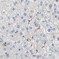


Comprehensive Human Protein Atlas IHC scoring per tissue (reliability: Uncertain — Presumed off target binding observed and disregarded. Caution, Splice and/or transcript discrepancy exists.). Rows are taken directly from the HPA tissue chart — click any row's HPA link to view the source.
| Tissue | Cell type | Level | Evidence | Source |
|---|---|---|---|---|
| Appendix | Lymphoid tissue | High | Protein (IHC) | HPA → |
| Colon | Glandular cells | High | Protein (IHC) | HPA → |
| Esophagus | Squamous epithelial cells | High | Protein (IHC) | HPA → |
| Vagina | Squamous epithelial cells | High | Protein (IHC) | HPA → |
| Bone marrow | Hematopoietic cells | Medium | Protein (IHC) | HPA → |
| Tissue | Cell type | Level | Evidence | Source |
|---|---|---|---|---|
| Adrenal gland | Glandular cells | Not detected | Protein (IHC) | HPA → |
| Breast | Adipocytes | Not detected | Protein (IHC) | HPA → |
| Heart muscle | Cardiomyocytes | Not detected | Protein (IHC) | HPA → |
| Hippocampus | Glial cells | Not detected | Protein (IHC) | HPA → |
| Liver | Cholangiocytes | Not detected | Protein (IHC) | HPA → |
Troubleshoot NDFIP2 staining in paraffin sections by checking retrieval, compartment-specific signal, controls, and cell-level interpretation.
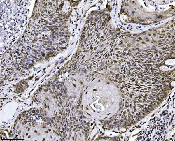
M08384 has IHC images from human paraffin sections of placenta, squamous cell lung carcinoma, breast cancer, and spleen, plus ICC/IF data in A431 cells (catalog image captions).
M08384 is listed for human IHC, with images from paraffin sections of placenta, squamous cell lung carcinoma, breast cancer, and spleen (catalog applications; IHC image captions). The same SKU has an ICC/IF image in A431 cells (catalog IF image caption).
Which to pick: Choose M08384 for human paraffin-section IHC: it is a mouse monoclonal, clone 10D6D7, with IHC listed and a paraffin-section image (catalog antibody record; IHC image captions); the fixative is unreported (catalog IHC image captions). For IF/ICC, M08384 is also listed and has an A431-cell image (catalog applications; IF image caption); use the separate IF/ICC guide for that application. No cross-species choice is supported here: M08384 lists human reactivity only (catalog antibody record).