This website uses cookies to ensure you get the best experience on our website.


- Table of Contents
Plan NDUFA13 IHC in paraffin sections using the granular cytoplasmic tissue pattern as a reference (HPA tissue IHC). The guide covers the IHC-validated antibody, staining controls and interpretation alongside its expected mitochondrial location (UniProt).

Expected localisation, validated protocols, controls and antibodies — the at-a-glance facts below, then the full design guide.
| Expected localisation | Granular cytoplasm (HPA tissue IHC); inner mitochondrial membrane expected (UniProt) | |
| Staining pattern | Ubiquitous granular cytoplasm; high in glandular cells (HPA tissue IHC) | |
| Antigen retrieval | EDTA pH 8.0 HIER, heat-mediated (datasheet A05981-2) | |
| Positive control | Adrenal gland+4 more · see all | |
| Negative control | None in HPA (detected in all 44 tissues); use no-primary + isotype controls |
| Fixation | Keep paraffin-section fixation consistent (standard IHC practice; not target-specific); Matched tissue-IHC evidence does not establish the fixation claim. Validate the specimen-specific method before use. (selected-SKU IHC image A05981-2) | |
| Caveat | Widespread staining limits negative-tissue controls (HPA tissue IHC) | |
| Regulation | Reduced in Crohn/UC intestinal mucosa (UniProt) | |
| Isoform / epitope | 2 isoforms; epitope coverage is unspecified (UniProt) |
The catalog antibody’s IHC protocol uses heat-mediated EDTA pH 8.0 retrieval (datasheet A05981-2). The published heart-section IHC protocol provides additional staining conditions (PMC13299234).
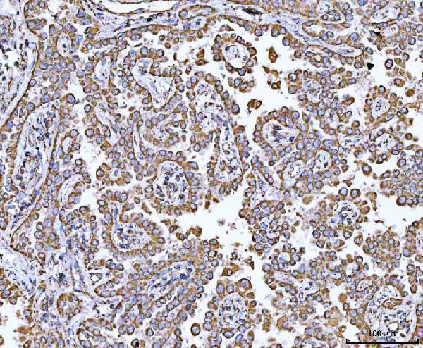
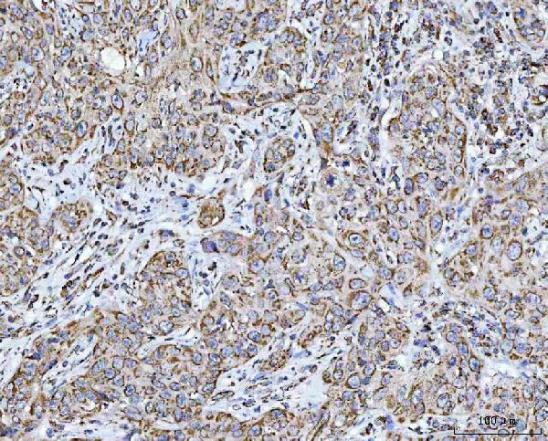
| Sample | Paraffin-embedded human lung cancer tissue; fixative not specified (datasheet A05981-2) |
| Fixation | Image fixative and duration unreported (datasheet A05981-2); verify before use. |
| Sectioning | 4–5 µm sections on charged slides (standard) |
| Deparaffinisation | Xylene, graded ethanol series to water (standard) |
| Antigen retrieval | Heat retrieval: EDTA pH 8.0 (datasheet A05981-2); 20 min, 95–100 °C (standard) |
| Peroxidase block | 3% H2O2, 10 min, room temperature (standard) |
| Blocking | 10% goat serum (datasheet A05981-2) |
| Primary antibody | Rabbit anti-NDUFA13, 2-5 μg/ml (datasheet A05981-2) |
| Primary incubation | Overnight at 4 °C (datasheet A05981-2) |
| Detection | HRP-conjugated secondary, DAB chromogen (datasheet A05981-2) |
| Counterstain | Hematoxylin, blue, dehydrate and mount (standard) |
| Expected result | NDUFA13-positive staining in glandular cells of adrenal gland (HPA tissue IHC: High). HPA tissue profile: Ubiquitous cytoplasmic expression with a granular pattern. No signal in the no-primary control. |
NDUFA13 is mainly mitochondrial and has one transmembrane segment at residues 30–51 in the inner membrane (UniProt Q9P0J0 topology/localisation). In paraffin IHC, expect granular cytoplasmic staining across many cell types, including strong staining in several glandular and neuronal populations (HPA: ubiquitous granular cytoplasmic profile; High in listed cells). The tissue IHC profile is Supported, with high consistency between antibody staining and RNA expression (HPA: reliability Supported).
| Granular cytoplasmic staining in colon glandular cells. | A convincing positive pattern: colon glandular cells are High by HPA tissue IHC, and the granular cytoplasm matches its overall profile (HPA: colon; tissue profile). Score cellular staining against adjacent background rather than treating every brown deposit as positive (general IHC practice). |
| Predominantly nuclear staining with little granular cytoplasm. | Question specificity or interpretation: the usual location is mitochondrial (UniProt Q9P0J0 localisation; HPA: mitochondria supported). UniProt allows possible nuclear translocation after IFN/RA treatment, so a documented treated sample needs separate interpretation (UniProt Q9P0J0 localisation). |
| A sharply restricted cell population stains while expected glandular cells do not. | Investigate cross-reactivity, endogenous detection activity, and tissue identification (general IHC practice). HPA reports broad expression and High glandular staining in several listed tissues; an unlisted cell type alone is not evidence of off-target binding (HPA: tissue profile; listed High cells). |
| Uniform haze or precipitate obscures cell boundaries. | This cannot establish NDUFA13 localisation. The supported tissue profile is granular cytoplasmic staining (HPA: tissue profile). Review blocking, washes, reagent concentration, and chromogen development as general IHC variables; compare a matched no-primary control (general IHC practice). |
| No staining in colon glandular cells or another HPA-listed High population. | Treat the run as inconclusive before calling the sample negative: HPA reports High staining in colon glandular cells, among other listed populations (HPA: colon and listed High cells). Check tissue preservation, retrieval, antibody and detection steps with an appropriate positive control (general IHC practice). |
| Compartment and resolution | NDUFA13 is an inner-membrane protein with one segment at residues 30–51 (UniProt Q9P0J0 topology). Routine chromogenic IHC supports a granular cytoplasmic readout, not visual assignment to the inner membrane itself (HPA: tissue profile; general IHC practice). |
| Choice of tissue comparator | HPA lists High glandular staining in colon, duodenum, appendix, breast and adrenal gland, and High staining in selected neuronal populations (HPA: listed High cells). Adipocytes are Low, not a validated negative control (HPA: adipose tissue Low; negative list empty). |
| Conditional nuclear localisation | UniProt says NDUFA13 localises mainly to mitochondria and may enter the nucleus after IFN/RA treatment (UniProt Q9P0J0 localisation). Record treatment context before interpreting nuclear staining; untreated tissue has no supplied target-specific nuclear expectation. |
| Isoforms and antibody validation | UniProt annotates 2 isoforms but supplies no isoform-specific staining pattern here (UniProt Q9P0J0 isoforms). HPA041213 is Supported for IHC and ICC; that status does not identify which isoform a given stain represents (HPA: HPA041213 validation). |
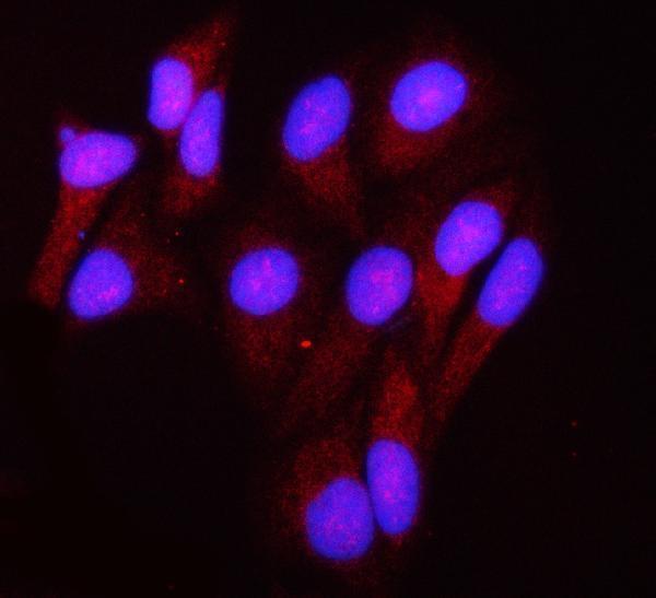
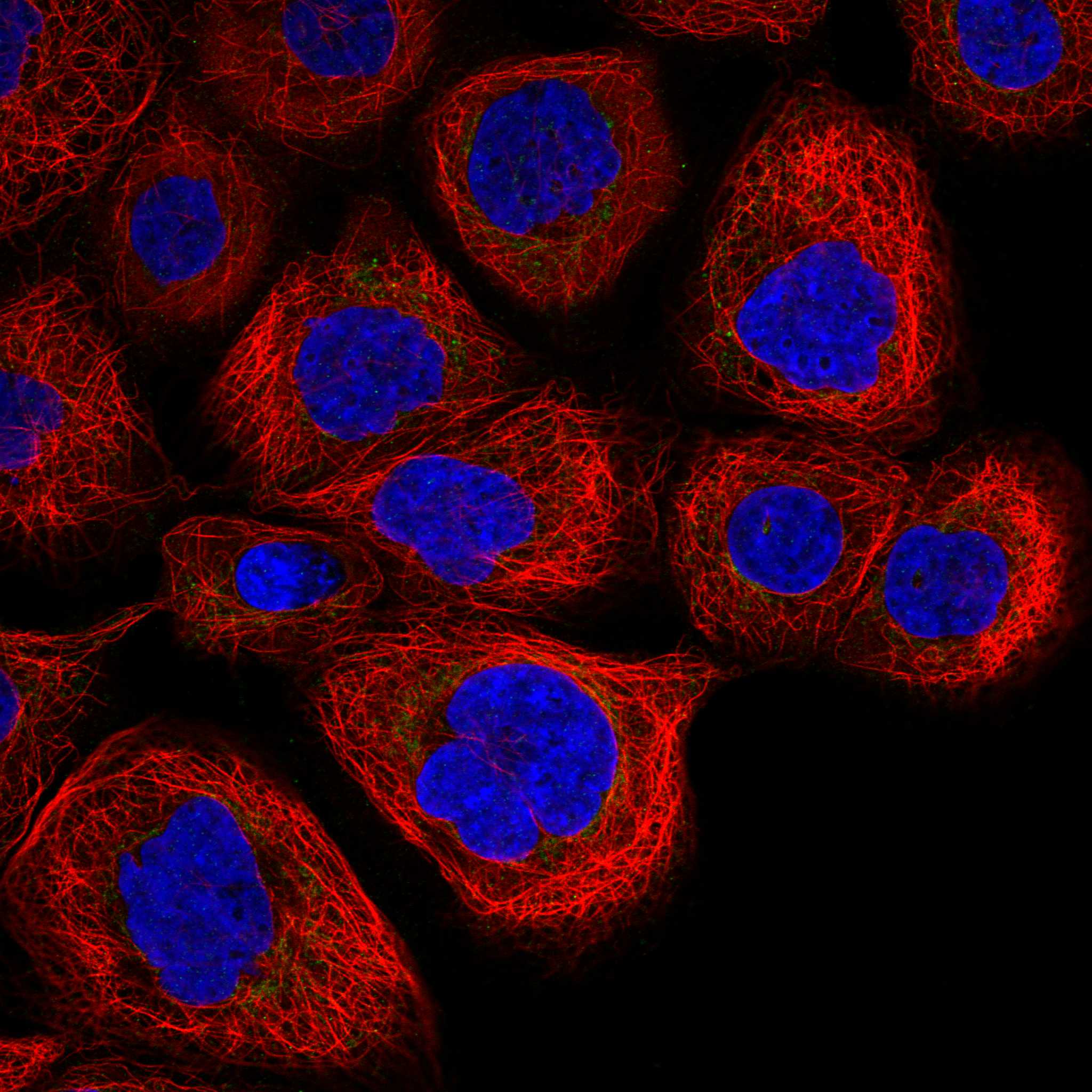
| IF/ICC Q&A: what should fluorescence show? | Expect mitochondrial localisation (HPA: ICC-IF mitochondria supported). HPA041213 is ICC Supported, with images listed for A-431, U-251MG, U2OS and sperm (HPA: ICC-IF; HPA041213 validation). Those ICC observations do not prescribe an IF protocol for paraffin sections. |
| Situation | Likely cause | Next action |
|---|---|---|
| A listed High tissue shows no cellular staining. | The result conflicts with HPA's High call for that specific cell population (HPA: listed High cells); a missed IHC step or inadequate assay performance is possible (general IHC practice). | Confirm the expected cell population is present, then review retrieval, primary dilution, detection and a working positive control (general IHC practice). Do not infer NDUFA13-specific fixation sensitivity from this failure. |
| Signal is diffuse rather than granular. | The observed pattern conflicts with HPA's granular cytoplasmic profile (HPA: tissue profile). Excess reagent, incomplete washing or detection background can blur localisation (general IHC practice). | Compare with a no-primary control; review dilution, washing and chromogen development (general IHC practice). Score only clearly cellular signal with interpretable morphology. |
| Brown signal persists without primary antibody. | Primary-independent colour may arise from endogenous detection activity or pigment (general chromogenic IHC practice); it does not demonstrate NDUFA13. | Inspect the no-primary slide in the same tissue region; apply the detection system's appropriate endogenous-activity control and distinguish pigment from reaction product (general IHC practice). |
| Only nuclei stain strongly. | This differs from the usual mitochondrial localisation (UniProt Q9P0J0 localisation; HPA: ICC-IF mitochondria supported), although IFN/RA-associated nuclear translocation is possible (UniProt Q9P0J0 localisation). | Check whether IFN/RA treatment is documented; compare granular cytoplasmic signal and controls before assigning the nuclear colour to NDUFA13 (UniProt Q9P0J0 localisation; general IHC practice). |
| Adipocytes are faint beside stained cells. | Faint adipocyte staining is compatible with HPA's Low call; HPA supplies no negative tissue in this record (HPA: adipose tissue Low; negative list empty). | Use an HPA-listed High cell population to judge whether the run worked; do not set the positivity threshold by assuming adipocytes must be blank (HPA: listed High cells; adipose tissue Low). |
| An unexpected cell type stains more strongly than nearby expected cells. | Broad expression means unexpected cell staining alone does not prove cross-reactivity (HPA: ubiquitous tissue profile). Endogenous activity or nonspecific staining remains possible (general IHC practice). | Identify both cell populations, compare their morphology and the no-primary control, and seek independent confirmation before calling a new cell-specific pattern (general IHC practice). |



Comprehensive Human Protein Atlas IHC scoring per tissue (reliability: Supported — High consistency between antibody staining and RNA expression data.). Rows are taken directly from the HPA tissue chart — click any row's HPA link to view the source.
| Tissue | Cell type | Level | Evidence | Source |
|---|---|---|---|---|
| Adrenal gland | Glandular cells | High | Protein (IHC) | HPA → |
| Appendix | Glandular cells | High | Protein (IHC) | HPA → |
| Breast | Glandular cells | High | Protein (IHC) | HPA → |
| Caudate | Neuronal cells | High | Protein (IHC) | HPA → |
| Cerebellum | Cells in granular layer | High | Protein (IHC) | HPA → |
| Tissue | Cell type | Level | Evidence | Source |
|---|---|---|---|---|
| None in HPA: NDUFA13 is detected in all 44 scored tissues. Use a no-primary (secondary-only) and an isotype control instead. | ||||
Use the catalog antibody’s paraffin-section IHC evidence to optimize retrieval and detection, then judge staining against NDUFA13’s expected mitochondrial distribution.
A05981-2 has IHC images from human paraffin sections and IF images from HeLa cells and human lung cancer tissue (catalog image captions); its listed reactivity covers Human, Mouse and Rat (catalog).
A05981-2 has IHC images from human lung cancer, skin squamous cell carcinoma and urothelial carcinoma paraffin sections, plus IF images from HeLa cells and human lung cancer tissue (catalog image captions). M05981-1 lists IHC and ICC/IF applications with Human reactivity, but provides no IHC or IF image captions (catalog).
Which to pick: Choose A05981-2 for tissue IHC because its own images document staining in human paraffin sections; the fixative is unreported (A05981-2 IHC image captions). Choose A05981-2 for IF/ICC when image evidence matters: its own IF captions show HeLa cells and human lung cancer tissue, while M05981-1 is a rabbit monoclonal, clone 19N74, with listed IHC and ICC/IF applications but no image captions (catalog). For cross-species work, A05981-2 lists Human, Mouse and Rat reactivity, versus Human for M05981-1; A05981-2’s IHC images document human samples only (catalog reactivity; A05981-2 IHC image captions).