This website uses cookies to ensure you get the best experience on our website.


- Table of Contents
Plan chromogenic NDUFB2 IHC in paraffin sections using the IHC-validated antibody A13719-1 (datasheet). Compare staining with the cytoplasmic and nuclear tissue pattern (HPA tissue IHC), while assessing its fit with the mitochondrial inner-membrane assignment (UniProt).

Expected localisation, validated protocols, controls and antibodies — the at-a-glance facts below, then the full design guide.
| Expected localisation | Cytoplasmic and nuclear tissue staining (HPA tissue IHC) | |
| Staining pattern | General cytoplasmic and nuclear staining; nucleoli in a few tissues (HPA tissue IHC) | |
| Antigen retrieval | EDTA pH 8.0 HIER, heat-mediated (datasheet A13719-1) | |
| Positive control | Ovary+4 more · see all | |
| Negative control | Bone marrow+1 more · see all |
| Fixation | Keep fixation consistent across sections. (standard IHC practice; not target-specific) | |
| Caveat | Nuclear staining differs from the inner-membrane assignment (HPA tissue IHC; UniProt) | |
| Regulation | Low tissue RNA specificity (HPA tissue RNA) | |
| Isoform / epitope | No isoforms annotated; map epitopes to processed chain 34–105 (UniProt) |
The catalog antibody’s IHC-P protocol (datasheet: EDTA pH 8.0 HIER) is accompanied by 1 published glioblastoma IHC protocol (PMC12777021).
| Sample | Paraffin-embedded human liver tissue; fixative not specified (datasheet A13719-1) |
| Fixation | Image fixative and duration unreported (datasheet A13719-1); verify before use. |
| Sectioning | 4–5 µm sections on charged slides (standard) |
| Deparaffinisation | Xylene, graded ethanol series to water (standard) |
| Antigen retrieval | Heat retrieval: EDTA pH 8.0 (datasheet A13719-1); 20 min, 95–100 °C (standard) |
| Peroxidase block | 3% H2O2, 10 min, room temperature (standard) |
| Blocking | 10% goat serum (datasheet A13719-1) |
| Primary antibody | Rabbit anti-NDUFB2, 2-5μg/ml (datasheet A13719-1) |
| Primary incubation | Overnight at 4 °C (datasheet A13719-1) |
| Detection | Streptavidin-biotin complex (SABC), DAB chromogen (datasheet A13719-1) |
| Counterstain | Hematoxylin, blue, dehydrate and mount (standard) |
| Expected result | NDUFB2-positive staining in follicle cells of ovary (HPA tissue IHC: High). HPA tissue profile: General cytoplasmic and nuclear expression with additional nucleoli in a few tissues. No signal in the no-primary control. |
NDUFB2 is an inner mitochondrial membrane Complex I subunit with no annotated transmembrane segment (UniProt O95178 topology). In paraffin-section IHC, expect staining in ovarian follicle cells and cells in testicular seminiferous ducts, where HPA reports high signal (HPA: High). HPA also describes general cytoplasmic and nuclear staining. Its tissue IHC reliability is Approved, with medium agreement with RNA data and external verification pending (HPA: reliability).
| Follicle cells or cells in seminiferous ducts stain strongly, with cytoplasmic signal (HPA: High in both). | This fits the strongest supplied tissue observations and the mitochondrial location (HPA: tissue IHC; UniProt O95178). Judge cell identity and compartment together: a strong brown deposit elsewhere in the section alone does not reproduce the reported positive pattern (HPA: tissue IHC). |
| Nuclear staining accompanies cytoplasmic staining, sometimes with nucleoli (HPA: tissue IHC profile). | HPA reports this pattern in tissue IHC, so nuclear signal alone cannot be called an artefact (HPA: tissue IHC profile). Interpret isolated, dominant nuclear staining cautiously: UniProt places NDUFB2 at the mitochondrial inner membrane, and HPA rates nucleoplasmic localization uncertain in ICC-IF (UniProt O95178; HPA: subcellular). |
| An unexpected cell population stains more strongly than the reported cells (HPA: tissue IHC). | First check the section’s cell identification and the matched negative control (general IHC practice). If the pattern persists, cross-reactivity or endogenous detection activity is possible (general IHC practice). HPA reports low tissue specificity, so staining outside the listed high-signal cells is not, by itself, proof of either problem (HPA: RNA specificity). |
| Brown colour spreads across tissue, stroma, or the whole section without clear cellular boundaries. | Treat this as background rather than a scored cellular pattern (general IHC practice). Compare a matched negative control, then review blocking, washes, detection chemistry, and section condition (general IHC practice). The supplied HPA observations describe cellular staining; they do not establish a target-specific cause for diffuse colour (HPA: tissue IHC). |
| A verified ovary or testis section shows no signal in the listed high-signal cells (HPA: High). | A failed positive control makes a negative result in a test specimen hard to interpret (general IHC practice). Confirm the expected cells are present, then check the validated antibody, reagent activity, retrieval, and detection run (general IHC practice). HPA’s Approved rating still carries medium RNA agreement and pending external verification (HPA: reliability). |
| Tissue and cell choice (HPA: tissue IHC) | Ovarian follicle cells and cells in seminiferous ducts are the supplied high-signal examples; adrenal and appendix glandular cells are medium, while bone marrow hematopoietic cells are not detected (HPA: tissue IHC). Match the comparison to the named cell population, not merely the organ (general IHC practice). |
| Compartment evidence (UniProt O95178; HPA: subcellular) | UniProt assigns NDUFB2 to the mitochondrial inner membrane and annotates no transmembrane segment (UniProt O95178 topology). HPA tissue IHC reports cytoplasmic and nuclear staining; ICC-IF adds approved mitochondrial localization but uncertain nucleoplasmic localization (HPA: tissue IHC; HPA: subcellular). Keep these evidence types distinct. |
| Antibody evidence (HPA: antibodies and reliability) | The listed rabbit polyclonal antibody, HPA051377, is Approved for IHC and ICC (HPA: antibodies). The tissue IHC assessment reports medium consistency with RNA expression and awaits external verification (HPA: reliability). An Approved label supports use of the observed pattern but does not settle every unexpected compartment or cell type. |
| Processing and epitope limits (UniProt O95178) | UniProt lists a chain spanning residues 34–105 and no annotated glycosylation sites, modified residues, or isoforms (UniProt O95178). The payload gives no antibody epitope or NDUFB2-specific retrieval response; it cannot predict which retrieval condition will recover staining or how fixation will affect this target. |
| Situation | Likely cause | Next action |
|---|---|---|
| No stain in the named high-signal cells of ovary or testis (HPA: High). | The run may have failed, or the expected cells may be absent from the examined section (general IHC practice). | Confirm cell identity and section quality; compare a known-positive section from the same run, then check retrieval and detection reagents (general IHC practice). Avoid scoring other specimens as NDUFB2-negative until the positive control works. |
| Broad, even brown staining obscures cell boundaries. | Background from blocking, washes, or detection chemistry is possible (general IHC practice). | Inspect the matched negative control and review blocking, wash, and detection steps (general IHC practice). Score only resolvable cellular staining; the HPA tissue profile describes cellular compartments, not diffuse section-wide colour (HPA: tissue IHC profile). |
| A supposedly negative comparison stains strongly. | The compared cells may differ from the HPA-listed population; cross-reactivity or endogenous detection activity is also possible (HPA: tissue IHC; general IHC practice). | Verify cell identity first. HPA reports bone marrow hematopoietic cells and parathyroid glandular cells as not detected, but does not establish every cell in those organs as negative (HPA: tissue IHC). Check a matched negative control if the discrepancy remains. |
| Staining is predominantly nuclear or nucleolar. | Some nuclear and occasional nucleolar tissue staining is reported, while nucleoplasmic ICC-IF localization is uncertain (HPA: tissue IHC profile; HPA: subcellular). | Document the compartment and compare cytoplasmic signal, expected cell types, and the negative control (HPA: tissue IHC; general IHC practice). Do not call nuclear signal alone definitive mitochondrial NDUFB2 localization (UniProt O95178; HPA: subcellular). |
| An unexpected cell type is the strongest stained population. | Cell identification, cross-reactivity, or endogenous detection activity may explain a mismatch (general IHC practice). HPA reports low tissue RNA specificity, so expression outside listed examples remains possible (HPA: RNA specificity). | Recheck morphology and compare the matched negative control (general IHC practice). Report the unexpected population separately from the HPA high-signal examples rather than assigning it the same expected intensity (HPA: tissue IHC). |
| Q: Does nuclear staining in ICC-IF resolve an IHC localization question? | A: HPA labels nucleoplasm uncertain and additional mitochondrial localization approved in ICC-IF; tissue IHC reports nuclear and cytoplasmic staining (HPA: subcellular; HPA: tissue IHC profile). | Use the ICC-IF observations as context, not a substitute for interpreting the paraffin section (HPA: subcellular; general IHC practice). Compare IHC cell type, compartment, and controls on its own terms; ICC-IF workflow belongs in its separate guide. |
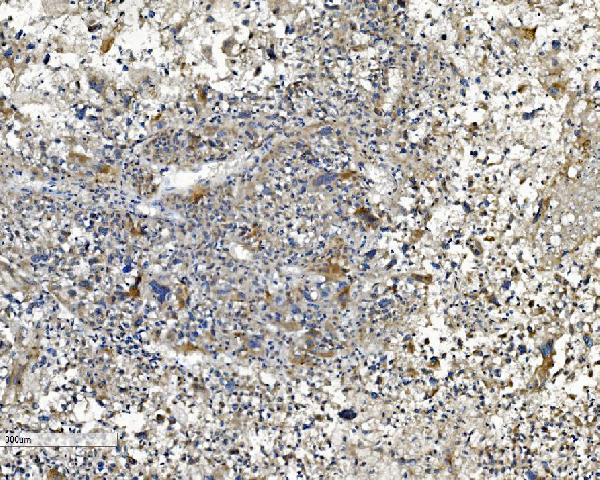
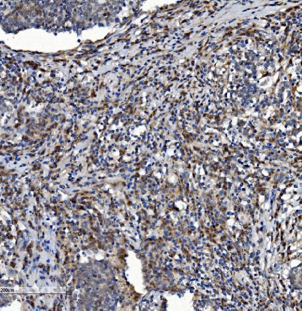


Comprehensive Human Protein Atlas IHC scoring per tissue (reliability: Approved — Medium consistency between antibody staining and RNA expression data. Pending external verification.). Rows are taken directly from the HPA tissue chart — click any row's HPA link to view the source.
| Tissue | Cell type | Level | Evidence | Source |
|---|---|---|---|---|
| Ovary | Follicle cells | High | Protein (IHC) | HPA → |
| Testis | Cells in seminiferous ducts | High | Protein (IHC) | HPA → |
| Adrenal gland | Glandular cells | Medium | Protein (IHC) | HPA → |
| Appendix | Glandular cells | Medium | Protein (IHC) | HPA → |
| Breast | Glandular cells | Medium | Protein (IHC) | HPA → |
Troubleshoot NDUFB2 staining in paraffin sections by checking retrieval, compartment, controls and scoring before interpreting signal.
A13719-1 has real IHC images from paraffin-embedded human liver, lung cancer, and rectal cancer sections; no IF image is supplied (catalog image captions and IF image list).
A13719-1 is listed for human IHC and shown on paraffin-embedded human liver sections (catalog applications, reactivity, and image captions). Its other IHC images show human lung cancer and rectal cancer sections (catalog image captions).
Which to pick: For tissue IHC, choose A13719-1; the catalog specifies 2–5 μg/ml for human paraffin sections and identifies the antibody as rabbit-hosted, with clonality unspecified (catalog dilution, reactivity, host, and clone fields). Its image captions describe heat retrieval in EDTA at pH 8.0, while the fixative is unreported (catalog image captions). No IF/ICC or cross-species SKU can be recommended from this catalog: A13719-1 has no listed IF/ICC application or IF image and lists human reactivity only (catalog applications, IF image list, and reactivity).