This website uses cookies to ensure you get the best experience on our website.


- Table of Contents
Plan NDUFB9 paraffin IHC around the granular cytoplasmic staining seen in most tissues (HPA tissue IHC). Cardiomyocytes and kidney tubule cells show high staining (HPA tissue IHC); the A08623-1 IHC-P starting concentration is 5 μg/mL (datasheet).

Expected localisation, validated protocols, controls and antibodies — the at-a-glance facts below, then the full design guide.
| Expected localisation | Granular cytoplasm in most tissues (HPA tissue IHC) | |
| Staining pattern | Granular cytoplasm in cardiomyocytes and kidney tubule cells (HPA tissue IHC) | |
| Antigen retrieval | Citrate pH 6.0 HIER, 95–98 °C, 20 min (rule: cytoplasmic / membrane antigen) | |
| Positive control | Adipose tissue+4 more · see all | |
| Negative control | Bone marrow+4 more · see all |
| Fixation | Keep fixation consistent across sections. (standard IHC practice; not target-specific) | |
| Caveat | Antibody staining has low consistency with RNA expression (HPA tissue IHC) | |
| Regulation | No stimulus-specific regulation annotated (UniProt) | |
| Isoform / epitope | No annotated isoforms; chain spans residues 2–179 (UniProt) |
Compare the catalog antibody’s IHC-P protocol with a published NDUFB9 staining workflow for FFPE slides (PMC13376752).
| Sample | Tissue sections; selected-image fixative not specified (standard IHC workflow) |
| Fixation | Image fixative and duration unreported (datasheet A08623-1); verify before use. |
| Sectioning | 4–5 µm sections on charged slides (standard) |
| Deparaffinisation | Xylene, graded ethanol series to water (standard) |
| Antigen retrieval | Heat-induced epitope retrieval in citrate buffer, pH 6.0, 20 min at 95–98 °C (standard rule: cytoplasmic / membrane antigen) |
| Peroxidase block | 3% H2O2, 10 min, room temperature (standard) |
| Blocking | 10% normal serum of the secondary host, 30 min, room temperature (standard) |
| Primary antibody | Rabbit anti-NDUFB9, 5 μg/mL (datasheet A08623-1) |
| Primary incubation | Overnight at 4 °C (standard) |
| Detection | HRP-polymer secondary, DAB chromogen 5–10 min (standard) |
| Counterstain | Hematoxylin, blue, dehydrate and mount (standard) |
| Expected result | NDUFB9-positive staining in adipocytes of adipose tissue (HPA tissue IHC: High). HPA tissue profile: Cytoplasmic expression with a granular expression in most tissues. No signal in the no-primary control. |
NDUFB9 is a mitochondrial inner-membrane Complex I accessory subunit with no annotated transmembrane segment (UniProt Q9Y6M9). In paraffin-section IHC, expect granular cytoplasmic staining in many tissues, including cardiomyocytes, kidney tubule cells, adipocytes and several glandular cell populations scored High by HPA (HPA: tissue IHC). HPA rates its tissue staining Approved but reports low consistency with RNA expression, so interpret intensity alongside controls and tissue morphology (HPA: reliability).
| Granular cytoplasmic staining in cardiomyocytes or kidney tubule cells, with preserved tissue detail. | This fits HPA's granular cytoplasmic profile and High staining scores for those cells (HPA: tissue IHC). It is compatible with NDUFB9's mitochondrial location (UniProt Q9Y6M9; HPA: subcellular IF). Chromogenic granules alone do not resolve individual mitochondria (general IHC practice). |
| Predominantly nuclear staining or a sharp cell-surface outline replaces cytoplasmic granules. | That compartment differs from the reported mitochondrial location and granular cytoplasmic IHC profile (UniProt Q9Y6M9; HPA: tissue IHC). Check controls, morphology and detection conditions before treating it as NDUFB9 localisation (general IHC practice). |
| Strong staining appears in bone-marrow hematopoietic cells or lymph-node germinal-center cells. | HPA reports NDUFB9 as Not detected in those specific cell populations (HPA: tissue IHC). Review cell identification and controls; cross-reactivity or endogenous detection activity is possible, but an HPA Not detected score does not prove every specimen must be negative (general IHC practice). |
| Color spreads diffusely across tissue, including spaces without recognizable cells. | That distribution does not match HPA's granular cytoplasmic profile (HPA: tissue IHC). Background from the detection workflow is a possibility; use the appropriate negative control and assess whether the color tracks tissue structure (general IHC practice). |
| No staining is visible in an otherwise intact heart-muscle or kidney section. | HPA scores cardiomyocytes and kidney tubule cells High, making them useful reference populations (HPA: tissue IHC). Recheck the assay and a concurrently stained positive control before interpreting the absence as biological; HPA staining is a reference observation, not a guarantee for every specimen (general IHC practice). |
| Where should chromogenic staining appear? | NDUFB9 is assigned to the mitochondrial inner membrane (UniProt Q9Y6M9), while tissue IHC shows a granular cytoplasmic pattern (HPA: tissue IHC). Interpret the chromogenic pattern at the resolution of the section; do not claim that individual granules prove inner-membrane localisation (general IHC practice). |
| How should tissue-to-tissue intensity be used? | HPA scores cardiomyocytes, kidney tubule cells and adipocytes High, breast glandular cells Medium, and liver hepatocytes Low (HPA: tissue IHC). These are observed cell-specific scores, useful for comparison within the documented context; they are not a universal intensity threshold for all specimens (general IHC practice). |
| How strong is the tissue-staining evidence? | HPA marks tissue IHC Approved while noting low consistency between antibody staining and RNA expression (HPA: reliability). The listed antibody HPA042768 is IHC Approved; no Enhanced IHC status is supplied (HPA: antibody validation). Keep unexpected staining provisional until controls and morphology support it (general IHC practice). |
| Do processing or sequence features predict the IHC pattern? | UniProt lists a chain spanning residues 2–179, no annotated transmembrane segment, and no annotated isoforms (UniProt Q9Y6M9). These facts do not identify the antibody epitope or establish antigen-retrieval requirements or fixation sensitivity; use them only as sequence context. |
| Does IF/ICC support the localisation? | Yes. HPA reports supported mitochondrial localisation by ICC-IF and lists A-431, U-251MG, U2OS and KOLF2.1J among cell lines with images (HPA: subcellular IF). This supports a mitochondrial interpretation; the IF/ICC staining procedure belongs in its separate guide. |
| Situation | Likely cause | Next action |
|---|---|---|
| A known-positive heart or kidney section has no visible signal. | The section conflicts with HPA's High scores for cardiomyocytes or kidney tubule cells; an assay failure is possible, but the image alone cannot identify its cause (HPA: tissue IHC; general IHC practice). | Check a concurrently stained positive control, antibody application, antigen-retrieval record, detection reagents and counterstain visibility; adjust workflow steps using the catalog antibody's IHC-P instructions (general IHC practice). |
| Color is predominantly nuclear or outlines cell membranes. | The compartment conflicts with mitochondrial localisation and HPA's granular cytoplasmic IHC profile (UniProt Q9Y6M9; HPA: tissue IHC). Nonspecific staining or misread morphology is possible (general IHC practice). | Reassess the cells at higher magnification, compare a negative control and review antibody and detection conditions before scoring NDUFB9 positivity (general IHC practice). |
| A population HPA scores Not detected stains strongly. | For example, HPA scores bone-marrow hematopoietic cells Not detected; cell misidentification, cross-reactivity or endogenous detection activity could explain a discrepancy (HPA: tissue IHC; general IHC practice). | Confirm the cell population morphologically, inspect a negative control and compare an HPA High reference population processed in the same run (HPA: tissue IHC; general IHC practice). |
| A diffuse haze obscures otherwise granular staining. | The haze differs from HPA's granular cytoplasmic profile; nonspecific binding or detection background may contribute (HPA: tissue IHC; general IHC practice). | Compare tissue-free areas and a negative control, then review blocking, washes and chromogen development within the validated IHC workflow (general IHC practice). |
| A low-scored tissue appears weaker than a high-scored tissue. | HPA scores liver hepatocytes Low and cardiomyocytes High, so a difference in these cells can reflect the reported tissue pattern (HPA: tissue IHC). Technical variation can also affect an IHC comparison (general IHC practice). | Compare the documented cell populations and run controls before assigning biological meaning to intensity; do not apply one tissue's HPA score to every cell in that tissue (HPA: tissue IHC; general IHC practice). |
| IHC granules appear less discrete than mitochondrial IF signal. | HPA reports granular cytoplasmic tissue IHC and supported mitochondrial ICC-IF localisation; the methods present localisation at different visual scales (HPA: tissue IHC; HPA: subcellular IF; general IHC practice). | Score the IHC section for the expected cytoplasmic pattern and cell type. Use the separate IF/ICC guide for fluorescence-specific interpretation (HPA: tissue IHC; HPA: subcellular IF). |


Comprehensive Human Protein Atlas IHC scoring per tissue (reliability: Approved — Low consistency between antibody staining and RNA expression data.). Rows are taken directly from the HPA tissue chart — click any row's HPA link to view the source.
| Tissue | Cell type | Level | Evidence | Source |
|---|---|---|---|---|
| Adipose tissue | Adipocytes | High | Protein (IHC) | HPA → |
| Appendix | Glandular cells | High | Protein (IHC) | HPA → |
| Duodenum | Glandular cells | High | Protein (IHC) | HPA → |
| Heart muscle | Cardiomyocytes | High | Protein (IHC) | HPA → |
| Kidney | Cells in tubules | High | Protein (IHC) | HPA → |
| Tissue | Cell type | Level | Evidence | Source |
|---|---|---|---|---|
| Bone marrow | Hematopoietic cells | Not detected | Protein (IHC) | HPA → |
| Bronchus | Basal cells | Not detected | Protein (IHC) | HPA → |
| Cervix | Glandular cells | Not detected | Protein (IHC) | HPA → |
| Epididymis | Glandular cells | Not detected | Protein (IHC) | HPA → |
| Lymph node | Germinal center cells | Not detected | Protein (IHC) | HPA → |
Troubleshoot NDUFB9 staining in paraffin sections by checking retrieval, compartment, tissue controls and scoring consistency (UniProt Q9Y6M9; HPA tissue IHC).
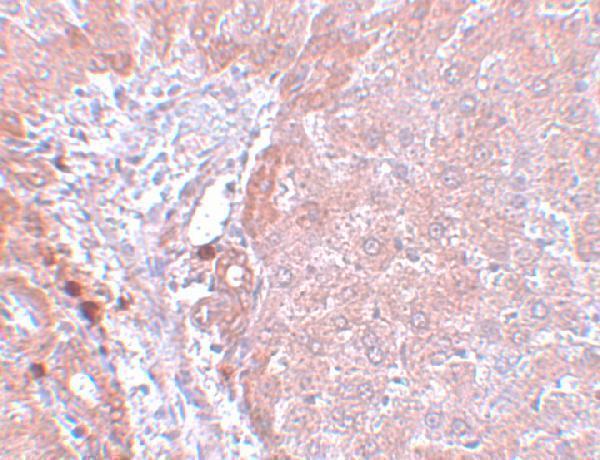
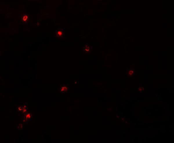
A08623-1 has rat liver IHC and IF images (catalog image captions); both antibodies list human, mouse and rat reactivity (catalog reactivity).
A08623-1 shows rat liver IHC at 5 μg/mL and IF at 20 μg/mL (catalog image captions). M08623-3 lists IHC and ICC/IF applications, but has no IHC or IF image in the payload (catalog applications and image captions).
Which to pick: Choose A08623-1 for paraffin-section IHC: it lists IHC-P and shows rat liver staining, although the fixative is unreported (catalog applications; A08623-1 IHC image caption). For IF/ICC, A08623-1 has a rat liver IF image, while M08623-3 lists ICC/IF and is monoclonal, with no image supplied (catalog applications, clone and image captions). Both list human, mouse and rat reactivity; the supplied tissue images document rat liver only (catalog reactivity; A08623-1 image captions).