This website uses cookies to ensure you get the best experience on our website.


- Table of Contents
Plan paraffin IHC for NDUFC1 with the catalog antibody at 1:100–1:300 (datasheet A15096-1). Use the cytoplasmic tissue profile and high staining in kidney tubules to assess the result (HPA tissue IHC).

Expected localisation, validated protocols, controls and antibodies — the at-a-glance facts below, then the full design guide.
| Expected localisation | Cytoplasmic staining in tissue (HPA tissue IHC) | |
| Staining pattern | Widespread cytoplasmic staining across cell types (HPA tissue IHC) | |
| Antigen retrieval | Tris-EDTA pH 9.0 HIER, heat-mediated (datasheet A15096-1) | |
| Positive control | Adrenal gland+4 more · see all | |
| Negative control | Bone marrow+4 more · see all |
| Fixation | Keep fixation consistent across sections. (standard IHC practice; not target-specific) | |
| Caveat | IHC staining has medium agreement with RNA data (HPA tissue IHC) | |
| Regulation | No expression regulation annotated (UniProt) | |
| Isoform / epitope | 0 isoforms; mature chain 28–76; epitope unknown (UniProt) |
The catalog antibody's IHC-P protocol (datasheet A15096-1) is accompanied by two published NDUFC1 IHC protocols (PMC8710466; PMC9479186).
| Sample | Paraffin-embedded human Colon cancer tissue; fixative not specified (datasheet A15096-1) |
| Fixation | Image fixative and duration unreported (datasheet A15096-1); verify before use. |
| Sectioning | 4–5 µm sections on charged slides (standard) |
| Deparaffinisation | Xylene, graded ethanol series to water (standard) |
| Antigen retrieval | Heat retrieval: Tris-EDTA pH 9.0 (datasheet A15096-1); 20 min, 95–100 °C (standard) |
| Peroxidase block | 3% H2O2, 10 min, room temperature (standard) |
| Blocking | 10% normal serum of the secondary host, 30 min, room temperature (standard) |
| Primary antibody | Rabbit anti-NDUFC1, 1:100-1:300 (datasheet A15096-1) |
| Primary incubation | Overnight at 4 °C (standard) |
| Detection | HRP-polymer secondary, DAB chromogen 5–10 min (standard) |
| Counterstain | Hematoxylin, blue, dehydrate and mount (standard) |
| Expected result | NDUFC1-positive staining in glandular cells of adrenal gland (HPA tissue IHC: High). HPA tissue profile: Ubiquitous cytoplasmic expression. No signal in the no-primary control. |
NDUFC1 is an inner mitochondrial membrane Complex I subunit with a transmembrane segment at residues 41–59 (UniProt O43677 topology). In paraffin IHC, expect cytoplasmic staining in cell populations reported positive by HPA, including kidney tubular and colon glandular cells (HPA tissue IHC: High). HPA calls the tissue pattern ubiquitous cytoplasmic, with Enhanced reliability, medium agreement with RNA expression, and external verification pending (HPA tissue IHC).
| Granular cytoplasmic staining in kidney tubular cells or colon glandular cells, with little distracting background. | This fits the mitochondrial location and the cell populations scored High by HPA (UniProt O43677 subcellular location; HPA tissue IHC: High in kidney tubular cells and colon glandular cells). Chromogenic IHC shows a cellular pattern; it cannot resolve staining specifically to the inner mitochondrial membrane (general IHC practice). |
| Predominantly nuclear or cell-surface staining in otherwise positive cells. | That compartment conflicts with the reported mitochondrial location (UniProt O43677 subcellular location; HPA subcellular ICC-IF: mitochondria). Treat it as a possible assay artefact and inspect controls before scoring it as NDUFC1 (general IHC practice). |
| Strong staining in a cell population HPA lists as not detected, such as bone marrow hematopoietic cells. | This is discordant with the reported cell-specific pattern (HPA tissue IHC: Not detected in bone marrow hematopoietic cells). Consider antibody cross-reactivity or endogenous chromogenic activity, and assess the staining against appropriate controls (general IHC practice). |
| Diffuse colour covers cells, extracellular spaces, or the whole section without a discernible cytoplasmic pattern. | The result cannot be interpreted as the reported cytoplasmic pattern (HPA tissue IHC: ubiquitous cytoplasmic expression). Uneven reagent deposition, nonspecific binding, or detection background are possible causes; compare with a detection control (general IHC practice). |
| No staining in kidney tubular cells or colon glandular cells on a run expected to show NDUFC1. | Those populations are reported High and can serve as positive comparators (HPA tissue IHC: High in kidney tubular cells and colon glandular cells). First check whether the run and tissue are interpretable; absence alone does not establish biological loss of NDUFC1 (general IHC practice). |
| Compartment and optical resolution | NDUFC1 occupies the mitochondrial inner membrane, with one annotated transmembrane segment at 41–59 (UniProt O43677 topology). Interpret chromogenic signal at the cytoplasmic level; the slide cannot establish membrane topology (general IHC practice). |
| Processing and epitope limits | UniProt annotates a mature chain at residues 28–76 and no glycosylation sites or modified residues (UniProt O43677 processing/PTMs). No antibody epitope is supplied, so these annotations cannot predict antigen retrieval needs or staining sensitivity. |
| Cell-specific tissue comparison | HPA scores kidney tubular and colon glandular cells High, but bone marrow hematopoietic cells and oral mucosal squamous cells Not detected (HPA tissue IHC). Compare the named cell populations, since a tissue-wide positive or negative label would overstate the evidence. |
| Strength of IHC evidence | Both listed antibodies, HPA044556 and HPA062427, have Enhanced IHC status (HPA antibodies). The tissue profile reports medium agreement with RNA and pending external verification (HPA tissue IHC); use the pattern as an expectation, not proof of specificity in every section. |
| IF/ICC Q: What localization should appear? | A: Mitochondria are the supported main location; HPA lists ICC-IF images in A-431, U-251MG, and U2OS and marks HPA044556 ICC Approved (HPA subcellular ICC-IF; HPA antibodies). This localization cross-check does not specify an IF protocol for this IHC guide. |
| Situation | Likely cause | Next action |
|---|---|---|
| Positive comparator has no convincing cytoplasmic signal. | The run may have weak detection, an unsuitable antibody concentration, or a missed processing step (general IHC practice); the expected populations are High (HPA tissue IHC: kidney tubular and colon glandular cells). | Check the positive control, primary-antibody application, detection reagents, and counterstain. Optimize retrieval and antibody concentration as general IHC variables; target-specific fixation or retrieval sensitivity is unreported in the supplied sources. |
| Nuclei dominate the staining. | The distribution conflicts with the mitochondrial assignment (UniProt O43677 subcellular location; HPA subcellular ICC-IF). Excess background or nonspecific binding may obscure the intended pattern (general IHC practice). | Compare with the primary-omission control, review antibody concentration and blocking, and score only a reproducible cytoplasmic pattern in an expected cell population (general IHC practice; HPA tissue IHC). |
| Strong signal appears in HPA Not detected cells. | Unexpected cell-specific staining may reflect cross-reactivity or endogenous detection activity (general IHC practice; HPA tissue IHC: Not detected in bone marrow hematopoietic cells). | Confirm which cells carry the colour, then inspect primary-omission and detection controls. If signal persists without primary antibody, address endogenous detection activity before interpreting the section (general IHC practice). |
| Diffuse colour obscures cell boundaries. | Nonspecific binding, inadequate washing, or excess detection product can obscure a cytoplasmic pattern (general IHC practice; HPA tissue IHC: ubiquitous cytoplasmic expression). | Review blocking and wash steps, antibody concentration, and chromogen development using the same run's controls. Reassess whether staining follows identifiable cells before assigning intensity (general IHC practice). |
| Only low-level or variable staining appears across candidate tissues. | HPA reports Low staining in several named populations, including thyroid glandular and bronchial respiratory epithelial cells; weak signal there is less informative than a High comparator (HPA tissue IHC). | Read the correct cell population alongside kidney tubules or colon glands, and avoid treating every faint deposit as positive. Record intensity and background together when scoring (HPA tissue IHC; general IHC practice). |
| A negative cell population is used to declare the entire section negative. | HPA levels refer to specified cell populations: ovarian stroma is Not detected, while the overall tissue profile is described as ubiquitous cytoplasmic expression (HPA tissue IHC). | Identify the cells being scored and report their staining separately. Use a named High population as the positive comparator before drawing a conclusion about assay performance (HPA tissue IHC; general IHC practice). |


Comprehensive Human Protein Atlas IHC scoring per tissue (reliability: Enhanced — Medium consistency between antibody staining and RNA expression data. Pending external verification.). Rows are taken directly from the HPA tissue chart — click any row's HPA link to view the source.
| Tissue | Cell type | Level | Evidence | Source |
|---|---|---|---|---|
| Adrenal gland | Glandular cells | High | Protein (IHC) | HPA → |
| Caudate | Glial cells | High | Protein (IHC) | HPA → |
| Cerebral cortex | Neuronal cells | High | Protein (IHC) | HPA → |
| Colon | Glandular cells | High | Protein (IHC) | HPA → |
| Duodenum | Glandular cells | High | Protein (IHC) | HPA → |
| Tissue | Cell type | Level | Evidence | Source |
|---|---|---|---|---|
| Bone marrow | Hematopoietic cells | Not detected | Protein (IHC) | HPA → |
| Nasopharynx | Respiratory epithelial cells | Not detected | Protein (IHC) | HPA → |
| Oral mucosa | Squamous epithelial cells | Not detected | Protein (IHC) | HPA → |
| Ovary | Ovarian stroma cells | Not detected | Protein (IHC) | HPA → |
| Parathyroid gland | Glandular cells | Not detected | Protein (IHC) | HPA → |
Troubleshoot NDUFC1 staining in paraffin sections by checking retrieval, compartment, controls and scoring before interpreting chromogenic signal.
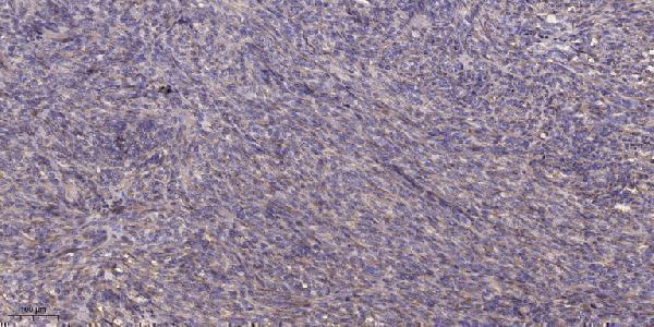
The catalog antibody has real IHC data from paraffin-embedded human colon cancer (catalog IHC image caption); IF is listed (catalog applications), with human and mouse reactivity (catalog reactivity).
A15096-1 is shown by IHC on paraffin-embedded human colon cancer using 1:200 primary dilution and Tris-EDTA pH 9.0 retrieval (catalog IHC image caption). A15096-1 also lists IF and human/mouse reactivity, but provides no IF image (catalog applications/reactivity; catalog IF image alts).
Which to pick: For tissue IHC, choose A15096-1 because its own image shows staining in paraffin-embedded human colon cancer; the fixative is unreported (catalog IHC image caption). For IF/ICC, A15096-1 lists IF at 1:50, but ICC-specific validation and an IF image are unreported (catalog applications/dilution; catalog IF image alts). For work across human and mouse, A15096-1 lists both species, although the supplied IHC image shows human tissue only and clonality is unreported (catalog reactivity; catalog IHC image caption; catalog clone field).