NDUFS1 / NADH-ubiquinone oxidoreductase 75 kDa subunit, mitochondrial · IHC design guide

Design Immunohistochemistry for NDUFS1

Plan paraffin-section NDUFS1 IHC using the cytoplasmic tissue pattern, most abundant in heart muscle (HPA tissue IHC), and mitochondrial inner-membrane localization as a molecular expectation (UniProt). This guide covers the catalog antibody’s 2–5 μg/ml IHC range (datasheet A04920-1), chromogenic detection and tissue controls.

Evidence assembled Oct 2026 · For research use; verify linked source records and product datasheet before use
Immunohistochemistry protocol sheet for NDUFS1 (IHC for NDUFS1): expected localisation Cytoplasmic staining (HPA tissue IHC); inner mitochondrial membrane expected (UniProt), antibody A04920-1, validated IHC image, and IHC protocol steps
Printable NDUFS1 IHC protocol sheet — expected localisation Cytoplasmic staining (HPA tissue IHC); inner mitochondrial membrane expected (UniProt), antibody A04920-1, controls and protocol steps. Open the full NDUFS1 IHC guide →

NDUFS1 Immunohistochemistry Experimental Design Guide

Expected localisation, validated protocols, controls and antibodies — the at-a-glance facts below, then the full design guide.

Must know before staining
Expected localisation Cytoplasmic staining (HPA tissue IHC); inner mitochondrial membrane expected (UniProt)
Staining pattern Cytoplasmic staining in cardiomyocytes and other tissues (HPA tissue IHC)
Antigen retrieval EDTA pH 8.0 HIER, heat-mediated (datasheet A04920-1)
Positive control ⓘ Adrenal gland+4 more · see all
Negative control ⓘ Adipose tissue+4 more · see all
Important caveats
Reasons your staining may differ from the expected pattern.
Fixation Keep fixation consistent across sections (standard IHC practice; not target-specific); Matched tissue-IHC evidence does not establish the fixation claim. Validate the specimen-specific method before use. (selected-SKU IHC image A04920-1)
Caveat Tumor staining may differ from low or undetected normal tissue (datasheet A04920-1; HPA tissue IHC)
Regulation Highest tissue expression in heart muscle (HPA tissue IHC)
Isoform / epitope 5 isoforms; mature chain spans residues 24–727; antibody epitope unspecified (UniProt; datasheet A04920-1)
Section 1

Recommended NDUFS1 IHC & IF Protocols

The catalog antibody’s IHC-P protocol is paired with published NDUFS1 IHC methods for prostate, gastric, and lung cancer samples (PMC8656993; PMC10575981; PMC4981865).

Recommended immunohistochemistry (IHC-P) protocol parameters
SampleParaffin-embedded human lung cancer tissue; fixative not specified (datasheet A04920-1)
FixationImage fixative and duration unreported (datasheet A04920-1); verify before use.
Sectioning4–5 µm sections on charged slides (standard)
DeparaffinisationXylene, graded ethanol series to water (standard)
Antigen retrievalHeat retrieval: EDTA pH 8.0 (datasheet A04920-1); 20 min, 95–100 °C (standard)
Peroxidase block3% H2O2, 10 min, room temperature (standard)
Blocking10% goat serum (datasheet A04920-1)
Primary antibodyRabbit anti-NDUFS1, 2-5 μg/ml (datasheet A04920-1)
Primary incubationOvernight at 4 °C (datasheet A04920-1)
DetectionHRP-conjugated secondary, DAB chromogen (datasheet A04920-1)
CounterstainHematoxylin, blue, dehydrate and mount (standard)
Expected resultNDUFS1-positive staining in glandular cells of adrenal gland (HPA tissue IHC: Medium). HPA tissue profile: Cytoplasmic expression in several tissues, most abundant in heart muscle. No signal in the no-primary control.
💡Decision noteStart with heat-mediated EDTA pH 8.0 retrieval for the catalog antibody (datasheet A04920-1); the published methods used other antibodies and retrieval conditions (PMC8656993; PMC4981865).
Section 2

What Is the Expected NDUFS1 Staining Pattern?

NDUFS1 is a mitochondrial inner-membrane Complex I subunit with no annotated transmembrane segment (UniProt P28331 topology). In paraffin-section IHC, expect cytoplasmic staining in cells reported positive by HPA, including cardiomyocytes and kidney tubular cells (HPA: tissue IHC). HPA describes the tissue profile as cytoplasmic and most abundant in heart muscle; its IHC reliability is Enhanced, with medium consistency between staining and RNA data (HPA: tissue IHC).

What am I looking at on my slide?
Cytoplasmic staining in cardiomyocytes or kidney tubular cells.This fits HPA's reported Medium staining in both cell types (HPA: tissue IHC). Cytoplasmic staining is compatible with mitochondrial localization; chromogenic IHC alone may not resolve individual mitochondria (UniProt P28331; general IHC practice).
Predominantly nuclear, extracellular, or sharply outlined cell-surface staining.That distribution conflicts with the mitochondrial inner-membrane assignment (UniProt P28331 topology). Treat it as suspect until controls exclude nonspecific antibody binding, chromogen precipitate, or a detection artefact (general IHC practice).
Strong staining in adipocytes, lung alveolar cells, or bone-marrow hematopoietic cells.HPA reports NDUFS1 as Not detected in those particular cell populations (HPA: tissue IHC). Recheck cell identity and controls; cross-reactivity or endogenous detection activity is possible, but an unexpected stain alone does not identify its cause (general IHC practice).
Diffuse color across tissue, stroma, or the whole slide, obscuring cell boundaries.An uninterpretable background pattern cannot establish NDUFS1 localization (general IHC practice). Compare a no-primary control, examine wash and blocking steps, and check for uneven reagent coverage before scoring cells (general IHC practice).
No detectable signal in a heart-muscle section expected to be positive.HPA reports Medium cardiomyocyte staining and describes heart muscle as the most abundant tissue in its IHC profile (HPA: tissue IHC). First verify tissue preservation and the run's positive control, then review antibody and detection conditions (general IHC practice).
💡Expected NDUFS1 appearanceCall the IHC result consistent when cardiomyocytes show discernible cytoplasmic, approximately Medium staining (HPA: tissue IHC); predominantly nuclear or extracellular color is suspect against the mitochondrial localization (UniProt P28331).
How each factor affects the staining
Compartment and optical resolutionNDUFS1 is assigned to the mitochondrial inner membrane without an annotated transmembrane segment (UniProt P28331 topology). HPA calls tissue IHC cytoplasmic (HPA: tissue IHC); a cytoplasmic chromogen pattern can be compatible with mitochondrial protein at light-microscope resolution (general IHC practice).
Choice of comparison tissue and cellHPA reports Medium staining in cardiomyocytes and kidney tubular cells, Low staining in hepatocytes, and Not detected in adipocytes (HPA: tissue IHC). Score the specified cell population rather than treating an entire organ as uniformly positive or negative (general IHC practice).
Antibody evidenceThe listed antibody CAB070844 has Enhanced IHC validation, while HPA064605 has Supported ICC validation and no listed IHC status (HPA: antibodies). Validation is application-specific; an ICC result does not establish how a different antibody performs in paraffin-section IHC (general IHC practice).
Sequence processing and isoformsUniProt lists five isoforms and a chain spanning residues 24–727, with no annotated signal peptide or propeptide (UniProt P28331). These entries alone do not identify the catalog antibody's epitope or predict isoform-specific staining; consult that antibody's documented antigen information before making such a claim.
IF/ICC Q&A: where should fluorescence appear?HPA reports supported mitochondrial localization in ICC-IF images from PC-3, Rh30, and U2OS cells (HPA: subcellular ICC-IF). This supports a mitochondrial interpretation for IF/ICC; its staining setup belongs in the separate IF/ICC guide.
Why is my staining missing, weak or wrong?
SituationLikely causeNext action
Heart-muscle cardiomyocytes are blank.HPA reports Medium cardiomyocyte staining, so an absent signal could reflect a failed IHC run or a tissue/sample issue; the observation alone cannot distinguish them (HPA: tissue IHC; general IHC practice).Inspect the positive control and tissue integrity, then check the catalog antibody's IHC-P instructions, retrieval, incubation, and detection steps against the run record (general IHC practice). No NDUFS1-specific fixation sensitivity is established by the supplied sources.
Only nuclei stain, or color lies outside cells.That location is inconsistent with the mitochondrial inner-membrane assignment (UniProt P28331 topology). Nonspecific binding, precipitate, or detection artefact are possibilities (general IHC practice).Review the no-primary control and slide for precipitate; repeat with clean detection reagents and the documented IHC-P conditions before interpreting the pattern (general IHC practice).
Adipocytes or lung alveolar cells stain strongly.Those cell populations are Not detected in the HPA tissue entries (HPA: tissue IHC). Unexpected color could reflect misidentified cells, cross-reactivity, or endogenous detection activity (general IHC practice).Confirm cell identity on the counterstained section and compare no-primary and positive controls. If background persists, review blocking and the detection system before assigning NDUFS1 positivity (general IHC practice).
Diffuse brown background hides cytoplasmic detail.Excess background can make compartment scoring unreliable (general IHC practice); HPA's cytoplasmic tissue profile does not justify calling diffuse stromal color a positive result (HPA: tissue IHC).Compare a no-primary control; check reagent coverage, washes, blocking, and chromogen development. Score only cells whose signal is distinguishable from background (general IHC practice).
A low-staining tissue appears weaker than heart muscle.HPA reports Low hepatocyte staining, Medium cardiomyocyte staining, and greatest abundance in heart muscle across its tissue profile (HPA: tissue IHC). Different visual strength can therefore fit the reported pattern.Compare like cell populations under matched staining and imaging conditions. Use the reported HPA levels as context, not as a calibrated intensity standard for a different antibody or run (general IHC practice).
An ICC-IF image is used to judge a paraffin-section IHC result.HPA supports mitochondrial localization in ICC-IF, while the listed antibodies have different application validation entries (HPA: subcellular ICC-IF; HPA: antibodies).Use ICC-IF to check the broad localization expectation, then judge IHC against tissue cell patterns, IHC-specific antibody evidence, and controls (HPA: tissue IHC; HPA: antibodies; general IHC practice).

Sample controls for NDUFS1 IHC & IF

🧪Run adrenal gland first and score glandular cells for NDUFS1 staining (HPA: adrenal gland glandular cells, Medium); use adipose tissue adipocytes as the negative comparison (HPA: adipocytes, Not detected). On the adrenal slide, neighboring cells without visible staining should show background-only chromogen, but no cell type there is established as an internal biological negative (HPA: only glandular cells are listed).
Positive control tissue: Adrenal gland (Glandular cells, HPA Medium)
Negative control tissue: Adipose tissue (HPA Not detected)
ICC-IF cell lines (HPA subcellular resource): HPA ICC-IF images show NDUFS1 in PC-3, Rh30, U2OS, with annotated localisation: Mitochondria (supported) (HPA subcellular).
Technical controls: Include a no-primary, secondary-only control; a concentration-matched rabbit control immunoglobulin appropriate to the primary antibody’s clonality; and an NDUFS1 knockout specimen, if available (caption: rabbit primary antibody; standard IHC controls). Quench endogenous peroxidase and assess background in the adrenal section before interpreting DAB staining (caption: peroxidase secondary and DAB; standard chromogenic IHC practice).
⚠️Feasibility: No target-specific fixation window or fixation effect is reported, and the selected A04920-1 paraffin-section caption does not state the fixative (caption: fixative not stated). The caption uses heat-mediated EDTA retrieval at pH 8.0, but does not establish that retrieval is required (caption: EDTA retrieval). The evidence does not establish whether frozen sections or IF/ICC would be easier, and reports no adrenal-specific artefact; evaluate granular staining against background controls when assessing the expected mitochondrial localization (HPA subcellular: mitochondria; UniProt P28331: mitochondrial inner membrane).

HPA tissue IHC evidence for NDUFS1

Comprehensive Human Protein Atlas IHC scoring per tissue (reliability: Enhanced — Medium consistency between antibody staining and RNA expression data.). Rows are taken directly from the HPA tissue chart — click any row's HPA link to view the source.

Positive expression · recommended positive controls

TissueCell typeLevelEvidenceSource
Adrenal gland Glandular cells Medium Protein (IHC) HPA →
Appendix Glandular cells Medium Protein (IHC) HPA →
Cerebellum Cells in granular layer Medium Protein (IHC) HPA →
Cerebral cortex Neuropil Medium Protein (IHC) HPA →
Duodenum Glandular cells Medium Protein (IHC) HPA →

Undetected expression · recommended negative controls

TissueCell typeLevelEvidenceSource
Adipose tissue Adipocytes Not detected Protein (IHC) HPA →
Bone marrow Hematopoietic cells Not detected Protein (IHC) HPA →
Caudate Glial cells Not detected Protein (IHC) HPA →
Epididymis Glandular cells Not detected Protein (IHC) HPA →
Esophagus Squamous epithelial cells Not detected Protein (IHC) HPA →
Section 3

Advanced NDUFS1 IHC Tips

Troubleshoot NDUFS1 chromogenic IHC in paraffin sections by checking retrieval, mitochondrial localisation, controls and scoring; IF considerations are covered separately.

What retrieval should I try first when NDUFS1 staining is weak?
Start with heat-mediated retrieval in EDTA pH 8.0 for paraffin sections (datasheet A04920-1). The selected tissue image used that retrieval before incubation with 2 μg/ml catalog antibody overnight at 4°C (datasheet A04920-1). If staining is weak, verify heating consistency, section adhesion and reagent performance before changing retrieval conditions (standard IHC practice). Compare any fallback buffer or retrieval time with the EDTA condition on adjacent sections, including a positive tissue control in each run (standard IHC practice; HPA: NDUFS1 staining in heart muscle). Judge improvement by stronger cellular staining without a parallel rise in diffuse background (standard IHC practice).
Could fixation explain weak or uneven NDUFS1 staining?
Target-specific sensitivity of NDUFS1 to fixation is unknown from the supplied evidence; the selected paraffin-section caption does not state a fixative (datasheet A04920-1). Record the fixative and fixation duration for each specimen, because differences can affect antigen accessibility and tissue morphology in IHC (standard IHC practice). Keep section thickness, EDTA pH 8.0 retrieval and antibody incubation consistent when comparing specimens (datasheet A04920-1; standard IHC practice). Examine adjacent sections for poor preservation or uneven staining before attributing a weak signal to NDUFS1 abundance (standard IHC practice). Use similarly processed positive-control sections to assess whether a staining failure affects the run (standard IHC practice).
Where should convincing NDUFS1 signal appear in a stained section?
Expect cellular staining compatible with mitochondria: NDUFS1 is assigned to the mitochondrial inner membrane, and tissue IHC is described as cytoplasmic (UniProt P28331 localisation; HPA: cytoplasmic expression). At chromogenic IHC resolution, assess the cytoplasmic distribution and cell morphology rather than claiming that DAB resolves the inner membrane (standard IHC practice). Heart muscle cardiomyocytes provide a documented positive reference, while alveolar cells in lung are reported as not detected (HPA: heart muscle and lung tissue IHC). Compare suspect regions with those reference patterns and with a matched negative control (standard IHC practice). Predominantly nuclear staining warrants scrutiny because it conflicts with the supported mitochondrial location (HPA: mitochondrial localisation).
How should isoforms and epitope placement affect a negative result?
NDUFS1 has 5 annotated isoforms and a processed chain spanning residues 24–727 (UniProt P28331 isoforms and processing). Without a mapped epitope for the catalog antibody in this payload, a negative IHC result cannot establish that every isoform is absent (standard antibody-validation practice). The protein has no annotated transmembrane segment, and several modified lysines are annotated; neither fact identifies this antibody’s binding site (UniProt P28331 topology and modified residues). Compare positive and negative controls under the same EDTA pH 8.0 retrieval condition, and check any available epitope documentation before making isoform-specific claims (datasheet A04920-1; standard IHC practice). Report staining as antibody-detected NDUFS1 unless isoform discrimination has been independently established (standard antibody-validation practice).
How can IF help assess an ambiguous NDUFS1 IHC pattern?
Use IF as a separate validation experiment and compare its distribution with the chromogenic IHC pattern; supported IF localisation is mitochondrial (HPA: subcellular localisation). Multiplex NDUFS1 with a validated marker for the cell population under study and, if needed, a mitochondrial marker to assess cellular identity and colocalisation (standard IF practice). Choose spectrally separated fluorophores after checking tissue autofluorescence and include single-label controls for bleed-through (standard IF practice). Select permeabilisation according to the antibody’s mapped epitope: an epitope facing the mitochondrial interior may require membrane access, but its orientation is not supplied here (standard IF practice; UniProt P28331 topology). Do not transfer the IHC caption’s 2 μg/ml incubation directly to IF without titration (datasheet A04920-1; standard IF practice).
How can I reduce diffuse or misleading brown background?
Check whether background persists in a section processed without the primary antibody, then inspect endogenous peroxidase activity and nonspecific secondary-antibody binding (standard IHC practice). The selected image used a 10% goat-serum block, a peroxidase-conjugated secondary and DAB development (datasheet A04920-1). Peroxidase blocking before detection and controlled DAB development are general chromogenic IHC steps, rather than evidence specific to NDUFS1 (standard IHC practice). Titrate the catalog antibody around the documented 2 μg/ml condition while comparing signal with background on adjacent sections (datasheet A04920-1; standard IHC practice). Review pigment, tissue edges and damaged areas separately from intact cells before calling diffuse colour positive (standard IHC practice).
How should I score NDUFS1 staining across tissue sections? ⚠ ANSWER MARKED FOR VERIFICATION
Define the cell population and anatomical region before scoring, then record the percentage of positive cells and staining intensity on a consistent scale (standard IHC quantification practice). An H-score can combine intensity with the percentage of cells at each intensity; retain the underlying counts so the score remains interpretable (standard IHC quantification practice). For spatial comparisons, report positive-cell density per mm² of viable, evaluable tissue and exclude folds, edges and necrotic regions (standard IHC quantification practice). Normalise comparisons to the same cell population, sampled tissue area, processing batch and control performance (standard IHC quantification practice). Document the EDTA pH 8.0 retrieval and 2 μg/ml antibody condition when those documented settings are used (datasheet A04920-1).
When is an NDUFS1-positive result credible rather than artefactual?
A credible result shows cellular staining compatible with mitochondrial localisation in an interpretable cell population and reproducible control performance (UniProt P28331 localisation; standard IHC practice). Heart muscle cardiomyocytes are reported positive, whereas lung alveolar cells are reported not detected; use those observations as context, not universal thresholds (HPA: tissue IHC). The selected image shows staining in a paraffin section of human lung cancer tissue, which does not establish positivity in normal alveolar cells (datasheet A04920-1; HPA: lung tissue IHC). Question predominantly nuclear colour, edge-only staining, necrotic deposits or signal retained without primary antibody (HPA: mitochondrial localisation; standard IHC practice). Check endogenous peroxidase when DAB signal appears in the negative control (standard IHC practice).
Boster reagents

Best NDUFS1 / NADH-ubiquinone oxidoreductase 75 kDa subunit, mitochondrial IHC Antibodies

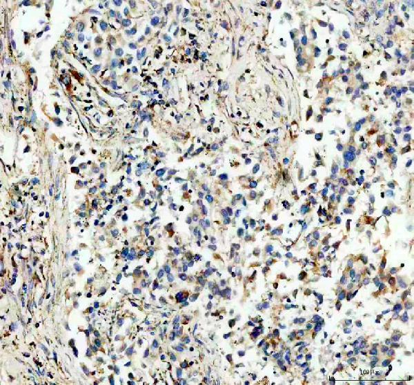
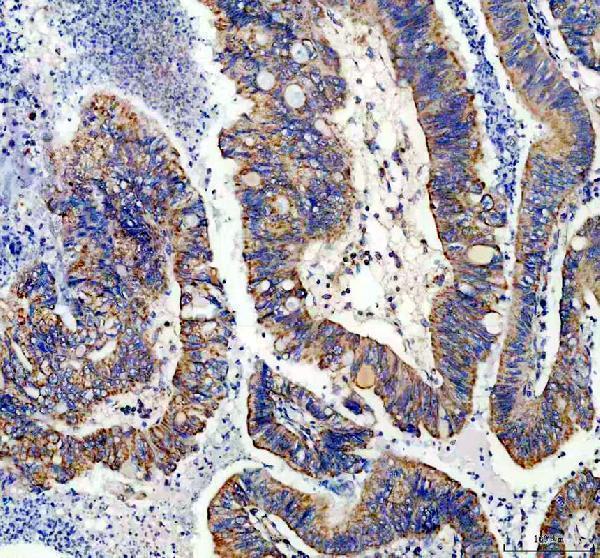
A04920-1 has IHC images from human lung and colon cancer paraffin sections (catalog image captions). The catalog lists human, mouse and rat reactivity, but provides no IF image (catalog reactivity; IF images).

Real IHC data IHC analysis of NDUFS1 using anti-NDUFS1 antibody (A04920-1). NDUFS1 was detected in a paraffin-embedded section of human lung cancer tissue. Heat mediated antigen retrieval was performed in EDTA buffer (pH 8.0, epitope retrieval solution). The tissue section was blocked with 10% goat serum. The tissue section was then incubated with 2 μg/ml rabbit anti-NDUFS1 Antibody (A04920-1) overnight at 4°C. Peroxidase Conjugated Goat Anti-rabbit IgG was used as secondary antibody and incubated for 30 minutes at 37°C. The tissue section was developed using HRP Conjugated Rabbit IgG Super Vision Assay Kit (Catalog # SV0002) with DAB as the chromogen.
Anti-NDUFS1 Antibody ®
Cat # A04920-1

A04920-1 will render with its human lung cancer paraffin-section IHC figure (card image caption). The catalog also reports human colon cancer paraffin-section IHC and lists IHC among its applications, with human, mouse and rat reactivity (catalog image captions; applications; reactivity).

Which to pick: For tissue IHC, choose A04920-1: its rabbit antibody was used at 2 μg/ml on human paraffin sections with EDTA retrieval and DAB detection; the fixative is unreported (catalog image captions). For IF/ICC, this payload offers no validated option because A04920-1 lists no IF application or IF image (catalog applications; IF images). For cross-species work, A04920-1 lists mouse and rat reactivity, but the supplied IHC images document human tissue only (catalog reactivity; image captions).

Each figure is that product's own IHC / IF validation image from its datasheet.

References

  1. UniProt Consortium. UniProt entry P28331 (NDUS1_HUMAN, NADH-ubiquinone oxidoreductase 75 kDa subunit, mitochondrial).
  2. Human Protein Atlas. NDUFS1 tissue IHC expression (reliability: Enhanced).
  3. Human Protein Atlas. NDUFS1 subcellular location (ICC-IF): Localized to the mitochondria..
  4. Human Protein Atlas. NDUFS1 antibody validation summary (2 antibodies).
  5. Proteomic Analysis Identifies NDUFS1 and ATP5O as Novel Markers for Survival Outcome in Prostate Cancer. Cancers 2021 — PMC8656993.
  6. First Experience with Hypothermic Oxygenated Perfusion in Human Uteri: Feasibility and Metabolic Characterization. Journal of clinical medicine 2026 — PMC13116741.
  7. Loss of NDUFS1 promotes gastric cancer progression by activating the mitochondrial ROS-HIF1α-FBLN5 signaling pathway. British journal of cancer 2023 — PMC10575981.
  8. The opposite prognostic effect of NDUFS1 and NDUFS8 in lung cancer reflects the oncojanus role of mitochondrial complex I. Scientific reports 2016 — PMC4981865.
  9. PubMed PMID:1935949 — UniProt-cited evidence.
  10. PubMed PMID:14702039 — UniProt-cited evidence.
  11. PubMed PMID:15815621 — UniProt-cited evidence.