This website uses cookies to ensure you get the best experience on our website.


- Table of Contents
Plan chromogenic NDUFS3 IHC in paraffin sections using the IHC-validated antibody at 2–5 μg/ml (datasheet A05867-2). Compare cytoplasmic staining in high-staining kidney tubules with undetected prostate glandular cells (HPA tissue IHC).

Expected localisation, validated protocols, controls and antibodies — the at-a-glance facts below, then the full design guide.
| Expected localisation | Cytoplasmic staining (HPA tissue IHC); mitochondrial inner membrane (UniProt) | |
| Staining pattern | General cytoplasmic staining across tissues (HPA tissue IHC) | |
| Antigen retrieval | EDTA pH 8.0 HIER, heat-mediated (datasheet A05867-2) | |
| Positive control | Appendix+4 more · see all | |
| Negative control | Oral mucosa+2 more · see all |
| Fixation | Keep fixation consistent across sections. (standard IHC practice; not target-specific) | |
| Caveat | Prostate glandular cells may show no staining (HPA tissue IHC) | |
| Regulation | Expression varies by tissue and cell type (HPA tissue IHC) | |
| Isoform / epitope | 2 isoforms; their epitope coverage is unspecified (UniProt) |
The catalog antibody protocol (datasheet A05867-2) is paired with published NDUFS3 IHC methods for melanoma tissue microarrays (PMC12669717) and formalin-fixed tissue sections (PMC6385215).
| Sample | Paraffin-embedded human breast cancer tissue; fixative not specified (datasheet A05867-2) |
| Fixation | Image fixative and duration unreported (datasheet A05867-2); verify before use. |
| Sectioning | 4–5 µm sections on charged slides (standard) |
| Deparaffinisation | Xylene, graded ethanol series to water (standard) |
| Antigen retrieval | Heat retrieval: EDTA pH 8.0 (datasheet A05867-2); 20 min, 95–100 °C (standard) |
| Peroxidase block | 3% H2O2, 10 min, room temperature (standard) |
| Blocking | 10% goat serum (datasheet A05867-2) |
| Primary antibody | Rabbit anti-NDUFS3, 2-5 μg/ml (datasheet A05867-2) |
| Primary incubation | Overnight at 4 °C (datasheet A05867-2) |
| Detection | HRP-conjugated secondary, DAB chromogen (datasheet A05867-2) |
| Counterstain | Hematoxylin, blue, dehydrate and mount (standard) |
| Expected result | NDUFS3-positive staining in glandular cells of appendix (HPA tissue IHC: High). HPA tissue profile: General cytoplasmic expression. No signal in the no-primary control. |
NDUFS3 is a core Complex I subunit of the mitochondrial inner membrane and has no transmembrane segment (UniProt O75489: function and topology). In paraffin-section IHC, expect cytoplasmic staining in documented positive cells, including cardiomyocytes, kidney tubule cells, and pancreatic exocrine glandular cells (HPA: High in each). HPA describes general cytoplasmic expression and rates its tissue IHC reliability Enhanced, with medium consistency between staining and RNA data (HPA: tissue IHC).
| Granular or finely distributed cytoplasmic chromogen in cardiomyocytes or kidney tubule cells (HPA: High in both). | This fits the expected mitochondrial-associated pattern (UniProt O75489: inner membrane; HPA: mainly mitochondrial in ICC-IF). Routine chromogenic IHC may show cytoplasmic signal without resolving individual mitochondria (general IHC practice). |
| Predominantly nuclear or cell-surface staining, with little cytoplasmic signal. | Treat this as discordant with the principal location (UniProt O75489: inner membrane; HPA: mitochondria supported). HPA also lists nuclear bodies as an uncertain additional ICC-IF location; that observation alone does not establish nuclear IHC as the expected pattern (HPA: subcellular). |
| Strong staining in prostate glandular cells while an adjacent documented positive control stains appropriately (HPA: prostate glandular cells Not detected). | Investigate nonspecific antibody binding or endogenous chromogen activity (general IHC practice). HPA's Not detected call applies to the listed cells and assay observations; it is not proof that every prostate specimen must be negative (HPA: tissue IHC). |
| Uniform color across stroma, empty spaces, and cells, obscuring cell boundaries. | Diffuse background limits interpretation of a cell-specific result (general IHC practice). Check the detection-only control and background-reduction steps before scoring intensity; HPA's cytoplasmic profile cannot validate a uniformly stained section (HPA: tissue IHC). |
| No signal in cardiomyocytes, kidney tubule cells, or pancreatic exocrine glandular cells (HPA: High in each). | A negative run is difficult to interpret until a known-positive section works. Review section integrity, retrieval, antibody application, and detection with matched controls (general IHC practice); HPA's Enhanced rating does not guarantee signal under every local assay condition (HPA: tissue IHC). |
| Compartment and optical resolution (UniProt O75489: inner membrane; HPA: general cytoplasmic IHC profile). | Score the chromogenic pattern at the cytoplasmic level; a paraffin section need not resolve an inner-membrane outline to agree with mitochondrial localization (general IHC practice). |
| Choice of comparison cells (HPA: High in heart cardiomyocytes and kidney tubules; Not detected in oral squamous cells). | Use a documented high-staining cell population to judge assay performance. Interpret a Not detected population as an HPA observation, not a universal biological absence (HPA: tissue IHC). |
| Antibody evidence (HPA: HPA004484 and CAB034894 IHC Enhanced; HPA: tissue IHC reliability Enhanced). | Enhanced supports the reported IHC pattern, while HPA notes only medium consistency with RNA expression. Check the actual antibody's validation before transferring a pattern claim to it (HPA: tissue IHC and antibodies). |
| Protein processing and variants (UniProt O75489: mature chain 37–264; two isoforms). | The record identifies processing and two isoforms, but supplies no antibody epitope or isoform-specific staining result. Do not assign an unexpected IHC pattern to either factor without antibody-specific evidence (UniProt O75489: processing and isoforms). |
| Situation | Likely cause | Next action |
|---|---|---|
| Known-positive cells are blank (HPA: High in cardiomyocytes or kidney tubule cells). | A failed staining run or inadequate assay sensitivity is possible (general IHC practice). | Confirm tissue preservation and detection controls; review the antibody's IHC-P instructions, retrieval, and working dilution as general assay checks. No NDUFS3-specific fixation sensitivity is established here (HPA: tissue IHC; general IHC practice). |
| Chromogen is predominantly nuclear (HPA: mitochondria supported in ICC-IF). | The pattern conflicts with the principal NDUFS3 compartment; HPA calls nuclear bodies an uncertain additional ICC-IF location (HPA: subcellular). | Compare a documented positive cell population and a detection-only control, then assess antibody specificity before calling nuclear IHC positive (HPA: tissue IHC; general IHC practice). |
| A nominally negative cell population stains strongly (HPA: prostate glandular cells Not detected). | Nonspecific binding or endogenous detection activity may contribute (general IHC practice). HPA's negative call is limited to its observed samples and cells (HPA: tissue IHC). | Check a detection-only control and the stained cell identity; compare with a documented positive cell population before interpreting the new signal (general IHC practice; HPA: tissue IHC). |
| Diffuse background obscures cytoplasmic detail. | Excess reagent, incomplete blocking, or endogenous detection activity can obscure localization (general IHC practice). | Assess detection-only and background controls, then optimize blocking, antibody concentration, and washes within the IHC-P workflow (general IHC practice). Score only when cellular signal can be distinguished from background. |
| Different tissues show unequal intensity (HPA: High in pancreas exocrine cells; Low in smooth muscle cells). | HPA reports different staining levels by cell population, so equal intensity is not an expected acceptance criterion (HPA: tissue IHC). | Score identified cell populations separately and compare each with its documented HPA category. Use a high-staining population to assess run performance before interpreting a low-staining one (HPA: tissue IHC; general IHC practice). |
| Q: Should an IF/ICC image match the chromogenic IHC appearance? | HPA supports mitochondria as the main ICC-IF location, while paraffin IHC is described as generally cytoplasmic (HPA: subcellular and tissue IHC). | A: Compare compartment-level localization, allowing for each method's resolution and detection readout (general IHC/IF practice). Treat HPA's nuclear-body and principal-piece ICC-IF locations as uncertain, not required IHC features (HPA: subcellular). |
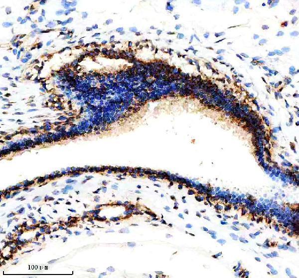
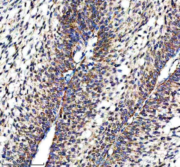
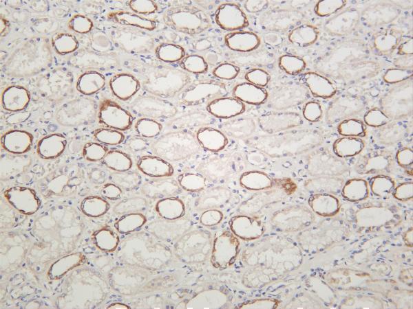


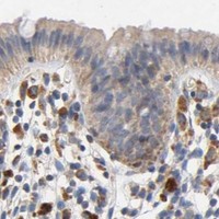


Comprehensive Human Protein Atlas IHC scoring per tissue (reliability: Enhanced — Medium consistency between antibody staining and RNA expression data.). Rows are taken directly from the HPA tissue chart — click any row's HPA link to view the source.
| Tissue | Cell type | Level | Evidence | Source |
|---|---|---|---|---|
| Appendix | Glandular cells | High | Protein (IHC) | HPA → |
| Cerebellum | Cells in granular layer | High | Protein (IHC) | HPA → |
| Cerebral cortex | Neuropil | High | Protein (IHC) | HPA → |
| Duodenum | Glandular cells | High | Protein (IHC) | HPA → |
| Heart muscle | Cardiomyocytes | High | Protein (IHC) | HPA → |
Troubleshoot NDUFS3 chromogenic IHC in paraffin sections by checking retrieval, compartment-specific staining, controls and scoring before interpreting biological differences.
Two anti-NDUFS3 antibodies have IHC images from human and rat tissue (catalog image captions); both list human, mouse, and rat reactivity, and one lists IF (catalog applications/reactivity).
A05867-2 has IHC images of paraffin-embedded human breast cancer, human ovarian cancer, and rat testis sections (A05867-2 image captions). M05867-2 has an IHC image of human kidney and lists IF among its applications; no IF image is supplied (M05867-2 image caption; catalog applications/images).
Which to pick: For paraffin-section IHC, choose A05867-2: its captions document 2 μg/ml antibody after heat retrieval in EDTA at pH 8.0; the fixative is unreported (A05867-2 image captions). For IF, choose M05867-2 (catalog applications); for ICC, M05867 lists that application but has no IHC or IF image in the payload (catalog applications/images). Both rendered SKUs list human, mouse, and rat reactivity, but the supplied IHC images show human and rat tissue for A05867-2 and human tissue for M05867-2 (catalog reactivity; image captions).