NDUFS4 / NADH dehydrogenase [ubiquinone] iron-sulfur protein 4, mitochondrial · IHC design guide

Design Immunohistochemistry for NDUFS4

Plan chromogenic NDUFS4 IHC in paraffin sections with the IHC-validated antibody A03608-3 (datasheet A03608-3). Use the reported mitochondrial tissue pattern and high cardiomyocyte staining to assess results (HPA tissue IHC).

Evidence assembled Oct 2026 · For research use; verify linked source records and product datasheet before use
Immunohistochemistry protocol sheet for NDUFS4 (IHC for NDUFS4): expected localisation Mitochondrial tissue staining (HPA tissue IHC); inner membrane location (UniProt), antibody A03608-3, validated IHC image, and IHC protocol steps
Printable NDUFS4 IHC protocol sheet — expected localisation Mitochondrial tissue staining (HPA tissue IHC); inner membrane location (UniProt), antibody A03608-3, controls and protocol steps. Open the full NDUFS4 IHC guide →

NDUFS4 Immunohistochemistry Experimental Design Guide

Expected localisation, validated protocols, controls and antibodies — the at-a-glance facts below, then the full design guide.

Must know before staining
Expected localisation Mitochondrial tissue staining (HPA tissue IHC); inner membrane location (UniProt)
Staining pattern Widespread mitochondrial staining; high in cardiomyocytes (HPA tissue IHC)
Antigen retrieval EDTA pH 8.0 HIER, heat-mediated (datasheet A03608-3)
Positive control ⓘ Appendix+4 more · see all
Negative control ⓘ Adipose tissue
Important caveats
Reasons your staining may differ from the expected pattern.
Fixation Keep fixation consistent across compared paraffin sections (standard IHC practice). (selected-SKU IHC image A03608-3)
Caveat Adipocytes show no detectable staining (HPA tissue IHC)
Regulation Staining-relevant regulation is not specified (UniProt)
Isoform / epitope No isoforms annotated; mature chain spans 43–175 (UniProt)
Section 1

Recommended NDUFS4 IHC & IF Protocols

The catalog antibody protocol is paired with 3 published NDUFS4 IHC protocols that report usable preparation or staining details (PMC10868282; PMC7248291; PMC2694433).

Recommended immunohistochemistry (IHC-P) protocol parameters
SampleParaffin-embedded human breast cancer tissue; fixative not specified (datasheet A03608-3)
FixationImage fixative and duration unreported (datasheet A03608-3); verify before use.
Sectioning4–5 µm sections on charged slides (standard)
DeparaffinisationXylene, graded ethanol series to water (standard)
Antigen retrievalHeat retrieval: EDTA pH 8.0 (datasheet A03608-3); 20 min, 95–100 °C (standard)
Peroxidase block3% H2O2, 10 min, room temperature (standard)
Blocking10% goat serum (datasheet A03608-3)
Primary antibodyRabbit anti-NDUFS4, 2-5 μg/ml (datasheet A03608-3)
Primary incubationOvernight at 4 °C (datasheet A03608-3)
DetectionHRP-conjugated secondary, DAB chromogen (datasheet A03608-3)
CounterstainHematoxylin, blue, dehydrate and mount (standard)
Expected resultNDUFS4-positive staining in glandular cells of appendix (HPA tissue IHC: High). HPA tissue profile: Ubiquitous mitochondrial expression. No signal in the no-primary control.
💡Decision noteStart with heat-mediated EDTA pH 8.0 retrieval (datasheet: A03608-3); a published thyroid tumour protocol used Tris–EDTA pH 9.0 (PMC2694433).
Section 2

What Is the Expected NDUFS4 Staining Pattern?

NDUFS4 is a complex I subunit of the mitochondrial inner membrane, with no transmembrane segment annotated (UniProt O43181). In paraffin section IHC, expect cytoplasmic staining compatible with mitochondria in many cell types, including cardiomyocytes and glandular cells (HPA: ubiquitous mitochondrial expression; High in heart muscle and appendix). HPA rates its tissue staining Supported, citing consistency with RNA expression (HPA: Supported).

What am I looking at on my slide?
Fine, granular cytoplasmic staining in cardiomyocytes or appendix glandular cells.This fits the expected mitochondrial compartment and HPA High staining in these cells (UniProt O43181; HPA: High in cardiomyocytes and appendix glandular cells). Judge the pattern within intact cells; chromogen intensity alone cannot establish subcellular specificity.
Predominantly nuclear, cell surface, or extracellular staining.This is discordant with the reported mitochondrial location (UniProt O43181; HPA: mitochondria supported by ICC-IF). Consider nonspecific staining or a detection artefact. Check a matched negative control and inspect whether any cytoplasmic granular signal remains.
Strong staining in adipocytes, especially when expected positive cells are faint.HPA reports NDUFS4 as Not detected in adipocytes, while several other cell types stain High (HPA: adipocytes Not detected; cardiomyocytes High). Treat this as a specificity warning, not proof of cross-reactivity: endogenous detection activity or section background could also contribute.
Diffuse chromogen across cells and extracellular spaces.A uniform haze does not resolve the expected mitochondrial distribution (UniProt O43181; HPA: ubiquitous mitochondrial expression). Review the no-primary control for detection background, and assess blocking, washes, and chromogen development as general IHC workflow checks.
No signal in a section containing well-preserved, expected positive cells.A negative result in cardiomyocytes or appendix glandular cells conflicts with their reported High staining (HPA: High in both). First verify tissue preservation and the IHC run with controls; a failed run cannot establish absence of NDUFS4 in the sample.
💡Expected NDUFS4 appearanceCall the result consistent with NDUFS4 when cytoplasmic, mitochondria-compatible granules appear in HPA High cell types such as cardiomyocytes; isolated nuclear or extracellular signal is suspect (UniProt O43181; HPA: High in cardiomyocytes, mitochondrial expression).
How each factor affects the staining
Cell and tissue contextHPA describes low tissue specificity, yet individual cells differ: cardiomyocytes stain High, follicle cells Low, and adipocytes Not detected (HPA: tissue IHC). Compare like cell types when judging intensity.
Subcellular targetNDUFS4 belongs to mitochondrial complex I at the inner membrane (UniProt O43181). An IHC section can support a mitochondria-compatible cytoplasmic pattern, but chromogenic staining alone cannot resolve the inner membrane.
Processing and epitope uncertaintyUniProt annotates a mature chain at residues 43–175 (UniProt O43181). The supplied record gives no antibody epitope, so it cannot predict whether processing changes this antibody's staining.
Antibody evidenceHPA rates tissue IHC Supported and lists HPA003884 as IHC Supported (HPA: tissue reliability; HPA: HPA003884). This supports interpretation of the reported pattern, without proving specificity in every specimen.
IF/ICC Q&A: What localisation should IF/ICC show?Mainly mitochondria (HPA: ICC-IF mitochondria Supported). HPA also reports equatorial segment, perinuclear theca, and calyx localisation (HPA: ICC-IF additional locations Approved). Those observations do not supply an IF/ICC protocol here.
Why is my staining missing, weak or wrong?
SituationLikely causeNext action
Expected positive cells show no chromogen.The IHC run or antibody detection may have failed; HPA reports High staining in cardiomyocytes and appendix glandular cells (HPA: tissue IHC).Inspect a known-positive section and run controls. As general IHC practice, review retrieval and antibody incubation before interpreting a sample as negative.
Most cells show flat, widespread colour.Diffuse background can obscure a mitochondrial pattern (UniProt O43181; HPA: ubiquitous mitochondrial expression). Excess detection signal or incomplete washing are general IHC possibilities.Compare with a no-primary control; review blocking, washes, and detection development. Look again for cell-associated granular staining after background is reduced.
Signal is mainly nuclear or extracellular.That distribution disagrees with the mitochondrial location (UniProt O43181; HPA: ICC-IF mitochondria Supported). It may reflect nonspecific binding or detection artefact.Compare the no-primary control and a known-positive section; score the result as uncertain until the expected cytoplasmic distribution is demonstrated.
Adipocytes stain strongly.HPA reports adipocytes as Not detected (HPA: adipocytes Not detected). Cross-reactivity or endogenous detection activity is possible, but the staining alone cannot distinguish them.Check a no-primary control and review the detection chemistry. Compare nearby expected positive cells before treating adipocyte colour as target signal.
Positive cells vary markedly between tissues.That can reflect reported cell-level variation: cardiomyocytes are High, while skeletal muscle myocytes are Low (HPA: tissue IHC). Differences alone do not establish assay failure.Compare each cell type with its own HPA expectation and use the same scoring criteria across sections; avoid requiring equal intensity in different tissues.
A granular result seems positive but its compartment is unclear.Chromogenic IHC has limited subcellular resolution; NDUFS4 is assigned to the mitochondrial inner membrane (UniProt O43181).Record it as mitochondria-compatible cytoplasmic staining when controls support it. Use the separate IF/ICC guide for a localisation experiment (HPA: ICC-IF mitochondria Supported).

Sample controls for NDUFS4 IHC & IF

🧪Run appendix first: glandular cells should show strong staining (HPA: High in appendix glandular cells). Use adipose tissue as the negative tissue (HPA: Not detected in adipocytes); on the appendix slide, internal negative cells should show counterstain without the granular cytoplasmic signal, but do not assume every nonglandular cell is negative.
Positive control tissue: Appendix (Glandular cells, HPA High)
Negative control tissue: Adipose tissue (HPA Not detected)
ICC-IF cell lines (HPA subcellular resource): HPA ICC-IF images show NDUFS4 in A-431, U-251MG, U2OS, Sperm, KOLF2.1J, with annotated localisation: Mitochondria (supported) (HPA subcellular).
Technical controls: Include a no-primary, secondary-only control; a concentration-matched rabbit IgG isotype control (selected-SKU caption: rabbit primary antibody); and matched NDUFS4-knockout material as a biological negative. Quench endogenous peroxidase and check for background DAB staining in the appendix section (standard chromogenic IHC practice).
⚠️Feasibility: A target-specific fixation window or fixation effect is unreported, and the fixative in the selected-SKU paraffin-section caption is unreported; paraffin embedding alone does not establish fixation. The reported IHC procedure uses heat retrieval in EDTA at pH 8.0, but retrieval dependence is untested (selected-SKU caption). No matched evidence establishes that frozen sections or IF are easier; for appendix, assess luminal debris separately from cellular staining because trapped chromogen can resemble a positive signal (standard IHC practice). The selected A03608-3 tissue-IHC caption documents paraffin sections, but does not specify the fixative (selected-SKU IHC image A03608-3).

HPA tissue IHC evidence for NDUFS4

Comprehensive Human Protein Atlas IHC scoring per tissue (reliability: Supported — High consistency between antibody staining and RNA expression data.). Rows are taken directly from the HPA tissue chart — click any row's HPA link to view the source.

Positive expression · recommended positive controls

TissueCell typeLevelEvidenceSource
Appendix Glandular cells High Protein (IHC) HPA →
Bronchus Respiratory epithelial cells High Protein (IHC) HPA →
Caudate Neuronal cells High Protein (IHC) HPA →
Cerebellum Purkinje cells High Protein (IHC) HPA →
Endometrium Cells in endometrial stroma High Protein (IHC) HPA →

Undetected expression · recommended negative controls

TissueCell typeLevelEvidenceSource
Adipose tissue Adipocytes Not detected Protein (IHC) HPA →
Section 3

Advanced NDUFS4 IHC Tips

Use compartment, cell type, and matched controls to troubleshoot NDUFS4 staining in paraffin sections; assess IF separately.

Which retrieval condition should I start with for weak NDUFS4 staining in paraffin sections?
Start with heat-mediated antigen retrieval in EDTA at pH 8.0 for paraffin sections stained with the catalog antibody (datasheet A03608-3). The tissue-IHC caption specifies that buffer but gives no heating temperature or duration, so record both and optimize them with a consistently processed reference section (datasheet A03608-3; standard IHC practice). If staining remains weak, adjust heating duration incrementally while checking tissue morphology and a no-primary control (standard IHC practice). Keep the documented primary concentration of 2 μg/ml and overnight incubation at 4°C initially, so retrieval changes can be judged independently (datasheet A03608-3; standard IHC practice).
How can I troubleshoot variable NDUFS4 staining across differently fixed specimens?
The catalog image identifies a paraffin section but does not report its fixative, so NDUFS4 sensitivity to a particular fixation condition is unknown here (datasheet A03608-3). Record fixative, fixation duration, processing history, and section age for each specimen, then compare sections processed alike before changing antibody conditions (standard IHC practice). Begin those comparisons with EDTA retrieval at pH 8.0 and 2 μg/ml primary antibody overnight at 4°C (datasheet A03608-3). Include a consistently processed reference section in each run and assess morphology alongside staining, because differences between specimens cannot establish a target-specific fixation effect on their own (standard IHC practice).
What staining pattern supports NDUFS4 localisation in tissue IHC?
NDUFS4 is assigned to the mitochondrial inner membrane, with no annotated transmembrane segment (UniProt O43181). In chromogenic sections, assess whether cytoplasmic staining has a fine granular distribution compatible with mitochondria, while recognizing that DAB alone cannot resolve the inner membrane (UniProt O43181; standard IHC practice). Compare the candidate pattern with well-preserved cells and a no-primary control, especially when diffuse nuclear or extracellular colour dominates (standard IHC practice). High staining is reported in heart cardiomyocytes and cerebellar Purkinje cells, whereas adipocytes are reported as not detected; these are useful contextual comparators when those tissues are available (HPA: tissue IHC).
Could processing or epitope accessibility explain inconsistent NDUFS4 staining?
The record annotates one mature chain spanning residues 43–175, no isoforms, and phosphorylation at Ser173 (UniProt O43181). An antibody epitope is not supplied here, so do not attribute weak tissue staining to cleavage or phosphorylation without epitope mapping or direct validation (UniProt O43181; standard IHC practice). Compare sections processed together using the documented EDTA retrieval at pH 8.0, then vary one retrieval condition at a time while retaining morphology controls (datasheet A03608-3; standard IHC practice). If the epitope is later mapped near either annotated site, evaluate that possibility with an independently validated reagent or orthogonal assay before interpreting staining differences as biology (UniProt O43181; standard IHC practice).
How should I design a complementary NDUFS4 immunofluorescence check?
Use IF as a complementary localisation check: the supported main subcellular location is mitochondria (HPA: subcellular). Multiplex NDUFS4 with a validated mitochondrial marker and, where relevant, a marker identifying the expected cell type; choose fluorophores and image controls after checking the specimen’s autofluorescence in each channel (HPA: subcellular; standard IF practice). Because the antibody epitope and its membrane-facing side are unspecified, compare mild and stronger permeabilisation conditions empirically instead of assuming access from the absence of an annotated transmembrane segment (UniProt O43181; standard IF practice). Keep the IHC condition of EDTA pH 8.0 and 2 μg/ml specific to the documented tissue-IHC experiment; optimize IF conditions independently (datasheet A03608-3; standard IF practice).
How can I separate NDUFS4 signal from chromogenic background?
Start with a no-primary control, matched tissue processing, and inspection of staining at section edges and damaged areas (standard IHC practice). For an HRP–DAB workflow, check endogenous peroxidase blocking and shorten development if the control also acquires colour; these are general detection checks, not NDUFS4-specific findings (standard IHC practice). The catalog example used 10% goat serum blocking, 2 μg/ml primary antibody overnight at 4°C, and a secondary incubation of 30 minutes at 37°C (datasheet A03608-3). If background persists, change one variable at a time while preserving a reference section and looking for the expected mitochondrial-pattern cytoplasmic signal (UniProt O43181; standard IHC practice).
How should I quantify NDUFS4 staining across tissue samples? ⚠ ANSWER MARKED FOR VERIFICATION
Define the cell population and tissue region before scoring, and exclude folds, necrosis, and section edges consistently (standard IHC practice). For cellular staining, report an H-score from intensity categories 0–3 weighted by the percentage of cells in each category, or report the percentage of positive cells using a fixed threshold (standard IHC practice). Normalize to the number of eligible cells or the viable tissue area, and retain the same staining and imaging settings across comparisons (standard IHC practice). Because HPA describes broadly distributed mitochondrial expression with cell-type differences, report results by cell type rather than treating one whole-section average as a universal NDUFS4 level (HPA: tissue IHC; standard IHC practice).
When should an apparent NDUFS4-positive section be considered suspect?
A convincing result should favour a mitochondrial-compatible cytoplasmic pattern in intact cells, consistent with NDUFS4’s inner-membrane assignment (UniProt O43181; standard IHC practice). Treat dominant nuclear, extracellular, edge-restricted, or necrotic staining as suspect and compare it with a no-primary control (standard IHC practice). Consider cell identity: HPA reports high staining in cardiomyocytes and Purkinje cells but no detection in adipocytes, although a single section should not be judged solely against those examples (HPA: tissue IHC; standard IHC practice). If colour persists without primary antibody, investigate endogenous enzyme activity or detection background before scoring; the documented assay used HRP with DAB (datasheet A03608-3; standard IHC practice).
Boster reagents

Best NDUFS4 / NADH dehydrogenase [ubiquinone] iron-sulfur protein 4, mitochondrial IHC Antibodies

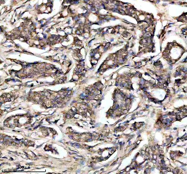
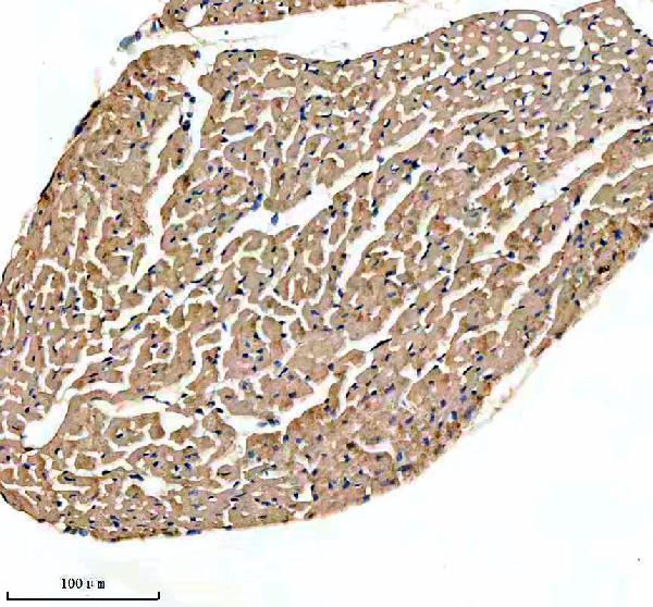
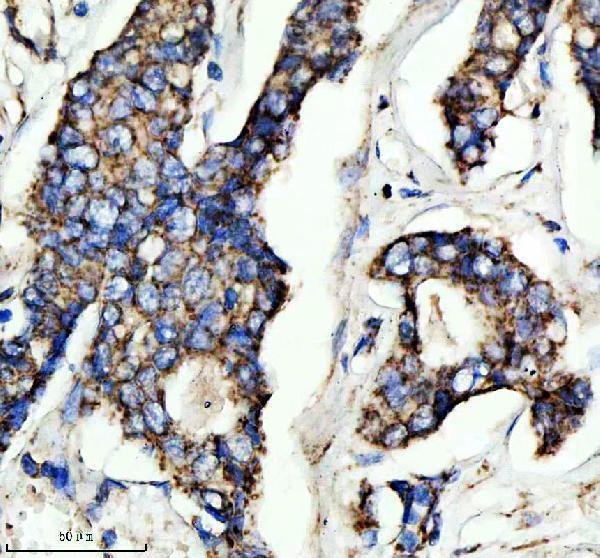
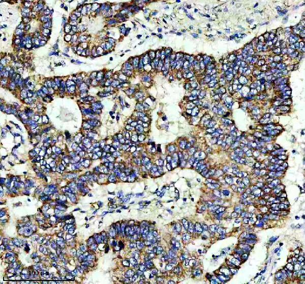
Catalog anti-NDUFS4 antibodies have paraffin-section IHC images from human breast, stomach, and lung cancer tissue and mouse and rat heart (catalog IHC captions); one is listed for IF/ICC (catalog applications).

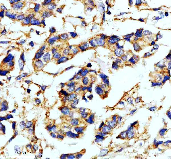
Real IHC data IHC analysis of NDUFS4 using anti-NDUFS4 antibody (A03608-3). NDUFS4 was detected in a paraffin-embedded section of human breast cancer tissue. Heat mediated antigen retrieval was performed in EDTA buffer (pH 8.0, epitope retrieval solution). The tissue section was blocked with 10% goat serum. The tissue section was then incubated with 2 μg/ml rabbit anti-NDUFS4 Antibody (A03608-3) overnight at 4°C. Peroxidase Conjugated Goat Anti-rabbit IgG was used as secondary antibody and incubated for 30 minutes at 37°C. The tissue section was developed using HRP Conjugated Rabbit IgG Super Vision Assay Kit (Catalog # SV0002) with DAB as the chromogen.
Anti-NDUFS4 Antibody ®
Cat # A03608-3
Real IHC data IHC analysis of NDUFS4 using anti-NDUFS4 antibody (M03608-2). NDUFS4 was detected in a paraffin-embedded section of human breast cancer tissue. Heat mediated antigen retrieval was performed in EDTA buffer (pH 8.0, epitope retrieval solution). The tissue section was blocked with 10% goat serum. The tissue section was then incubated with 1:50 rabbit anti-NDUFS4 Antibody (M03608-2) overnight at 4°C. Peroxidase Conjugated Goat Anti-rabbit IgG was used as secondary antibody and incubated for 30 minutes at 37°C. The tissue section was developed using HRP Conjugated Rabbit IgG Super Vision Assay Kit (Catalog # SV0002) with DAB as the chromogen.
Anti-NDUFS4 Rabbit Monoclonal Antibody
Cat # M03608-2
Real IHC data IHC analysis of NDUFS4 using anti-NDUFS4 antibody (M03608-1). NDUFS4 was detected in a paraffin-embedded section of human lung cancer tissue. Heat mediated antigen retrieval was performed in EDTA buffer (pH 8.0, epitope retrieval solution). The tissue section was blocked with 10% goat serum. The tissue section was then incubated with 1:50 rabbit anti-NDUFS4 Antibody (M03608-1) overnight at 4°C. Peroxidase Conjugated Goat Anti-rabbit IgG was used as secondary antibody and incubated for 30 minutes at 37°C. The tissue section was developed using HRP Conjugated Rabbit IgG Super Vision Assay Kit (Catalog # SV0002) with DAB as the chromogen.
Anti-Ndufs4 Rabbit Monoclonal Antibody
Cat # M03608-1

A03608-3 has IHC images from human breast and stomach cancer tissue and mouse and rat heart; M03608-2 has IHC images from those same sample types (each SKU’s IHC captions). M03608-1 has an IHC image from human lung cancer tissue (M03608-1 IHC caption).

Which to pick: For paraffin-section IHC, choose A03608-3 at 2–5 μg/ml or monoclonal M03608-2 at 1:50; each has its own EDTA pH 8.0 retrieval and DAB image caption (catalog dilutions and each SKU’s IHC caption). Choose M03608-2 for IF/ICC because that application is listed at 1:50, although the payload provides no IF image (M03608-2 applications and IF dilution; catalog IF image alts). For cross-species IHC, A03608-3 and M03608-2 each have human, mouse, and rat images; their captions specify paraffin sections but do not report the fixative (each SKU’s IHC captions).

Each figure is that product's own IHC / IF validation image from its datasheet.

References

  1. UniProt Consortium. UniProt entry O43181 (NDUS4_HUMAN, NADH dehydrogenase [ubiquinone] iron-sulfur protein 4, mitochondrial).
  2. Human Protein Atlas. NDUFS4 tissue IHC expression (reliability: Supported).
  3. Human Protein Atlas. NDUFS4 subcellular location (ICC-IF): Mainly localized to the mitochondria. In addition localized to the equatorial segment, perinuclear theca and calyx..
  4. Human Protein Atlas. NDUFS4 antibody validation summary (1 antibodies).
  5. Targeting oxidative phosphorylation to increase the efficacy of immune-combination therapy in renal cell carcinoma. Journal for immunotherapy of cancer 2024 — PMC10868282.
  6. Papillary thyroid carcinoma tall cell variant shares accumulation of mitochondria, mitochondrial DNA mutations, and loss of oxidative phosphorylation complex I integrity with oncocytic tumors. The journal of pathology. Clinical research 2022 — PMC8822387.
  7. A Single Intravenous Injection of AAV-PHP.B-hNDUFS4 Ameliorates the Phenotype of Ndufs4 (-/-) Mice. Molecular therapy. Methods & clinical development 2020 — PMC7248291.
  8. Lack of complex I is associated with oncocytic thyroid tumours. British journal of cancer 2009 — PMC2694433.
  9. PubMed PMID:9463323 — UniProt-cited evidence.
  10. PubMed PMID:15489334 — UniProt-cited evidence.
  11. PubMed PMID:11181577 — UniProt-cited evidence.