This website uses cookies to ensure you get the best experience on our website.


- Table of Contents
Plan chromogenic IHC in paraffin sections with the IHC-validated antibody (datasheet A09082-2). Use glandular cells as positive controls and assess granular cytoplasmic staining (HPA tissue IHC), while treating the mitochondrial inner-membrane location as a molecular annotation (UniProt).

Expected localisation, validated protocols, controls and antibodies — the at-a-glance facts below, then the full design guide.
| Expected localisation | Tissue: granular cytoplasm (HPA tissue IHC); protein: mitochondrial inner membrane (UniProt) | |
| Staining pattern | General granular cytoplasmic staining (HPA tissue IHC) | |
| Antigen retrieval | EDTA pH 8.0 HIER, heat-mediated (datasheet A09082-2) | |
| Positive control | Adrenal gland+4 more · see all | |
| Negative control | Adipose tissue+1 more · see all |
| Fixation | Keep fixation consistent across sections. (standard IHC practice; not target-specific) | |
| Caveat | Adipocytes are unstained despite broad tissue expression (HPA tissue IHC) | |
| Regulation | Tissue-enhanced RNA in skeletal muscle (HPA tissue RNA) | |
| Isoform / epitope | No isoforms annotated; mature chain spans 29–124 (UniProt) |
The catalog antibody’s IHC-P protocol is paired with four published NDUFS6 protocols for ovarian, neuroblastoma, cardiac, and other paraffin-embedded tissue sections (datasheet A09082-2; PMC12635173; PMC13263444; PMC12095822; PMC12148850).
| Sample | Paraffin-embedded human breast cancer tissue; fixative not specified (datasheet A09082-2) |
| Fixation | Image fixative and duration unreported (datasheet A09082-2); verify before use. |
| Sectioning | 4–5 µm sections on charged slides (standard) |
| Deparaffinisation | Xylene, graded ethanol series to water (standard) |
| Antigen retrieval | Heat retrieval: EDTA pH 8.0 (datasheet A09082-2); 20 min, 95–100 °C (standard) |
| Peroxidase block | 3% H2O2, 10 min, room temperature (standard) |
| Blocking | 10% goat serum (datasheet A09082-2) |
| Primary antibody | Rabbit anti-NDUFS6, 2-5 μg/ml (datasheet A09082-2) |
| Primary incubation | Overnight at 4 °C (datasheet A09082-2) |
| Detection | HRP-conjugated secondary, DAB chromogen (datasheet A09082-2) |
| Counterstain | Hematoxylin, blue, dehydrate and mount (standard) |
| Expected result | NDUFS6-positive staining in glandular cells of adrenal gland (HPA tissue IHC: High). HPA tissue profile: General cytoplasmic expression with a granular pattern. No signal in the no-primary control. |
NDUFS6 is a mitochondrial inner-membrane Complex I subunit with no predicted transmembrane segment (UniProt O75380 topology). In paraffin-section IHC, expect granular cytoplasmic staining in cells such as colon glandular cells, bronchial respiratory epithelial cells, and cerebral cortical neurons (HPA: tissue IHC). HPA describes the overall pattern as general granular cytoplasmic expression and rates its tissue IHC evidence Supported, with medium consistency between staining and RNA data (HPA: tissue IHC).
| Granular cytoplasmic stain in colon glandular cells or cerebral cortical neurons (HPA: High in both). | This fits the reported tissue pattern (HPA: granular cytoplasm) and mitochondrial location (UniProt O75380). Assess the named cells rather than treating every cell in the section as an equally strong positive control (HPA: cell-specific tissue IHC). |
| Predominantly nuclear, surface, or extracellular stain in otherwise positive cells. | That distribution does not fit the granular cytoplasmic IHC profile (HPA: tissue IHC) or inner-membrane location (UniProt O75380). Treat it as suspect staining; inspect morphology, detection controls, and the antibody conditions before scoring it as NDUFS6 (standard IHC practice). |
| Strong stain in adipocytes or parathyroid glandular cells (HPA: Not detected in both). | This conflicts with the reported cell-level negatives (HPA: tissue IHC). Cross-reactivity or endogenous detection activity is possible; compare a no-primary control and the positive tissue processed in the same run before assigning a cause (standard IHC practice). |
| Uniform haze across tissue, stroma, and cells, obscuring granules. | A diffuse field is hard to reconcile with the reported granular cytoplasmic profile (HPA: tissue IHC). Review background controls, blocking, washes, and chromogen development; broad color alone cannot establish target localisation (standard IHC practice). |
| No stain in colon glandular cells or bronchial respiratory epithelial cells (HPA: High in both). | The run lacks its expected positive readout (HPA: tissue IHC). Check tissue identity and cell preservation, then review antibody dilution, retrieval, and detection controls before calling the sample negative (standard IHC practice). HPA's Supported rating is not a guarantee for every section (HPA: reliability). |
| Cell-level tissue contrast | HPA reports High staining in adrenal, appendix, breast, colon, and duodenum glandular cells, but Not detected in parathyroid glandular cells (HPA: tissue IHC). Choose and score controls by the specified cell population, not by the organ name alone (standard IHC practice). |
| Protein location and processing | NDUFS6 resides at the mitochondrial inner membrane, has no annotated transmembrane segment, and has a mature chain at residues 29–124 (UniProt O75380). These annotations support a cytoplasmic, organelle-associated reading; they do not identify this antibody's epitope or prescribe retrieval conditions (UniProt O75380; standard IHC practice). |
| Strength of IHC evidence | The HPA tissue profile is Supported with medium staining–RNA consistency; HPA064221 is listed as IHC Supported (HPA: tissue IHC; HPA: antibody validation). Use the observed pattern as a reference and retain run controls when judging an unexpected result (standard IHC practice). |
| IF evidence boundary | HPA gives no main ICC-IF location and lists no cell lines with ICC-IF images; HPA064221 has no ICC status in the supplied record (HPA: subcellular; HPA: antibody validation). Can this establish an IF pattern? No: assess IF on its own guide page, without treating the IHC pattern as ICC-IF validation. |
| Situation | Likely cause | Next action |
|---|---|---|
| Known-positive cells remain unstained. | The run may have a tissue, retrieval, antibody, or detection problem; colon glandular cells are reported High (HPA: tissue IHC; standard IHC practice). | Confirm the expected cells are present, then review retrieval, dilution, reagent performance, and a run-matched positive control (standard IHC practice). No NDUFS6-specific retrieval setting is supplied. |
| The entire section develops brown color. | Broad color may reflect endogenous peroxidase activity or detection background rather than granular cytoplasmic NDUFS6 (standard IHC practice; HPA: tissue IHC). | Compare no-primary and detection controls; review peroxidase blocking, washes, and development time before interpreting cell-level stain (standard IHC practice). |
| Adipocytes or parathyroid glandular cells stain strongly. | Those populations are reported Not detected (HPA: tissue IHC); cross-reactivity or detection background is possible (standard IHC practice). | Inspect a no-primary control and an HPA High population from the same run; score only a credible granular cytoplasmic pattern (HPA: tissue IHC; standard IHC practice). |
| Nuclear staining dominates a positive tissue. | A nuclear pattern disagrees with granular cytoplasmic IHC and mitochondrial localisation (HPA: tissue IHC; UniProt O75380). | Check counterstain separation and no-primary controls, then reassess antibody and detection conditions before assigning target-specific signal (standard IHC practice). |
| Positive tissue shows weak, patchy granules. | HPA assigns High staining to particular cells, not every cell in an organ; uneven section quality or run performance can complicate scoring (HPA: tissue IHC; standard IHC practice). | Score the named cell population, compare a run-matched positive control, and inspect tissue preservation and detection consistency (standard IHC practice). |
| An IF image is requested as proof of the IHC result. | The supplied HPA subcellular record has no ICC-IF images or assigned main location, and the antibody has no listed ICC status (HPA: subcellular; HPA: antibody validation). | Use the IHC tissue pattern and IHC controls for this callout (HPA: tissue IHC; standard IHC practice). Evaluate IF separately on its own guide page; this record supplies no ICC-IF validation. |
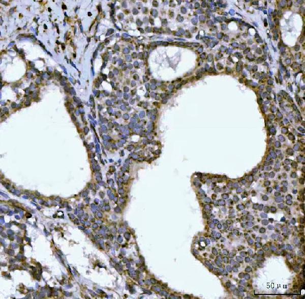
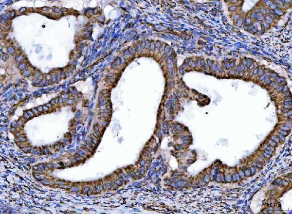


Comprehensive Human Protein Atlas IHC scoring per tissue (reliability: Supported — Medium consistency between antibody staining and RNA expression data.). Rows are taken directly from the HPA tissue chart — click any row's HPA link to view the source.
| Tissue | Cell type | Level | Evidence | Source |
|---|---|---|---|---|
| Adrenal gland | Glandular cells | High | Protein (IHC) | HPA → |
| Appendix | Glandular cells | High | Protein (IHC) | HPA → |
| Breast | Glandular cells | High | Protein (IHC) | HPA → |
| Bronchus | Respiratory epithelial cells | High | Protein (IHC) | HPA → |
| Cerebellum | Cells in granular layer | High | Protein (IHC) | HPA → |
Use the catalog antibody’s paraffin-section IHC conditions as the starting point, then assess granular cytoplasmic staining against tissue controls and morphology.
A09082-2 has paraffin-section IHC images from human breast and ovarian cancer tissue and mouse heart (catalog image captions). No IF image is supplied (catalog payload).
A09082-2 is listed for IHC in human and mouse samples (catalog: IHC dilution listing), with images from paraffin sections of human breast cancer, human ovarian cancer, and mouse heart (catalog image captions). Its general reactivity list also includes rat, but the supplied IHC images do not show rat tissue (catalog: reactivity list; image captions).
Which to pick: For tissue IHC, choose A09082-2 at the listed 2–5 μg/ml range (datasheet: IHC dilution); its captions document paraffin sections with EDTA retrieval, while the fixative is unreported (catalog image captions). No SKU in this payload is validated for IF/ICC, so there is no IF/ICC pick (catalog: application list; IF image alts). For cross-species planning, A09082-2 lists human, mouse, and rat reactivity, with IHC image evidence for human and mouse only; its clone is unreported.