NEBL / Nebulette · IHC design guide

Design Immunohistochemistry for NEBL

Use heart muscle cardiomyocytes as a high-staining reference for NEBL paraffin IHC (HPA tissue IHC). This guide covers the catalog antibody's paraffin IHC conditions (datasheet A08817-2) and interpretation of cytoplasmic staining (HPA tissue IHC).

Evidence assembled Oct 2026 · For research use; verify linked source records and product datasheet before use
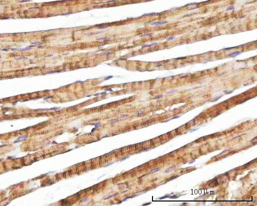
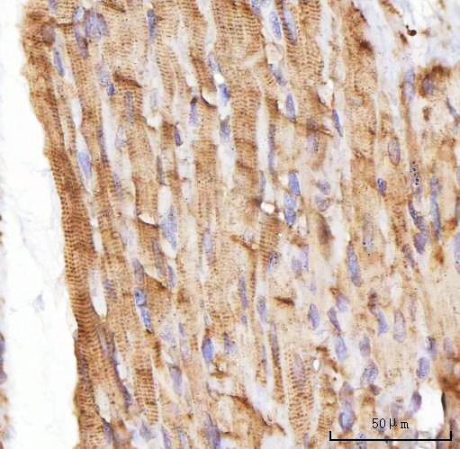
Immunohistochemistry protocol sheet for NEBL (IHC for NEBL): expected localisation Cytoplasmic staining, most abundant in heart muscle (HPA tissue IHC), antibody A08817-2, validated IHC image, and IHC protocol steps
Printable NEBL IHC protocol sheet — expected localisation Cytoplasmic staining, most abundant in heart muscle (HPA tissue IHC), antibody A08817-2, controls and protocol steps. Open the full NEBL IHC guide →

NEBL Immunohistochemistry Experimental Design Guide

Expected localisation, validated protocols, controls and antibodies — the at-a-glance facts below, then the full design guide.

Must know before staining
Expected localisation Cytoplasmic staining, most abundant in heart muscle (HPA tissue IHC)
Staining pattern High cytoplasmic staining in cardiomyocytes (HPA tissue IHC)
Antigen retrieval EDTA pH 8.0 HIER, heat-mediated (datasheet A08817-2)
Positive control ⓘ Heart muscle+4 more · see all
Negative control ⓘ Adipose tissue+4 more · see all
Important caveats
Reasons your staining may differ from the expected pattern.
Fixation Keep fixation consistent across sections (standard IHC practice; not target-specific)
Caveat Antibody staining and RNA expression have low consistency (HPA tissue IHC)
Regulation Abundant in cardiac muscle (UniProt)
Isoform / epitope 2 isoforms; epitope coverage is unspecified (UniProt)
Section 1

Recommended NEBL IHC & IF Protocols

The catalog IHC-P protocol (datasheet A08817-2) is accompanied by published NEBL IHC methods using paraffin sections, a tissue microarray, and frozen cardiac muscle sections (PMC10879697; PMC9310551; PMC3683841).

Recommended immunohistochemistry (IHC-P) protocol parameters
SampleParaffin-embedded mouse heart tissue; fixative not specified (datasheet A08817-2)
FixationImage fixative and duration unreported (datasheet A08817-2); verify before use.
Sectioning4–5 µm sections on charged slides (standard)
DeparaffinisationXylene, graded ethanol series to water (standard)
Antigen retrievalHeat retrieval: EDTA pH 8.0 (datasheet A08817-2); 20 min, 95–100 °C (standard)
Peroxidase block3% H2O2, 10 min, room temperature (standard)
Blocking10% goat serum (datasheet A08817-2)
Primary antibodyRabbit anti-NEBL, 2-5 μg/ml (datasheet A08817-2)
Primary incubationOvernight at 4 °C (datasheet A08817-2)
DetectionHRP-conjugated secondary, DAB chromogen (datasheet A08817-2)
CounterstainHematoxylin, blue, dehydrate and mount (standard)
Expected resultNEBL-positive staining in cardiomyocytes of heart muscle (HPA tissue IHC: High). HPA tissue profile: Cytoplasmic expression in several different tissues, most abundant in heart muscle. No signal in the no-primary control.
💡Decision noteStart with heat retrieval in EDTA pH 8.0 (datasheet A08817-2). A published paraffin-section method used buffered citrate at 100°C for 15 min (PMC10879697).
Section 2

What Is the Expected NEBL Staining Pattern?

NEBL is a cytoplasmic actin-binding protein that localizes to Z-lines in cardiac cells (UniProt O76041). In paraffin-section IHC, expect the strongest staining in cardiomyocytes, with high staining also reported in spermatogonia and medium staining in several glandular cell populations (HPA tissue IHC). Interpret those additional sites cautiously: HPA rates its tissue IHC “Approved” but reports low consistency between antibody staining and RNA expression (HPA tissue IHC). NEBL has no transmembrane segment (UniProt O76041 topology).

What am I looking at on my slide?
Strong cytoplasmic staining in cardiomyocytes, potentially with a striated appearance.This fits the high cardiomyocyte signal reported by HPA and NEBL localization to cardiac Z-lines (HPA tissue IHC; UniProt O76041). A visible striated pattern can support interpretation, but routine chromogenic sections may not resolve individual Z-lines (general IHC practice).
Predominantly nuclear or crisp cell-surface staining in cardiomyocytes.These compartments conflict with the reported cytoplasmic localization and lack of a transmembrane segment (UniProt O76041; UniProt O76041 topology). Treat such signal as suspect until controls and a repeat stain support it (general IHC practice).
Strong staining in adipocytes or bone-marrow hematopoietic cells.HPA reports NEBL as not detected in those cell populations (HPA tissue IHC). Unexpected signal may reflect cross-reactivity or endogenous detection activity; compare with a no-primary control and the cardiomyocyte positive control (general IHC practice).
Diffuse color across stroma, empty spaces, and multiple cell types.A pattern that does not follow cell boundaries is difficult to score as NEBL expression (general IHC practice). Broad background may arise from nonspecific reagent binding, residual endogenous enzyme activity, or excessive chromogen development (general IHC practice).
No cardiomyocyte signal in a heart-muscle control.This conflicts with HPA's high cardiomyocyte staining and UniProt's abundant cardiac-muscle expression (HPA tissue IHC; UniProt O76041). Check the staining run before interpreting a test specimen as negative; one failed control cannot establish absent NEBL (general IHC practice).
💡Expected NEBL appearanceCall a section positive when cardiomyocytes show clearly above-background cytoplasmic staining, often strong in heart muscle; isolated nuclear, cell-surface, or acellular color is suspect (HPA tissue IHC; UniProt O76041; general IHC practice).
How each factor affects the staining
Tissue and cell contextHeart-muscle cardiomyocytes are high, while spermatogonia are also high and several glandular populations are medium (HPA tissue IHC). Do not classify every noncardiac positive cell as an artefact; assess its specific HPA cell-level entry (HPA tissue IHC).
Antibody evidenceTwo listed rabbit polyclonal antibodies, HPA013994 and HPA013995, have IHC status “Approved,” with no ICC status supplied (HPA antibodies). The tissue profile carries a low staining-to-RNA consistency caveat, so a positive chromogenic result needs controls (HPA tissue IHC; general IHC practice).
Isoform and compartmentNEBL has two isoforms; isoform 2 is reported in non-muscle cells, including fibroblasts (UniProt O76041). Both the UniProt cytoplasmic annotation and HPA's tissue-wide cytoplasmic profile guide compartment scoring; this record does not establish isoform-specific IHC staining (UniProt O76041; HPA tissue IHC).
IF/ICC interpretation?HPA supplies no main ICC-IF location or imaged cell lines, and the listed antibodies have no ICC status (HPA subcellular; HPA antibodies). For IF/ICC, use its own guide and validate the compartmental pattern there; this IHC section provides no IF protocol (general IHC practice).
Why is my staining missing, weak or wrong?
SituationLikely causeNext action
Heart-muscle control is blank.A failed IHC run, unsuitable antibody conditions, or ineffective detection can erase an expected high signal (HPA tissue IHC; general IHC practice).Confirm that control tissue is present and staining reagents worked; review the catalog antibody's IHC-P instructions and repeat with an appropriate positive control (general IHC practice). Avoid assigning a biological negative from this run.
Heart-muscle signal is faint or uneven.Section quality, inconsistent reagent coverage, or suboptimal retrieval can affect routine paraffin IHC (general IHC practice). No NEBL-specific fixation or retrieval sensitivity is established by the supplied sources.Check section integrity and reagent coverage, then optimize retrieval and antibody dilution within the catalog IHC-P guidance using the same heart-muscle control (general IHC practice). Compare runs at matched development times.
Strong nuclear or membrane-edge color dominates.That pattern disagrees with NEBL's cytoplasmic location and absence of a transmembrane segment (UniProt O76041; UniProt O76041 topology). Nonspecific staining or detection artefact is possible (general IHC practice).Review a no-primary control, inspect counterstain and chromogen separately, and repeat with the IHC-validated antibody under controlled detection conditions (general IHC practice). Score only reproducible, cell-associated cytoplasmic signal.
Adipocytes or marrow hematopoietic cells stain strongly.HPA lists those cell populations as not detected (HPA tissue IHC). Cross-reactivity or endogenous enzyme activity can mimic a positive chromogenic stain (general IHC practice).Compare a no-primary control and an appropriate detection-system control; confirm endogenous-activity blocking and assess the suspect cells alongside heart-muscle controls (general IHC practice). Keep unexpected staining separate from the NEBL score.
Color is widespread, including spaces without cells.Diffuse deposition is more consistent with assay background than the cytoplasmic, cell-associated NEBL pattern (UniProt O76041; HPA tissue IHC; general IHC practice).Inspect control slides, blocking, washes, antibody concentration, and chromogen development; adjust one general IHC variable at a time while retaining the positive control (general IHC practice).
A noncardiac tissue is positive despite heart-enriched RNA.HPA reports high spermatogonial and medium glandular-cell staining, but flags low consistency between antibody staining and RNA expression (HPA tissue IHC). RNA enrichment alone cannot settle a cell-level IHC call (general IHC practice).Check the exact stained cell type against the HPA tissue entry, require a clean negative control and cytoplasmic localization, and report the HPA reliability caveat with the observation (HPA tissue IHC; UniProt O76041; general IHC practice).

Sample controls for NEBL IHC & IF

🧪Run heart muscle first: cardiomyocytes should stain strongly (HPA: High in cardiomyocytes). Use adipose tissue adipocytes as the negative tissue (HPA: Not detected in adipocytes); on the heart slide, cells without a cardiomyocyte Z-line pattern should show only background, but fibroblasts cannot be presumed negative because they may express isoform 2 (UniProt O76041 tissue specificity).
Positive control tissue: Heart muscle (Cardiomyocytes, HPA High)
Negative control tissue: Adipose tissue (HPA Not detected)
ICC-IF cell lines (HPA subcellular resource): HPA carries no ICC-IF cell line for NEBL; derive a cell-line control from the positive tissue's cell type (Cardiomyocytes) and confirm it by RNA or western blot first.
Technical controls: Include a no-primary, secondary-only control; host- and clonality-matched nonimmune IgG; and, if available, a NEBL knockout section or immunizing-peptide block (standard IHC controls; selected A08817-2 caption: rabbit primary). Quench endogenous peroxidase in heart sections before chromogenic detection (selected A08817-2 caption: HRP/DAB detection; standard IHC practice).
⚠️Feasibility: A target-specific fixation window and fixation effects are unreported; the selected paraffin-section caption does not state a fixative (selected A08817-2 caption: fixative not stated). The reported heart IHC workflow used heat retrieval in EDTA at pH 8.0, so test retrieval dependence for the specimens used (selected A08817-2 caption: EDTA retrieval). The supplied evidence does not establish frozen sections or IF/ICC as easier, and no ICC-IF image-bearing cell line is listed (HPA subcellular: no ICC-IF images); endogenous peroxidase in blood-rich heart tissue can produce chromogenic background (standard IHC practice).

HPA tissue IHC evidence for NEBL

Comprehensive Human Protein Atlas IHC scoring per tissue (reliability: Approved — Low consistency between antibody staining and RNA expression data.). Rows are taken directly from the HPA tissue chart — click any row's HPA link to view the source.

Positive expression · recommended positive controls

TissueCell typeLevelEvidenceSource
Heart muscle Cardiomyocytes High Protein (IHC) HPA →
Testis Spermatogonia cells High Protein (IHC) HPA →
Appendix Glandular cells Medium Protein (IHC) HPA →
Colon Glandular cells Medium Protein (IHC) HPA →
Duodenum Glandular cells Medium Protein (IHC) HPA →

Undetected expression · recommended negative controls

TissueCell typeLevelEvidenceSource
Adipose tissue Adipocytes Not detected Protein (IHC) HPA →
Bone marrow Hematopoietic cells Not detected Protein (IHC) HPA →
Breast Adipocytes Not detected Protein (IHC) HPA →
Caudate Glial cells Not detected Protein (IHC) HPA →
Cerebellum Cells in granular layer Not detected Protein (IHC) HPA →
Section 3

Advanced NEBL IHC Tips

Troubleshoot NEBL staining in paraffin sections by checking retrieval, cytoplasmic pattern, assay controls, and agreement with cardiac muscle expression.

Which retrieval conditions should I try first for NEBL in paraffin sections?
Start with heat mediated retrieval in EDTA, pH 8.0, for paraffin sections (datasheet A08817-2). The selected mouse heart example used this retrieval before 10% goat serum blocking and overnight incubation at 4°C with 2 μg/ml primary antibody (datasheet A08817-2). If staining is weak, confirm that sections were fully deparaffinised and that the retrieval bath reached and maintained its intended temperature across the batch (standard IHC practice). Compare any adjusted heating time on adjacent sections, keeping antibody concentration and DAB development consistent, because changing several steps together makes the source of improvement difficult to identify (standard IHC practice).
How should I troubleshoot variable NEBL staining after fixation?
The selected caption identifies a paraffin embedded mouse heart section but does not state its fixative or fixation duration (datasheet A08817-2). Target specific sensitivity to fixation is therefore unknown; record the fixative, tissue thickness, time before fixation, and fixation duration for each specimen before comparing staining (standard IHC practice). Process a consistently handled heart section alongside the affected samples and apply the documented EDTA, pH 8.0 retrieval and 2 μg/ml primary concentration identically (datasheet A08817-2). If signal varies between specimens, inspect morphology and compare adjacent sections before attributing the difference to fixation or to NEBL expression (standard IHC practice).
What NEBL pattern should I expect in cardiac tissue?
Expect cytoplasmic staining in cardiomyocytes, with a Z line associated pattern if section quality and resolution permit its assessment (UniProt O76041 localisation; HPA: High in heart muscle cardiomyocytes). NEBL binds actin and contributes to Z disk assembly, while its annotated topology has no transmembrane segment (UniProt O76041 function and topology). Evaluate the pattern within intact muscle fibres and compare it with a negative control and adjacent morphology, since diffuse DAB deposition alone does not establish Z line localisation (standard IHC practice). The selected mouse heart image used EDTA, pH 8.0 retrieval and DAB detection, providing a directly documented staining workflow for comparison (datasheet A08817-2).
Could NEBL isoforms or epitope accessibility explain unexpected staining?
NEBL has 2 annotated isoforms; isoform 2 is reported in nonmuscle cells such as fibroblasts (UniProt O76041 isoforms and tissue specificity). Its SH3 domain occupies residues 954–1014, and methylarginine modifications are annotated at residues 96 and 795 (UniProt O76041 domains and modified residues). The supplied caption does not identify the catalog antibody's binding epitope, so staining cannot be assigned to either isoform or linked to a particular modification from this IHC result (datasheet A08817-2; UniProt O76041). Ask for the epitope information and compare validated reagents targeting distinct regions on matched sections if isoform interpretation is essential (standard IHC practice).
How can I adapt NEBL assessment to multiplex immunofluorescence?
For a separate IF assay, pair NEBL with a validated cardiomyocyte marker to identify the expected cells in heart tissue (HPA: High in heart muscle cardiomyocytes; standard IF practice). Choose spectrally separated fluorophores after checking unstained tissue for autofluorescence, and include single stained controls to set exposure and assess bleed through (standard IF practice). NEBL is cytoplasmic and has no transmembrane segment, so use permeabilisation suited to accessing an intracellular epitope, then optimise it against preserved cell structure (UniProt O76041 localisation and topology; standard IF practice). The paraffin section's EDTA, pH 8.0 retrieval and DAB result document IHC conditions, while IF fixation and antibody performance need their own validation (datasheet A08817-2; standard IF practice).
How do I distinguish NEBL staining from chromogenic background?
The documented mouse heart workflow used 10% goat serum blocking, 2 μg/ml rabbit primary overnight at 4°C, and a peroxidase linked secondary before DAB development (datasheet A08817-2). Include a no primary control, block endogenous peroxidase, and compare DAB development times across sections to identify background from detection steps (standard IHC practice). If diffuse staining persists, check wash quality, section drying, and nonspecific secondary binding before changing the documented primary concentration (standard IHC practice). Judge residual signal against cardiomyocyte cytoplasm and the expected Z line association, since NEBL is annotated in cardiac Z lines (UniProt O76041 tissue specificity).
How should I score NEBL staining across heart sections? ⚠ ANSWER MARKED FOR VERIFICATION
Define cardiomyocytes as the analysis population before scoring, because heart muscle cardiomyocytes show high NEBL staining in the supplied tissue profile (HPA: High in heart muscle cardiomyocytes). Record the percentage of positive cardiomyocytes and staining intensity, then calculate an H score with the same intensity categories and threshold for every section (standard IHC practice). Normalise counts to the number of evaluable cardiomyocytes, or report positive area against evaluable myocardial area in mm², excluding folds and damaged regions (standard IHC practice). Keep EDTA, pH 8.0 retrieval, antibody concentration, and DAB development matched across batches, and include a reference section to monitor staining drift (datasheet A08817-2; standard IHC practice).
When should I doubt an apparent NEBL positive result?
A convincing heart result places staining in cardiomyocyte cytoplasm and, where resolvable, along Z lines (HPA: High in heart muscle cardiomyocytes; UniProt O76041 tissue specificity). Treat isolated nuclear or membrane rim signal cautiously because NEBL is annotated as cytoplasmic and has no transmembrane segment (UniProt O76041 localisation and topology). Review edge concentrated staining, necrotic regions, and signal retained in a no primary control for section artefact or endogenous peroxidase before calling tissue positive (standard IHC practice). Interpret other tissue staining with care: the HPA profile reports broader cytoplasmic expression but also describes low consistency between antibody staining and RNA data (HPA tissue IHC profile and reliability description).
Boster reagents

Best NEBL / Nebulette IHC Antibodies

A08817-2 has real IHC images from paraffin-embedded human, mouse, and rat heart sections (catalog IHC image captions). No IF/ICC data are listed (catalog applications).

Real IHC data IHC analysis of NEBL using anti-NEBL antibody (A08817-2). NEBL was detected in a paraffin-embedded section of mouse heart tissue. Heat mediated antigen retrieval was performed in EDTA buffer (pH 8.0, epitope retrieval solution). The tissue section was blocked with 10% goat serum. The tissue section was then incubated with 2 μg/ml rabbit anti-NEBL Antibody (A08817-2) overnight at 4°C. Peroxidase Conjugated Goat Anti-rabbit IgG was used as secondary antibody and incubated for 30 minutes at 37°C. The tissue section was developed using HRP Conjugated Rabbit IgG Super Vision Assay Kit (Catalog # SV0002) with DAB as the chromogen.
Anti-NEBL Antibody ®
Cat # A08817-2

A08817-2 is a rabbit antibody listed for IHC with human, mouse, and rat reactivity (catalog host/applications/reactivity). Its IHC images show paraffin-embedded heart sections from all three species (catalog IHC image captions).

Which to pick: For tissue IHC, choose A08817-2: its paraffin-section images used 2 μg/ml antibody after EDTA retrieval at pH 8.0 (catalog IHC image captions). The same SKU has IHC images for human, mouse, and rat heart, but the fixative is unreported (catalog IHC image captions). No listed SKU has IF/ICC validation, so there is no supported IF/ICC pick (catalog applications).

Each figure is that product's own IHC / IF validation image from its datasheet.

References

  1. UniProt Consortium. UniProt entry O76041 (NEBL_HUMAN, Nebulette).
  2. Human Protein Atlas. NEBL tissue IHC expression (reliability: Approved).
  3. Human Protein Atlas. NEBL subcellular location (ICC-IF): Highest expression in SH-SY5Y: 6.8 nTPM.
  4. Human Protein Atlas. NEBL antibody validation summary (2 antibodies).
  5. Upregulated α-actinin-1 impairs endometrial epithelial cell adhesion by downregulating NEBL in recurrent implantation failure. iScience 2024 — PMC10879697.
  6. Bioinformatics Profiling and Experimental Validation of 4 Differentially-Expressed LIM Genes in the Course of Colorectal-Adenoma-Carcinoma. Medical science monitor : international medical journal of experimental and clinical research 2022 — PMC9310551.
  7. Altered regional cardiac wall mechanics are associated with differential cardiomyocyte calcium handling due to nebulette mutations in preclinical inherited dilated cardiomyopathy. Journal of molecular and cellular cardiology 2013 — PMC3683841.
  8. PubMed PMID:9733644 — UniProt-cited evidence.
  9. PubMed PMID:10470015 — UniProt-cited evidence.
  10. PubMed PMID:15004028 — UniProt-cited evidence.