NECTIN2 / Nectin-2 · IHC design guide

Design Immunohistochemistry for NECTIN2

Plan NECTIN2 staining in paraffin sections using the observed cytoplasmic and membranous tissue pattern (HPA tissue IHC). The guide covers an IHC-validated antibody, tissue controls and interpretation of staining intensity (datasheet A08081-2; HPA tissue IHC).

Evidence assembled Oct 2026 · For research use; verify linked source records and product datasheet before use
Immunohistochemistry protocol sheet for NECTIN2 (IHC for NECTIN2): expected localisation Cytoplasmic and membranous staining (HPA tissue IHC), antibody A08081-2, validated IHC image, and IHC protocol steps
Printable NECTIN2 IHC protocol sheet — expected localisation Cytoplasmic and membranous staining (HPA tissue IHC), antibody A08081-2, controls and protocol steps. Open the full NECTIN2 IHC guide →

NECTIN2 Immunohistochemistry Experimental Design Guide

Expected localisation, validated protocols, controls and antibodies — the at-a-glance facts below, then the full design guide.

Must know before staining
Expected localisation Cytoplasmic and membranous staining (HPA tissue IHC)
Staining pattern Cytoplasmic and membranous staining in most tissues (HPA tissue IHC)
Antigen retrieval EDTA pH 8.0 HIER, heat-mediated (datasheet A08081-2)
Positive control ⓘ Testis+4 more · see all
Negative control ⓘ Adipose tissue+4 more · see all
Important caveats
Reasons your staining may differ from the expected pattern.
Fixation Keep fixation consistent across compared paraffin sections. (selected-SKU IHC image A08081-2)
Caveat Staining has medium agreement with RNA expression (HPA tissue IHC)
Regulation No specific expression regulator reported (UniProt)
Isoform / epitope 2 isoforms; map the epitope to extracellular or cytoplasmic regions (UniProt)
Section 1

Recommended NECTIN2 IHC & IF Protocols

The catalog antibody’s IHC-P protocol (datasheet: A08081-2) is accompanied by published GIST (PMC10914684), pancreatic cancer (PMC12379552), and lung tumor (PMC12077904) protocols.

Recommended immunohistochemistry (IHC-P) protocol parameters
SampleParaffin-embedded human mammary cancer tissue; fixative not specified (datasheet A08081-2)
FixationImage fixative and duration unreported (datasheet A08081-2); verify before use.
Sectioning4–5 µm sections on charged slides (standard)
DeparaffinisationXylene, graded ethanol series to water (standard)
Antigen retrievalHeat retrieval: EDTA pH 8.0 (datasheet A08081-2); 20 min, 95–100 °C (standard)
Peroxidase block3% H2O2, 10 min, room temperature (standard)
Blocking10% goat serum (datasheet A08081-2)
Primary antibodyRabbit anti-NECTIN2, 0.5-1μg/ml (datasheet A08081-2)
Primary incubationOvernight at 4 °C (datasheet A08081-2)
DetectionStreptavidin-biotin complex (SABC), DAB chromogen (datasheet A08081-2)
CounterstainHematoxylin, blue, dehydrate and mount (standard)
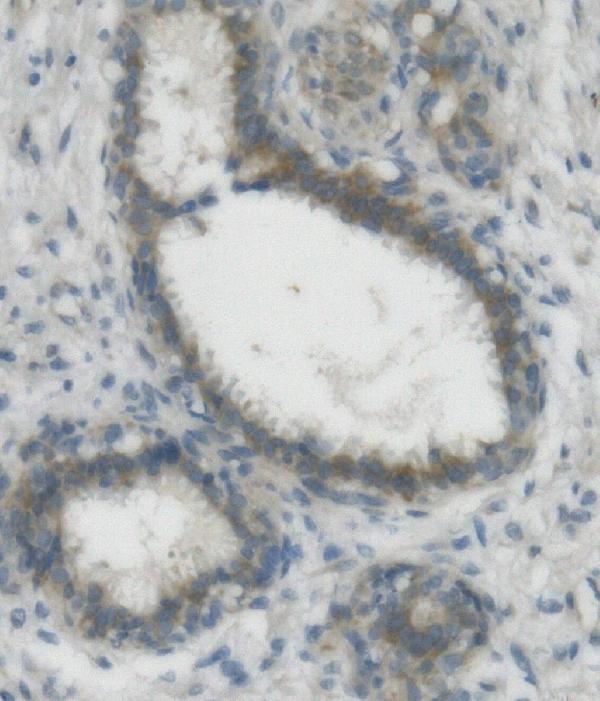
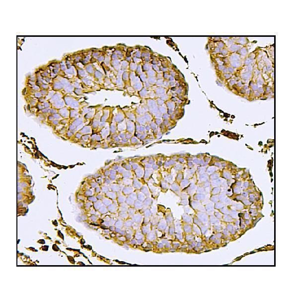
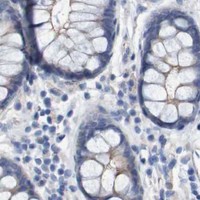
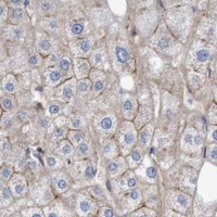
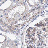
Expected resultNECTIN2-positive staining in sertoli cells of testis (HPA tissue IHC: High). HPA tissue profile: Cytoplasmic and membranous expression in most tissues. No signal in the no-primary control.
💡Decision noteStart with EDTA pH 8.0 heat retrieval for the catalog antibody (datasheet: A08081-2); the GIST study reports microwave retrieval without a buffer specification (PMC10914684).
Section 2

What Is the Expected NECTIN2 Staining Pattern?

NECTIN2 is a single-pass cell-membrane protein with an extracellular region at residues 32–360 and a cytoplasmic region at 382–538 (UniProt Q92692 topology). In paraffin-section IHC, expect membranous and cytoplasmic staining in many tissues, with particularly strong staining in testicular Sertoli cells (HPA tissue IHC: High in Sertoli cells). HPA rates the tissue IHC evidence Enhanced, while noting medium consistency between antibody staining and RNA expression (HPA tissue IHC: reliability).

What am I looking at on my slide?
Membranous staining with some cytoplasmic signal in the expected cells.This fits HPA's IHC description of cytoplasmic and membranous expression in most tissues (HPA tissue IHC: profile). The membrane component is consistent with NECTIN2 topology (UniProt Q92692 topology). Assess the named cell population, not the tissue as a whole: HPA levels are assigned to specific cells (HPA tissue IHC: cell-level entries).
Staining is confined to nuclei, with no convincing membranous or cytoplasmic pattern.Treat an isolated nuclear pattern as suspect because the supplied IHC profile is membranous and cytoplasmic (HPA tissue IHC: profile). Check its distribution against a known-positive section and detection controls before interpreting it as NECTIN2 (standard IHC practice).
Strong staining appears mainly in an HPA-listed undetected cell type, such as adipocytes.HPA reports NECTIN2 as not detected in adipocytes (HPA tissue IHC: adipocytes). Unexpected staining can reflect antibody cross-reactivity or endogenous detection activity; compare it with a negative reagent control and the expected positive cells on the same run (standard IHC practice).
Color is diffuse across tissue, including areas without a clear cell-specific pattern.Diffuse background does not establish NECTIN2 expression. The HPA result describes staining in identifiable cell populations (HPA tissue IHC: cell-level entries). Review blocking, antibody concentration, washing and detection controls as general IHC troubleshooting steps (standard IHC practice).
Sertoli cells show no signal in an otherwise evaluable testis section.A negative result conflicts with HPA's reported High Sertoli-cell staining (HPA tissue IHC: testis). First verify that Sertoli cells are present and that the IHC run worked; then review the antibody's IHC-P conditions before treating this section as biologically negative (standard IHC practice).
💡Expected NECTIN2 appearanceA convincing positive shows identifiable membranous and cytoplasmic staining in expected cells, especially High staining in Sertoli cells; isolated nuclear color or strong adipocyte staining is suspect (HPA tissue IHC: profile, Sertoli cells and adipocytes).
How each factor affects the staining
Tissue and cell selectionHPA reports High Sertoli-cell staining, Medium glandular-cell staining in adrenal gland, appendix, cervix, colon, duodenum, endometrium and epididymis, and no detected adipocyte staining (HPA tissue IHC: cell-level entries). These are cell-specific comparison points, not guarantees for every section.
Compartment and epitope locationNECTIN2 has extracellular residues 32–360, a membrane span at 361–381 and cytoplasmic residues 382–538 (UniProt Q92692 topology). Interpret staining with the antibody's stated immunogen or epitope in mind if available; the supplied record does not locate the antibody epitope.
Evidence strengthHPA labels tissue IHC reliability Enhanced but describes only medium consistency with RNA expression (HPA tissue IHC: reliability). An Enhanced rating supports the reported pattern; it does not establish the identity of every stained structure in a new specimen (standard IHC interpretation).
IF/ICC Q: Should vesicular fluorescence replace the IHC expectation?A: HPA reports vesicles as the main approved ICC-IF location and plasma membrane as an additional approved location (HPA subcellular: ICC-IF). Use that observation when interpreting IF/ICC; for paraffin-section IHC, use the separate HPA membranous and cytoplasmic tissue profile (HPA tissue IHC: profile).
Fixation and retrieval evidenceThe supplied UniProt and HPA records give no target-specific fixation sensitivity or antigen-retrieval condition. Choose retrieval using the IHC-validated antibody's protocol and run controls; do not infer a NECTIN2 fixation effect from its topology or HPA staining levels (standard IHC practice).
Why is my staining missing, weak or wrong?
SituationLikely causeNext action
Expected Sertoli-cell staining is absent.The run may have failed, the relevant cells may be missing, or the chosen IHC conditions may be unsuitable; HPA reports High Sertoli-cell staining (HPA tissue IHC: testis).Confirm cell identity and section quality, inspect a run control, then check retrieval, dilution and detection against the IHC-validated antibody's instructions (standard IHC practice).
Only nuclear staining is visible.An isolated nuclear signal does not match HPA's cytoplasmic and membranous tissue profile (HPA tissue IHC: profile).Compare with a known-positive section and a negative reagent control; review detection background before scoring the nuclear signal as NECTIN2 (standard IHC practice).
Adipocytes stain strongly.HPA lists adipocytes as not detected, so cross-reactivity or endogenous detection activity is plausible (HPA tissue IHC: adipocytes; standard IHC practice).Check a negative reagent control and whether expected positive cells stain in the same run; review blocking and detection chemistry (standard IHC practice).
The whole section has diffuse chromogenic color.Poor signal-to-background separation can obscure the cell-specific pattern described by HPA (HPA tissue IHC: profile; standard IHC practice).Inspect negative controls, then review antibody dilution, blocking, washes and detection development using the selected IHC-P workflow (standard IHC practice).
A low-staining tissue is judged negative from a faint section.HPA reports Low staining in respiratory epithelial cells of nasopharynx and bronchus, among other listed cells (HPA tissue IHC: low entries). A weak signal is harder to classify than the High Sertoli-cell reference (HPA tissue IHC: testis).Score the specified cell type and compartment; compare it with the positive control and background on the same run before calling the tissue negative (standard IHC practice).
Cytoplasmic IHC staining is dismissed because NECTIN2 is a membrane protein.UniProt assigns NECTIN2 to the cell membrane, while HPA also observes cytoplasmic staining in tissue IHC (UniProt Q92692 subcellular location; HPA tissue IHC: profile).Assess cytoplasmic signal alongside cell identity, membranous staining and controls; record the compartments separately when scoring (standard IHC practice).

Sample controls for NECTIN2 IHC & IF

🧪Run testis first: Sertoli cells should stain strongly (HPA: High in Sertoli cells). Use adipose tissue as a negative comparator, since adipocytes are listed as not detected (HPA: Not detected in adipocytes); on the testis slide, neighboring cells without specific staining can show background, but the supplied HPA rows do not establish a negative testicular cell type (HPA: testis row).
Positive control tissue: Testis (Sertoli cells, HPA High)
Negative control tissue: Adipose tissue (HPA Not detected)
ICC-IF cell lines (HPA subcellular resource): HPA ICC-IF images show NECTIN2 in CACO-2, PODO/TERT256, U2OS, with annotated localisation: Vesicles (approved) (HPA subcellular).
Technical controls: Include a no-primary, secondary-only control and a rabbit IgG isotype control matched to the catalog antibody’s clonality (caption: rabbit primary antibody); process NECTIN2 knockout material in parallel as a biological negative. For chromogenic testis IHC, check endogenous peroxidase and biotin signal when using the reported biotin-based detection system (caption: biotinylated secondary, SABC and DAB).
⚠️Feasibility: A target-specific fixation window or fixation effect is unreported, and the selected paraffin-section caption does not state its fixative (caption: fixative not stated). Start retrieval optimization with heat-mediated EDTA at pH 8.0, the condition reported for the catalog antibody; that caption does not establish that retrieval is required under all conditions (caption: heat-mediated EDTA retrieval). The supplied evidence does not establish whether frozen sections or IF are easier, and it reports no testis-specific artefact (HPA: testis tissue row; caption: paraffin-section IHC).

HPA tissue IHC evidence for NECTIN2

Comprehensive Human Protein Atlas IHC scoring per tissue (reliability: Enhanced — Medium consistency between antibody staining and RNA expression data.). Rows are taken directly from the HPA tissue chart — click any row's HPA link to view the source.

Positive expression · recommended positive controls

TissueCell typeLevelEvidenceSource
Testis Sertoli cells High Protein (IHC) HPA →
Adrenal gland Glandular cells Medium Protein (IHC) HPA →
Appendix Glandular cells Medium Protein (IHC) HPA →
Cervix Glandular cells Medium Protein (IHC) HPA →
Colon Glandular cells Medium Protein (IHC) HPA →

Undetected expression · recommended negative controls

TissueCell typeLevelEvidenceSource
Adipose tissue Adipocytes Not detected Protein (IHC) HPA →
Caudate Glial cells Not detected Protein (IHC) HPA →
Cerebellum Cells in granular layer Not detected Protein (IHC) HPA →
Hippocampus Glial cells Not detected Protein (IHC) HPA →
Oral mucosa Squamous epithelial cells Not detected Protein (IHC) HPA →
Section 3

Advanced NECTIN2 IHC Tips

Troubleshoot NECTIN2 staining in paraffin section IHC by checking retrieval, compartment, cell identity and background before comparing samples.

Which retrieval condition should I start with for NECTIN2 paraffin section IHC?
Start with heat-mediated retrieval in EDTA at pH 8.0 for the catalog antibody (datasheet A08081-2). The selected paraffin section image used this condition before overnight incubation at 4°C with 1 μg/mL primary antibody (datasheet A08081-2). If staining is weak, compare a modest adjustment in heating time on adjacent sections while holding detection and exposure to chromogen constant (standard IHC practice). Include a known expressing tissue and a no-primary control in the same run so improved signal can be distinguished from increased background (HPA: high in testis Sertoli cells; standard IHC practice).
How should I troubleshoot weak staining when fixation history is uncertain?
Target-specific sensitivity of NECTIN2 to fixation is unknown from the supplied evidence, and the selected paraffin section caption does not state a fixative (datasheet A08081-2). Record the available fixation and processing history, then compare sections processed together before changing antibody concentration or retrieval conditions (standard IHC practice). Use the reported EDTA pH 8.0 retrieval as the starting point for this antibody, with 1 μg/mL primary antibody incubated overnight at 4°C (datasheet A08081-2). A weak result in one block cannot establish a NECTIN2-specific fixation effect; evaluate morphology, controls and staining in matched material first (standard IHC practice).
Where should convincing NECTIN2 staining appear in tissue sections?
Assess cell borders alongside cytoplasmic staining: NECTIN2 is a cell membrane protein with an extracellular region at residues 32–360 and a cytoplasmic region at 382–538 (UniProt Q92692 topology). Tissue IHC reports cytoplasmic and membranous expression in most tissues, so cytoplasmic signal alone warrants careful comparison with controls (HPA: tissue profile). Vesicles are the main approved subcellular location in cell imaging, with plasma membrane also approved; tissue sections may not resolve these structures equally well (HPA: subcellular summary; standard microscopy practice). Check whether border staining follows intact cells rather than folds or damaged edges, and assess cytoplasmic signal against the no-primary control (standard IHC practice).
Can this antibody distinguish NECTIN2 isoforms or extracellular from intracellular epitopes?
NECTIN2 has Delta and Alpha isoforms, but the supplied product caption does not identify this antibody's epitope or establish isoform selectivity (UniProt Q92692 isoforms; datasheet A08081-2). The extracellular region spans residues 32–360, the membrane segment 361–381, and the cytoplasmic region 382–538 (UniProt Q92692 topology). Glycosylation sites at 137 and 324 and reported cytoplasmic phosphorylation sites provide reasons to check epitope documentation before attributing altered staining to a specific molecular form (UniProt Q92692 modifications). Score the observed tissue pattern as NECTIN2 immunoreactivity unless independent epitope mapping or isoform-specific validation supports a narrower claim (standard IHC interpretation practice).
How can IF help clarify an ambiguous NECTIN2 IHC pattern?
On a separate IF/ICC workflow, multiplex NECTIN2 with a marker for the cell population under examination, selecting that marker from independently validated evidence for the specimen (standard IF practice). NECTIN2 has an extracellular region at 32–360 and a cytoplasmic region at 382–538; choose permeabilisation according to the antibody's documented epitope side, which is unspecified here (UniProt Q92692 topology; datasheet A08081-2). Select fluorophores in channels with low specimen autofluorescence and inspect single-channel and no-primary controls before accepting overlap (standard IF practice). Compare the resulting membrane or vesicular pattern with cell imaging evidence while keeping conclusions about paraffin section chromogenic IHC tied to its own controls (HPA: approved plasma membrane and vesicle locations; standard IHC practice).
What should I check when DAB obscures cell borders?
Compare the stained section with a no-primary section to locate signal arising from the detection reagents or tissue rather than primary antibody binding (standard IHC practice). The selected image used a biotinylated secondary, streptavidin–biotin complex and DAB, so assess background from that detection sequence in the same tissue (datasheet A08081-2; standard IHC practice). Confirm that blocking and washing are adequate; the reported example blocked with 10% goat serum before 1 μg/mL primary antibody overnight at 4°C (datasheet A08081-2). If diffuse colour persists, assess endogenous peroxidase and endogenous biotin controls, then adjust detection conditions while preserving interpretable tissue morphology (standard IHC practice).
How should I score NECTIN2 staining across comparable sections? ⚠ ANSWER MARKED FOR VERIFICATION
Define the scored cell population and compartment before analysis, because tissue IHC reports both cytoplasmic and membranous NECTIN2 staining (HPA: tissue profile). For a defined population, report the percentage of positive cells and an H-score based on the proportions at each intensity; score membrane and cytoplasm separately when the question requires it (standard IHC scoring practice). If counting spatially distributed positive cells, report density per mm² of viable tissue and normalise counts to the measured viable area rather than total section area (standard IHC quantification practice). Apply identical staining, imaging, threshold and region-selection rules across samples, and retain no-primary controls to document background (standard IHC practice).
How do I distinguish credible NECTIN2 staining from artefact?
Give greatest weight to staining in intact cells with a plausible membrane or cytoplasmic pattern, interpreted alongside the no-primary control (UniProt Q92692 cell membrane; HPA: tissue profile; standard IHC practice). Compare candidate positive cells with documented examples such as high staining in testis Sertoli cells, while recognizing that tissue context determines which cells are present (HPA: high in Sertoli cells). Isolated nuclear colour is unsupported by the supplied localisation evidence; inspect section edges, folds and necrotic regions for staining that tracks damage instead of cell boundaries (UniProt Q92692 localisation; standard IHC practice). Check endogenous peroxidase and detection controls when DAB appears in unexpected cells, particularly before assigning a biological difference between samples (standard IHC practice).
Boster reagents

Best NECTIN2 / Nectin-2 IHC Antibodies

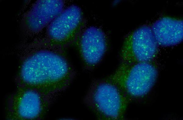
A08081-2 has IHC images from human mammary cancer and rat testis paraffin sections, plus an IF image from HeLa cells (catalog image captions).

Real IHC data IHC analysis of Nectin 2/NECTIN2 using anti-Nectin 2/NECTIN2 antibody (A08081-2). Nectin 2/NECTIN2 was detected in paraffin-embedded section of human mammary cancer tissue. Heat mediated antigen retrieval was performed in EDTA buffer (pH8.0, epitope retrieval solution). The tissue section was blocked with 10% goat serum. The tissue section was then incubated with 1μg/ml rabbit anti-Nectin 2/NECTIN2 Antibody (A08081-2) overnight at 4°C. Biotinylated goat anti-rabbit IgG was used as secondary antibody and incubated for 30 minutes at 37°C. The tissue section was developed using Strepavidin-Biotin-Complex (SABC) (Catalog # SA1022) with DAB as the chromogen.
Anti-Nectin 2/NECTIN2 Antibody ®
Cat # A08081-2

A08081-2 will render with an IHC figure from human mammary cancer paraffin tissue; its catalog also shows rat testis paraffin IHC (A08081-2 IHC captions). Its IF caption documents staining in HeLa cells at 2 μg/mL (A08081-2 IF caption).

Which to pick: Choose A08081-2 for paraffin-section IHC: its caption documents EDTA retrieval at pH 8.0 and primary antibody at 1 μg/mL; the fixative is unreported (A08081-2 IHC caption). For IF/ICC, A08081-2 has an image from HeLa cells, while M08081-2 lists ICC/IF without an image in the payload (catalog IF caption; M08081-2 application list). Consider M08081-2 when mouse reactivity is needed: it is rabbit monoclonal clone 20N15 with listed human, mouse and rat reactivity, but IHC validation is unreported (M08081-2 catalog entry).

Each figure is that product's own IHC / IF validation image from its datasheet.

References

  1. UniProt Consortium. UniProt entry Q92692 (NECT2_HUMAN, Nectin-2).
  2. Human Protein Atlas. NECTIN2 tissue IHC expression (reliability: Enhanced).
  3. Human Protein Atlas. NECTIN2 subcellular location (ICC-IF): Mainly localized to vesicles. In addition localized to the plasma membrane and mitotic spindle..
  4. Human Protein Atlas. NECTIN2 antibody validation summary (3 antibodies).
  5. Deciphering the tumor immune microenvironment of imatinib-resistance in advanced gastrointestinal stromal tumors at single-cell resolution. Cell death & disease 2024 — PMC10914684.
  6. Functional tumor-reactive CD8 + T cells in pancreatic cancer. Journal of experimental & clinical cancer research : CR 2025 — PMC12379552.
  7. ST6GalNAc-I regulates tumor cell sialylation via NECTIN2/MUC5AC-mediated immunosuppression and angiogenesis in non-small cell lung cancer. The Journal of clinical investigation 2025 — PMC12077904.
  8. Integrative analysis of neuroblastoma by single-cell RNA sequencing identifies the NECTIN2-TIGIT axis as a target for immunotherapy. Cancer cell 2024 — PMC10864003.
  9. PubMed PMID:7622062 — UniProt-cited evidence.
  10. PubMed PMID:9657005 — UniProt-cited evidence.
  11. PubMed PMID:14702039 — UniProt-cited evidence.