This website uses cookies to ensure you get the best experience on our website.


- Table of Contents
Plan NECTIN3 staining in paraffin sections using its reported cytoplasmic and membranous tissue pattern (HPA tissue IHC). Compare strong staining in placental trophoblastic cells and testicular Leydig cells with a negative control such as esophageal squamous epithelium (HPA tissue IHC).

Expected localisation, validated protocols, controls and antibodies — the at-a-glance facts below, then the full design guide.
| Expected localisation | Cytoplasmic and membranous staining in most tissues (HPA tissue IHC) | |
| Staining pattern | Most tissues show cytoplasmic and membranous staining (HPA tissue IHC) | |
| Antigen retrieval | EDTA pH 8.0 HIER, heat-mediated (datasheet A09633-3) | |
| Positive control | Placenta+4 more · see all | |
| Negative control | Esophagus+2 more · see all |
| Fixation | Keep fixation consistent across sections. (standard IHC practice; not target-specific) | |
| Caveat | Tissue staining awaits external verification (HPA tissue IHC) | |
| Regulation | Regulation is not specified (UniProt) | |
| Isoform / epitope | 3 isoforms; map the epitope to the extracellular or cytoplasmic region (UniProt) |
The catalog antibody’s IHC-P protocol is accompanied by two published chromogenic IHC protocols for NECTIN3 (PMC13562484; PMC9688614).
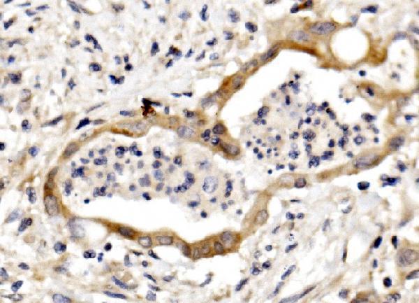
| Sample | Paraffin-embedded human rectal cancer tissue; fixative not specified (datasheet A09633-3) |
| Fixation | Image fixative and duration unreported (datasheet A09633-3); verify before use. |
| Sectioning | 4–5 µm sections on charged slides (standard) |
| Deparaffinisation | Xylene, graded ethanol series to water (standard) |
| Antigen retrieval | Heat retrieval: EDTA pH 8.0 (datasheet A09633-3); 20 min, 95–100 °C (standard) |
| Peroxidase block | 3% H2O2, 10 min, room temperature (standard) |
| Blocking | 10% goat serum (datasheet A09633-3) |
| Primary antibody | Rabbit anti-NECTIN3, 0.5-1μg/ml (datasheet A09633-3) |
| Primary incubation | Overnight at 4 °C (datasheet A09633-3) |
| Detection | Streptavidin-biotin complex (SABC), DAB chromogen (datasheet A09633-3) |
| Counterstain | Hematoxylin, blue, dehydrate and mount (standard) |
| Expected result | NECTIN3-positive staining in trophoblastic cells of placenta (HPA tissue IHC: High). HPA tissue profile: Cytoplasmic and membranous expression in most tisssues. No signal in the no-primary control. |
NECTIN3 is a single-pass cell-surface protein associated with adherens junctions (UniProt Q9NQS3 topology and localisation). In paraffin-section IHC, expect membranous and some cytoplasmic staining (HPA: tissue IHC profile), including high staining in placental trophoblasts, testicular Leydig cells and thyroid glandular cells (HPA: tissue IHC). HPA rates the tissue profile Approved, with external verification pending (HPA: reliability).
| Cell borders stain, with some cytoplasmic signal, in trophoblasts, Leydig cells or thyroid glandular cells (HPA: tissue IHC). | This fits the reported high staining in those cells (HPA: tissue IHC) and NECTIN3's membrane and junction localisation (UniProt Q9NQS3). Cytoplasmic signal can be compatible with the HPA tissue profile; a perfectly sharp border is not required. |
| Strong staining is confined to nuclei, with little membrane or cytoplasmic staining. | An isolated nuclear pattern does not match the reported tissue pattern (HPA: cytoplasmic and membranous) or annotated localisation (UniProt Q9NQS3: membrane and junctions). Treat it as suspect and review morphology, controls and detection background before scoring it as NECTIN3. |
| Squamous epithelial cells in esophagus or vagina stain strongly, although the expected positive cells also stain. | HPA reports NECTIN3 as not detected in those squamous cells (HPA: tissue IHC). Review antibody specificity and, where applicable, endogenous chromogenic detection activity (general IHC practice). A positive control alone cannot establish that staining in an unexpected cell type is specific. |
| Color spreads across stroma, lumens or most cells without recognizable cell borders. | Diffuse staining is hard to reconcile with a cell-associated membrane and cytoplasmic pattern (HPA: tissue IHC; UniProt Q9NQS3 localisation). Consider nonspecific antibody binding or detection background (general IHC practice), and compare tissue architecture and control sections before interpreting weak cells. |
| Placental trophoblasts or testicular Leydig cells show no convincing staining. | Both are reported high in the HPA tissue survey (HPA: tissue IHC), so a blank result warrants a technical review. Confirm that the sampled cells are present, then check antibody dilution, retrieval, detection and control performance (general IHC practice). HPA's Approved profile still awaits external verification (HPA: reliability). |
| Topology and epitope location (UniProt Q9NQS3 topology) | NECTIN3 has an extracellular region at residues 58–404, a transmembrane segment at 405–425 and a cytoplasmic tail at 426–549 (UniProt Q9NQS3). The supplied record does not locate the IHC antibody's epitope, so topology alone cannot predict which retrieval condition will work. |
| Processing and glycosylation (UniProt Q9NQS3) | A signal peptide spans residues 1–57; the annotated mature chain spans 58–549, with six listed glycosylation sites (UniProt Q9NQS3). These features describe the target but do not establish a fixation effect, a retrieval requirement or shedding in these sections. |
| Tissue choice and validation (HPA: tissue IHC; HPA: antibodies) | Placental trophoblasts, testicular Leydig cells and thyroid glandular cells are reported high; esophageal and vaginal squamous cells are reported not detected (HPA: tissue IHC). HPA lists two IHC antibodies as Approved, while the tissue profile remains pending external verification (HPA: antibodies and reliability). |
| IF/ICC Q&A: what localisation is reported? (HPA: subcellular) | HPA reports cell junction and cytosol localisation in ICC-IF images from U2OS, but labels both locations uncertain (HPA: subcellular). That observation offers a comparison for localisation; it does not establish an IF protocol or override the paraffin-section tissue IHC pattern. |
| Situation | Likely cause | Next action |
|---|---|---|
| Known-positive cells are blank (HPA: high trophoblasts or Leydig cells). | The section may lack the named cells, or an IHC workflow step may have failed (general IHC practice). The supplied sources give no NECTIN3-specific fixation sensitivity. | Verify cell identity on the section and compare a positive control processed alongside it. Check the recorded retrieval, primary-antibody dilution and detection steps (general IHC practice); avoid assigning the failure to fixation without evidence. |
| Only nuclei stain in otherwise intact tissue. | This compartment disagrees with HPA's membranous and cytoplasmic tissue profile and UniProt's membrane and junction annotations (HPA: tissue IHC; UniProt Q9NQS3). | Withhold a positive NECTIN3 call. Compare a no-primary control and a known-positive tissue, then review blocking and detection background (general IHC practice). |
| Esophageal or vaginal squamous cells show prominent color. | Those cells are reported not detected (HPA: tissue IHC); cross-reactivity or endogenous detection activity is possible (general IHC practice). | Compare a no-primary control and the expected positive cell types. If using an enzyme-based chromogen, check the appropriate endogenous-activity control (general IHC practice) before attributing color to NECTIN3. |
| The entire section has diffuse color that obscures cell borders. | Nonspecific binding or detection background can obscure the cell-associated pattern (general IHC practice; HPA: tissue IHC pattern). | Inspect the no-primary control and tissue morphology. Review blocking, washes, primary-antibody concentration and chromogen development under the assay's general IHC workflow. |
| A weak result appears in a tissue listed as low or medium. | HPA reports a range of levels across tissues and cell types (HPA: tissue IHC); weak staining there gives less assurance than a high positive reference. | Compare the named cell type with an HPA high reference and the assay controls before calling the weak signal absent or specific (HPA: tissue IHC; general IHC practice). |
| ICC-IF appears cytosolic or junctional, while IHC shows broader cytoplasmic staining. | HPA marks both ICC-IF locations uncertain, whereas its tissue IHC profile describes cytoplasmic and membranous expression (HPA: subcellular and tissue IHC). | Interpret each assay against its own controls and reported pattern. Do not use the uncertain ICC-IF localisation as a strict acceptance rule for paraffin-section IHC (HPA: subcellular and tissue IHC). |
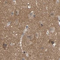


Comprehensive Human Protein Atlas IHC scoring per tissue (reliability: Approved — Pending external verification.). Rows are taken directly from the HPA tissue chart — click any row's HPA link to view the source.
| Tissue | Cell type | Level | Evidence | Source |
|---|---|---|---|---|
| Placenta | Trophoblastic cells | High | Protein (IHC) | HPA → |
| Testis | Leydig cells | High | Protein (IHC) | HPA → |
| Thyroid gland | Glandular cells | High | Protein (IHC) | HPA → |
| Adipose tissue | Adipocytes | Medium | Protein (IHC) | HPA → |
| Adrenal gland | Glandular cells | Medium | Protein (IHC) | HPA → |
Use compartment-aware controls and the documented paraffin-section method to troubleshoot NECTIN3 chromogenic IHC; assess IF separately.
The catalog includes one human-reactive NECTIN3 antibody with chromogenic IHC data from a paraffin-embedded rectal cancer section (catalog: reactivity and applications; A09633-3 image caption). No IF data are supplied (catalog).
A09633-3 is listed for human IHC, with an image showing staining of a paraffin-embedded human rectal cancer section (catalog: applications and reactivity; A09633-3 image caption). Its catalog entry supplies no IF image or IF dilution (catalog).
Which to pick: For tissue IHC, choose A09633-3: its rabbit antibody is listed for human IHC at 0.5–1 μg/ml, and its chromogenic example uses a paraffin-embedded section with EDTA retrieval at pH 8.0 (catalog: host, applications, reactivity and IHC dilution; A09633-3 image caption). The caption does not report the fixative (A09633-3 image caption). There is no supported IF/ICC or cross-species pick in this payload: A09633-3 has no listed IF/ICC application or IF data, is listed for human reactivity only, and has no reported clone designation (catalog: applications, IF fields, reactivity and clone).