This website uses cookies to ensure you get the best experience on our website.


- Table of Contents
Plan paraffin-section NEDD4 IHC around the general cytoplasmic tissue pattern (HPA tissue IHC). This guide highlights cell-type variation when choosing controls and interpreting chromogenic staining (HPA tissue IHC).

Expected localisation, validated protocols, controls and antibodies — the at-a-glance facts below, then the full design guide.
| Expected localisation | Cytoplasmic tissue staining observed; nuclear location reported (HPA tissue IHC; UniProt) | |
| Staining pattern | General cytoplasmic staining across tissue cell types (HPA tissue IHC) | |
| Antigen retrieval | EDTA pH 8.0 HIER, heat-mediated (datasheet A00984-3) | |
| Positive control | Skin+4 more · see all | |
| Negative control | Adipose tissue+4 more · see all |
| Fixation | Keep tissue fixation consistent across samples (standard IHC practice; not target-specific) | |
| Caveat | Glandular staining ranges from high to undetected by organ (HPA tissue IHC) | |
| Regulation | Expression regulation is not specified (UniProt) | |
| Isoform / epitope | Four isoforms; check whether the epitope is shared (UniProt) |
The catalog antibody protocol uses EDTA pH 8.0 retrieval (datasheet A00984-3). Three published NEDD4 IHC protocols provide tissue-specific starting conditions (PMC6923956; PMC12820133; PMC5817799).
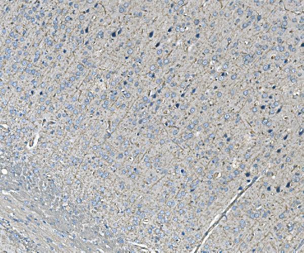
| Sample | Paraffin-embedded rat brain tissue; fixative not specified (datasheet A00984-3) |
| Fixation | Image fixative and duration unreported (datasheet A00984-3); verify before use. |
| Sectioning | 4–5 µm sections on charged slides (standard) |
| Deparaffinisation | Xylene, graded ethanol series to water (standard) |
| Antigen retrieval | Heat retrieval: EDTA pH 8.0 (datasheet A00984-3); 20 min, 95–100 °C (standard) |
| Peroxidase block | 3% H2O2, 10 min, room temperature (standard) |
| Blocking | 10% goat serum (datasheet A00984-3) |
| Primary antibody | Rabbit anti-NEDD4, 2-5 μg/ml (datasheet A00984-3) |
| Primary incubation | Overnight at 4 °C (datasheet A00984-3) |
| Detection | HRP-conjugated secondary, DAB chromogen (datasheet A00984-3) |
| Counterstain | Hematoxylin, blue, dehydrate and mount (standard) |
| Expected result | NEDD4-positive staining in fibroblasts of skin (HPA tissue IHC: Medium). HPA tissue profile: General cytoplasmic expression. No signal in the no-primary control. |
NEDD4 should stain predominantly in the cytoplasm, with possible nuclear staining; it has no transmembrane segment (UniProt P46934: localization and topology). Thyroid glandular cells and vaginal squamous epithelial cells are strong tissue examples (HPA: High in both). Interpret intensity by cell type: HPA describes general cytoplasmic expression, but its tissue IHC rating is Approved with medium consistency between staining and RNA data (HPA: tissue IHC).
| Cytoplasmic staining in thyroid glandular cells or vaginal squamous epithelial cells, with clear cellular boundaries. | This fits the strongest listed tissue examples (HPA: High in both cell types) and NEDD4's predominant cytoplasmic localization (UniProt P46934: subcellular location). Some nuclear signal can be plausible (UniProt P46934: nucleus); evaluate its distribution alongside the cytoplasmic pattern. |
| Signal is exclusively extracellular, or appears as a continuous outline around every cell while cytoplasm is blank. | Treat this as a compartment mismatch requiring investigation: NEDD4 is predominantly cytoplasmic and has no transmembrane segment (UniProt P46934: localization and topology). Membrane recruitment is possible (UniProt P46934: GRB10 interaction), so a localized membrane signal alone does not prove an artefact. |
| The strongest staining is in adipocytes, liver cholangiocytes, or lymph node germinal center cells. | These cell types were not detected in the supplied tissue survey (HPA: Not detected in each). Check for cross-reactivity or endogenous detection activity before calling them positive. The HPA result is an observed pattern, not proof that every specimen of those cell types must be negative. |
| Brown signal coats tissue broadly, with little distinction between cells, compartments, and blank areas. | This is diffuse background rather than a convincing cellular pattern (general IHC practice). Assess blocking, washing, secondary or detection reagents, and the no-primary control; HPA's general cytoplasmic profile does not validate uniform staining across a section (HPA: tissue IHC). |
| No specific signal appears in thyroid glandular cells or vaginal squamous epithelial cells. | A negative result in these listed High examples warrants a workflow check (HPA: High in both). Verify section integrity, retrieval and detection controls, then review the antibody's IHC validation. A failed run cannot establish that NEDD4 is absent from the specimen (general IHC practice). |
| Compartment and topology | Predominant cytoplasmic signal, possible nuclear signal, and context-dependent plasma membrane recruitment fit the protein record (UniProt P46934: localization; no transmembrane segment). Do not require a membrane rim to score tissue IHC positive. |
| Choice of tissue and cell type | Use the named cells, rather than an entire organ, to judge a pattern: thyroid glandular cells and vaginal squamous epithelial cells are High; skin fibroblasts are Medium (HPA: tissue IHC). Adipocytes and liver cholangiocytes are listed as Not detected (HPA: tissue IHC). |
| Antibody evidence | HPA lists IHC Approved for HPA039883, CAB001991, and CAB072833; HPA046793 has ICC Supported but no listed IHC status (HPA: antibody validation). Approved is accompanied by medium staining–RNA consistency for the tissue profile (HPA: tissue IHC), so compare the observed cell type and compartment before scoring. |
| Isoforms and epitope coverage | UniProt lists four NEDD4 isoforms and a WW-domain region plus a HECT domain (UniProt P46934: isoforms and domains). The supplied evidence gives no antibody epitope or isoform coverage, so an unexpected pattern cannot be assigned to a particular isoform from these records. |
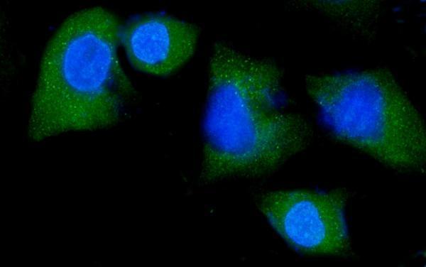
| IF/ICC expectation? | For IF/ICC, HPA supports nucleoplasm and cytosol localization and marks additional plasma membrane localization uncertain (HPA: subcellular ICC-IF). This is an interpretation cue for that separate application, not an IHC staining requirement; the supplied record gives no IF/ICC protocol. |
| Situation | Likely cause | Next action |
|---|---|---|
| Known High tissue is blank. | The run may have failed at retrieval, primary incubation, or detection (general IHC practice); HPA reports High staining in thyroid glandular and vaginal squamous epithelial cells (HPA: tissue IHC). | Check an on-run positive section and detection controls, then review the catalog antibody's IHC instructions before interpreting the specimen as negative (general IHC practice). |
| All tissue structures and empty spaces look brown. | Diffuse chromogen or detection background can obscure a cellular signal (general IHC practice); it does not match HPA's general cytoplasmic profile (HPA: tissue IHC). | Inspect a no-primary control, wash and block conditions, and detection reagent exposure; repeat scoring only when cells can be distinguished (general IHC practice). |
| Unexpected cells stain as strongly as the positive comparator. | Cross-reactivity or endogenous detection activity is possible (general IHC practice), especially when the cells are listed as Not detected (HPA: adipocytes, cholangiocytes, germinal center cells). | Compare matched cell types on the same run and review a no-primary control; treat discordance as unresolved until antibody specificity and detection background are assessed (general IHC practice). |
| Only nuclei stain in tissue IHC. | Nuclear localization is possible, but exclusively nuclear staining departs from the predominant cytoplasmic distribution (UniProt P46934: localization; HPA: general cytoplasmic expression). | Recheck cytoplasmic signal in a High reference cell type and inspect controls; report the nuclear-only pattern as discordant unless independently supported. |
| A sharp membrane rim dominates the section. | NEDD4 can be recruited to the plasma membrane, but its predominant location is cytoplasmic and it lacks a transmembrane segment (UniProt P46934: localization and topology). | Look for accompanying cytoplasmic staining and cell-type agreement; investigate a uniform rim across unrelated cells as possible background (general IHC practice). |
| IF/ICC shows cytosolic and nuclear fluorescence, while tissue IHC looks mainly cytoplasmic. | The observations can coexist: HPA supports nucleoplasm and cytosol in ICC-IF, while its tissue IHC profile is generally cytoplasmic (HPA: subcellular ICC-IF; tissue IHC). | Interpret each application against its own HPA evidence; use the IF/ICC guide for that assay's controls and settings, and do not impose an ICC-IF nuclear pattern on IHC. |
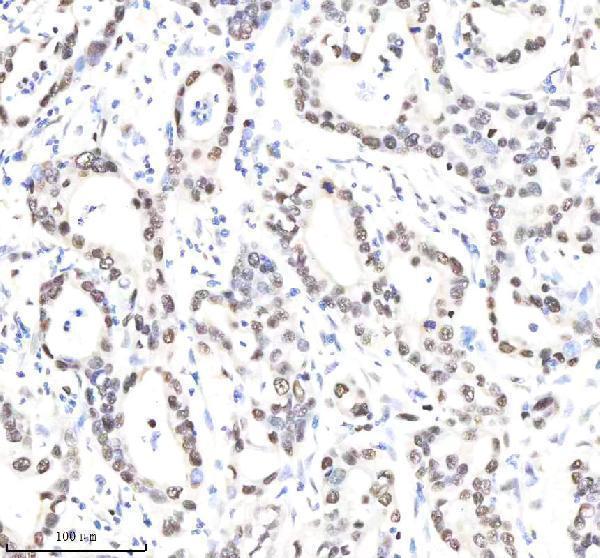
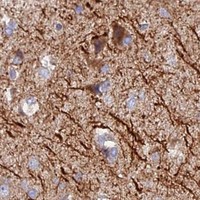
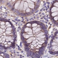
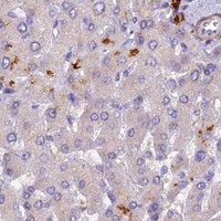
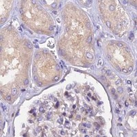


Comprehensive Human Protein Atlas IHC scoring per tissue (reliability: Approved — Medium consistency between antibody staining and RNA expression data.). Rows are taken directly from the HPA tissue chart — click any row's HPA link to view the source.
| Tissue | Cell type | Level | Evidence | Source |
|---|---|---|---|---|
| Skin | Fibroblasts | Medium | Protein (IHC) | HPA → |
| Thyroid gland | Glandular cells | High | Protein (IHC) | HPA → |
| Vagina | Squamous epithelial cells | High | Protein (IHC) | HPA → |
| Adrenal gland | Glandular cells | Medium | Protein (IHC) | HPA → |
| Appendix | Glandular cells | Medium | Protein (IHC) | HPA → |
| Tissue | Cell type | Level | Evidence | Source |
|---|---|---|---|---|
| Adipose tissue | Adipocytes | Not detected | Protein (IHC) | HPA → |
| Duodenum | Glandular cells | Not detected | Protein (IHC) | HPA → |
| Liver | Cholangiocytes | Not detected | Protein (IHC) | HPA → |
| Lymph node | Germinal center cells | Not detected | Protein (IHC) | HPA → |
| Ovary | Ovarian stroma cells | Not detected | Protein (IHC) | HPA → |
Use compartment-aware controls and the catalog antibody’s documented paraffin-section conditions to troubleshoot NEDD4 chromogenic IHC.
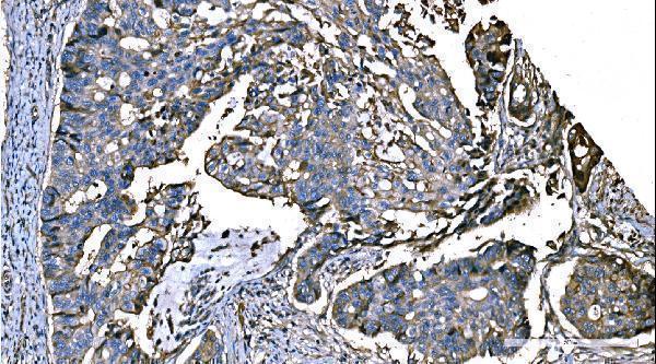
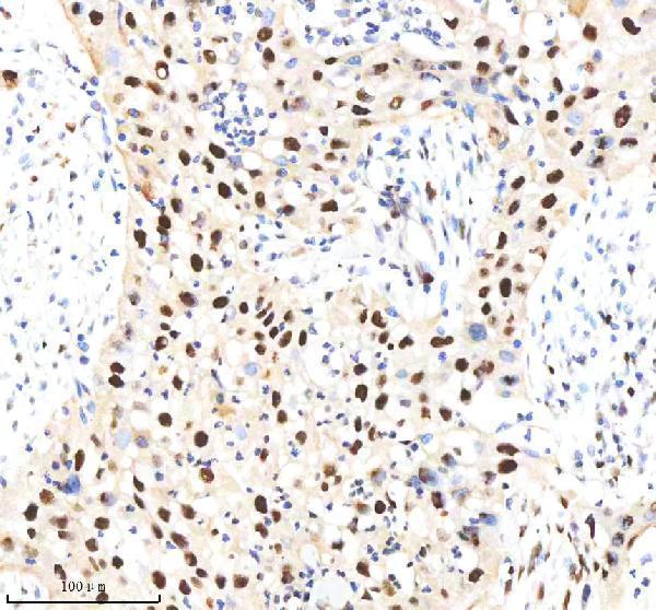
The catalog includes paraffin-section IHC images from rat brain and human tumors (catalog IHC captions), plus an IF/ICC image from A549 cells (A00984-3 IF caption).
A00984-3 has IHC images from rat brain and human colon, liver, and lung cancers, plus an IF/ICC image from A549 cells (A00984-3 image captions). M00984 has IHC images from human bladder and pancreas cancers (M00984 IHC captions).
Which to pick: For paraffin-section IHC, start with A00984-3 at 2 μg/ml for rat brain or the pictured human tissues (A00984-3 IHC captions), or rabbit monoclonal M00984 at 1:50 for the pictured human tumors (catalog: monoclonal; M00984 IHC captions); both images report EDTA retrieval at pH 8.0 (catalog IHC captions). For IF/ICC, choose A00984-3 at 5 μg/ml, as shown in A549 cells (A00984-3 IF caption); M00984 has no listed IF/ICC application (catalog applications). For studies spanning species, M00984 lists human, mouse, and rat reactivity, while A00984-3 lists human and rat (catalog reactivity); the IHC captions describe paraffin sections but do not report the fixative (catalog IHC captions).