This website uses cookies to ensure you get the best experience on our website.


- Table of Contents
Plan NEDD8 IHC on paraffin sections using the reported nuclear and cytoplasmic tissue pattern, with respiratory epithelium as a high staining reference (HPA tissue IHC). Compare sections with consistent fixation and interpret intensity cautiously because tissue staining reliability is uncertain (HPA tissue IHC).

Expected localisation, validated protocols, controls and antibodies — the at-a-glance facts below, then the full design guide.
| Expected localisation | Nuclear and cytoplasmic staining in most tissues (HPA tissue IHC) | |
| Staining pattern | Most tissues show nuclear and cytoplasmic signal (HPA tissue IHC) | |
| Antigen retrieval | Citrate pH 6 HIER, heat-mediated (datasheet A00547) | |
| Positive control | Bronchus+4 more · see all | |
| Negative control | None in HPA (detected in all 45 tissues); use no-primary + isotype controls |
| Fixation | Keep fixation consistent across sections (standard IHC practice; not target-specific) | |
| Caveat | Tissue staining has uncertain reliability (HPA tissue IHC) | |
| Regulation | Expression regulation is not established (UniProt) | |
| Isoform / epitope | No isoforms annotated; residues 77–81 are removed during maturation (UniProt) |
Compare the catalog antibody’s IHC-P protocol (datasheet: A00547) with published NEDD8 chromogenic IHC on frozen synovium, tissue sections, and paraffin breast arrays (PMC12960655; PMC6501157; PMC9830515).
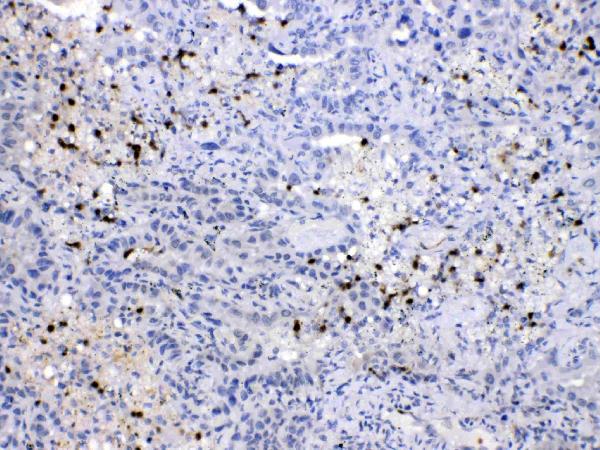
| Sample | Paraffin-embedded human lung cancer tissues; fixative not specified (datasheet A00547) |
| Fixation | Image fixative and duration unreported (datasheet A00547); verify before use. |
| Sectioning | 4–5 µm sections on charged slides (standard) |
| Deparaffinisation | Xylene, graded ethanol series to water (standard) |
| Antigen retrieval | Heat retrieval: Citrate pH 6, 20 min (datasheet A00547) |
| Peroxidase block | 3% H2O2, 10 min, room temperature (standard) |
| Blocking | 10% goat serum (datasheet A00547) |
| Primary antibody | Rabbit anti-NEDD8, 0.5-1μg/ml (datasheet A00547) |
| Primary incubation | Overnight at 4 °C (datasheet A00547) |
| Detection | Streptavidin-biotin complex (SABC), DAB chromogen (datasheet A00547) |
| Counterstain | Hematoxylin, blue, dehydrate and mount (standard) |
| Expected result | NEDD8-positive staining in respiratory epithelial cells of bronchus (HPA tissue IHC: High). HPA tissue profile: Cytoplasmic and nuclear expression in most tissues. No signal in the no-primary control. |
NEDD8 is mainly nuclear (UniProt Q15843: subcellular location) and has no transmembrane segment (UniProt Q15843: topology). In paraffin sections, expect nuclear and cytoplasmic staining in many tissues, with strong signal in selected epithelial, neuronal and glandular cells (HPA: tissue IHC). Treat this as a reference pattern, not a definitive specificity test: HPA rates the tissue IHC profile Uncertain because staining and RNA show only medium consistency (HPA: tissue IHC reliability).
| Nuclear staining, with some cytoplasmic staining, in respiratory epithelium. | This fits the reported tissue pattern (HPA: cytoplasmic and nuclear expression in most tissues). Respiratory epithelial cells stain High in bronchus and nasopharynx (HPA: tissue IHC). Judge intensity in the named cells, since an entire section need not stain uniformly (standard IHC interpretation). |
| Signal appears confined to an unexpected compartment across the section. | A pattern lacking the expected nuclear component warrants review because NEDD8 is mainly nuclear (UniProt Q15843: subcellular location). Check morphology, counterstain and detection controls before calling it artefact (standard IHC practice); cytoplasmic signal alone is not inherently false because HPA reports it in tissue IHC (HPA: tissue IHC profile). |
| The strongest signal is in cells other than the expected positive population. | For example, strong alveolar staining would conflict with the reported Low level in alveolar cells, while bronchial respiratory epithelium is High (HPA: tissue IHC). Review cell identity and controls; antibody cross-reactivity or endogenous detection activity are possible explanations, not diagnoses from one slide (standard IHC practice). |
| Brown deposit spreads across cells, stroma or the section edge. | Diffuse signal without a coherent cellular pattern cannot establish NEDD8 localization (standard IHC interpretation). Compare a control processed without primary antibody and inspect reagent and wash conditions for nonspecific staining or endogenous detection activity (standard IHC practice). |
| Little or no signal appears in a selected positive tissue. | Check that the expected cells are present: Purkinje cells in cerebellum and glandular cells in parathyroid are reported High (HPA: tissue IHC). A blank result may reflect the assay or sampling, and one blank section cannot overturn an HPA pattern rated Uncertain (HPA: tissue IHC reliability; standard IHC interpretation). |
| Choice of positive and lower-signal cells | Bronchus and nasopharynx respiratory epithelium, cerebellar Purkinje cells, cerebral cortical neurons and parathyroid glandular cells are High (HPA: tissue IHC). Alveolar cells, cholangiocytes, cardiomyocytes, myocytes and fibroblasts are Low comparators, not established negatives (HPA: tissue IHC). |
| Cell type within the same tissue | Interpret named cells rather than assigning one score to an organ: bronchial respiratory epithelial cells are High, whereas lung alveolar cells are Low (HPA: tissue IHC). A difference between those compartments can therefore be expected without treating either tissue as an absolute positive or negative control (HPA: tissue IHC). |
| NEDD8 form and localization | UniProt annotates an 81-aa precursor, removal of residues 77–81, and a mature 1–76 chain that conjugates to cellular proteins (UniProt Q15843: processing and function). The payload gives no antibody epitope, so it cannot establish which NEDD8 forms the IHC stain detects (UniProt Q15843: processing; supplied antibody data). |
| Strength of antibody evidence | The HPA tissue profile is Uncertain, and CAB004082 has Uncertain IHC validation; the other listed antibodies have no IHC status in the supplied record (HPA: tissue IHC reliability; HPA: antibody validation). Use morphology and controls when interpreting a surprising pattern (standard IHC practice). |
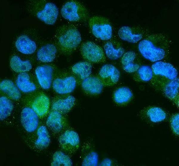
| IF/ICC Q: Where should fluorescence appear? | A: Mainly in the nucleoplasm (Supported); Golgi apparatus and cytosol are additional Uncertain locations (HPA: subcellular ICC-IF). This cellular imaging evidence helps interpret localization, while tissue intensity calls come from the separate HPA tissue IHC profile (HPA: subcellular ICC-IF; HPA: tissue IHC). |
| Situation | Likely cause | Next action |
|---|---|---|
| No stain in a selected positive section | The named positive cells may be absent from the sampled area, or the staining run may have failed (HPA: tissue IHC; standard IHC practice). | Confirm the relevant cells on the counterstained section, then review the run's positive control, primary antibody, retrieval and detection steps (standard IHC practice). |
| Widespread diffuse brown background | Nonspecific reagent binding or endogenous detection activity can produce deposit that does not follow cell anatomy (standard IHC practice). | Inspect a control without primary antibody; review blocking, washes and the detection system before scoring NEDD8 (standard IHC practice). |
| Predominantly cytoplasmic staining with weak nuclei | Cytoplasmic tissue staining is reported, but a consistently absent nuclear component differs from NEDD8's main localization (HPA: tissue IHC profile; UniProt Q15843: subcellular location). | Check nuclear morphology and counterstain, then compare another section and appropriate controls; avoid assigning specificity from compartment alone (standard IHC practice). |
| Strong staining in a reported lower-signal cell population | Cell misidentification, cross-reactivity or endogenous detection activity may explain a mismatch with an HPA Low call (HPA: tissue IHC; standard IHC practice). | Verify cell identity and compare nearby expected positive cells plus a control without primary antibody; report the mismatch if it persists (standard IHC practice). |
| Weak signal in heart or skeletal muscle despite UniProt expression | UniProt reports high tissue expression, while HPA calls cardiomyocyte and skeletal myocyte IHC staining Low (UniProt Q15843: tissue specificity; HPA: tissue IHC). | Use the cell-level HPA calls for slide interpretation; check a reported High IHC cell population before judging the run (HPA: tissue IHC; standard IHC practice). |
| Different fields or sections yield conflicting scores | The field may contain different cell populations, and the HPA tissue IHC profile has Uncertain reliability (HPA: tissue IHC reliability; standard IHC interpretation). | Score identified cell types in comparable fields, document the compartment and intensity, and keep the uncertainty attached to conclusions (standard IHC practice; HPA: tissue IHC reliability). |
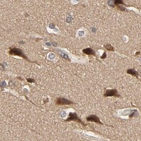


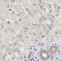


Comprehensive Human Protein Atlas IHC scoring per tissue (reliability: Uncertain — Medium consistency between antibody staining and RNA expression data. Pending external verification.). Rows are taken directly from the HPA tissue chart — click any row's HPA link to view the source.
| Tissue | Cell type | Level | Evidence | Source |
|---|---|---|---|---|
| Bronchus | Respiratory epithelial cells | High | Protein (IHC) | HPA → |
| Cerebellum | Purkinje cells | High | Protein (IHC) | HPA → |
| Cerebral cortex | Neuronal cells | High | Protein (IHC) | HPA → |
| Nasopharynx | Respiratory epithelial cells | High | Protein (IHC) | HPA → |
| Parathyroid gland | Glandular cells | High | Protein (IHC) | HPA → |
| Tissue | Cell type | Level | Evidence | Source |
|---|---|---|---|---|
| None in HPA: NEDD8 is detected in all 45 scored tissues. Use a no-primary (secondary-only) and an isotype control instead. | ||||
Troubleshoot NEDD8 staining in paraffin sections by checking retrieval, compartment, cell type and detection controls before interpreting signal.
A00547 has IHC data from human lung cancer paraffin sections and IF/ICC data from A431 cells (A00547 image captions). Both antibodies list Human, Mouse, and Rat reactivity (catalog reactivity).
A00547 lists IHC and IF/ICC, with images from human lung cancer paraffin sections and A431 cells, respectively (catalog applications; A00547 image captions). M00547 lists IHC and IF/ICC and Human, Mouse, and Rat reactivity, but has no supplied IHC or IF image (catalog applications, reactivity, and image alts).
Which to pick: Choose A00547 for tissue IHC: its caption documents human lung cancer paraffin sections, citrate pH 6 retrieval, and 1 μg/ml antibody; the fixative is unreported (A00547 IHC caption). For IF/ICC, A00547 has A431 cell data at 2 μg/ml, while M00547 lists IF/ICC without a supplied image (A00547 IF caption; M00547 catalog applications and image alts). Both list Mouse and Rat reactivity, although the supplied images do not show those species; M00547 is the rabbit monoclonal option, clone AOAE-14 (catalog reactivity, host, clone, and image alts).