NEFH / Neurofilament heavy polypeptide · IHC design guide

Design Immunohistochemistry for NEFH

Plan NEFH IHC in paraffin sections using neuronal cells and cerebellar granular-layer processes as expected staining sites (HPA tissue IHC). This guide covers fixation, staining controls and interpretation of cell-specific signal, including prostate glandular staining (HPA tissue IHC).

Evidence assembled Oct 2026 · For research use; verify linked source records and product datasheet before use
Immunohistochemistry protocol sheet for NEFH (IHC for NEFH): expected localisation Axonal cytoskeleton (UniProt); neuronal staining (HPA tissue IHC), antibody M05307-9, validated IHC image, and IHC protocol steps
Printable NEFH IHC protocol sheet — expected localisation Axonal cytoskeleton (UniProt); neuronal staining (HPA tissue IHC), antibody M05307-9, controls and protocol steps. Open the full NEFH IHC guide →

NEFH Immunohistochemistry Experimental Design Guide

Expected localisation, validated protocols, controls and antibodies — the at-a-glance facts below, then the full design guide.

Must know before staining
Expected localisation Axonal cytoskeleton (UniProt); neuronal staining (HPA tissue IHC)
Staining pattern Neuronal cells and cerebellar granular-layer processes (HPA tissue IHC)
Antigen retrieval No retrieval / mild HIER, citrate pH 6.0 if weak (rule: intermediate filament)
Positive control ⓘ Caudate+4 more · see all
Negative control ⓘ Adipose tissue+4 more · see all
Important caveats
Reasons your staining may differ from the expected pattern.
Fixation Keep fixation consistent across sections (standard IHC practice; not target-specific)
Caveat Prostate glandular cells also stain (HPA tissue IHC)
Regulation No staining-linked regulator annotated (UniProt)
Isoform / epitope 2 isoforms; epitope differences unspecified (UniProt)
Section 1

Recommended NEFH IHC & IF Protocols

The catalog antibody’s IHC-P protocol is accompanied by 2 published NEFH IHC protocols for rat optic nerve and esophageal tissue arrays (PMC12536187; PMC2815775).

Recommended immunohistochemistry (IHC-P) protocol parameters
SampleTissue sections; selected-image fixative not specified (standard IHC workflow)
FixationImage fixative and duration unreported (datasheet M05307-9); verify before use.
Sectioning4–5 µm sections on charged slides (standard)
DeparaffinisationXylene, graded ethanol series to water (standard)
Antigen retrievalNo retrieval needed for most intermediate-filament epitopes; if staining is weak, mild HIER in citrate buffer, pH 6.0, 10 min at 95 °C (standard rule: intermediate filament)
Peroxidase block3% H2O2, 10 min, room temperature (standard)
Blocking10% normal serum of the secondary host, 30 min, room temperature (standard)
Primary antibodyRabbit anti-NEFH, 1:200-1:1000 (datasheet M05307-9)
Primary incubationOvernight at 4 °C (standard)
DetectionHRP-polymer secondary, DAB chromogen 5–10 min (standard)
CounterstainHematoxylin, blue, dehydrate and mount (standard)
Expected resultNEFH-positive staining in neuronal cells of caudate (HPA tissue IHC: High). HPA tissue profile: Selective expression in CNS, peripheral nerves and glandular cells of prostate. No signal in the no-primary control.
💡Decision noteStart without retrieval; if staining is weak, try mild citrate HIER at pH 6.0 (page retrieval rule: intermediate filament).
Section 2

What Is the Expected NEFH Staining Pattern?

NEFH is an axonal cytoskeletal protein with no transmembrane segment (UniProt P12036). In IHC, expect strong staining in CNS neurons and cerebellar processes; prostate glandular cells may show medium staining (HPA tissue IHC, Enhanced).

What am I looking at on my slide?
Neuronal and cerebellar process staining (HPA tissue IHC)Expected; cerebellar granular-layer processes stain high (HPA tissue IHC).
Predominantly nuclear tissue staining (UniProt P12036 axonal location)Atypical for tissue IHC; assess controls before interpreting it as NEFH (UniProt P12036).
Strong adipocyte staining (HPA: adipocytes not detected)Consider cross-reactivity or endogenous detection activity (HPA tissue IHC; standard IHC practice).
Diffuse brown haze across the section (standard IHC practice)Suggests background from nonspecific binding or detection activity (standard IHC practice).
No signal in caudate neurons (HPA: High)Suggests an assay problem; check the positive control and detection steps (standard IHC practice).
💡Expected NEFH appearanceHigh neuronal/process signal is expected; adipocyte signal is suspect (HPA tissue IHC; UniProt P12036).
How each factor affects the staining
Phosphorylation (UniProt P12036)Multiple phosphoserines are annotated; epitope effects need antibody-specific evidence (UniProt P12036).
Isoforms (UniProt P12036)Two isoforms are annotated; recognition depends on the antibody epitope (UniProt P12036).
IF/ICC Q: same location? (HPA subcellular)ICC-IF reports approved centrosome/basal-body signal; nuclear-body signal is uncertain (HPA subcellular).
Chromogenic detection (standard IHC practice)Endogenous peroxidase can create background; assess it with controls (standard IHC practice).
Why is my staining missing, weak or wrong?
SituationLikely causeNext action
No caudate neuronal signal (HPA: High)Positive-control failure is possible (HPA tissue IHC; standard IHC practice).Check antibody dilution, retrieval and detection with a positive control (standard IHC practice).
Strong adipocyte signal (HPA: not detected)Cross-reactivity or endogenous activity is possible (standard IHC practice).Review a no-primary control and peroxidase blocking (standard IHC practice).
Nuclear-only tissue signal (UniProt P12036 axonal location)Nonspecific staining is possible (standard IHC practice).Compare with neuronal processes and a no-primary control (UniProt P12036; standard IHC practice).
Diffuse brown haze (standard IHC practice)Nonspecific binding or detection background is possible (standard IHC practice).Review blocking, washes and a no-primary control (standard IHC practice).
Weak prostate glandular signal (HPA: Medium)Prostate staining is lower than CNS neuronal staining (HPA tissue IHC).Compare with a high-staining CNS control (HPA tissue IHC; standard IHC practice).

Sample controls for NEFH IHC & IF

🧪Run caudate first: neuronal cells should stain strongly (HPA: High in caudate neuronal cells). Use adipose tissue as the negative tissue (HPA: Not detected in adipocytes); on the caudate slide, non-neuronal cells should lack specific staining, while axonal processes may stain (UniProt P12036: axonal localization).
Positive control tissue: Caudate (Neuronal cells, HPA High)
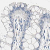
Negative control tissue: Adipose tissue (HPA Not detected)
ICC-IF cell lines (HPA subcellular resource): HPA ICC-IF images show NEFH in HEK293, SiHa, U2OS, ASC52telo, RPTEC/TERT1, hTERT-RPE1 (serum starved), with annotated localisation: Nuclear bodies (uncertain), Centrosome (approved), Basal body (approved) (HPA subcellular).
Technical controls: Include a no-primary, secondary-only control; a rabbit IgG isotype control matched to the primary antibody’s clonality; and, if available, NEFH-knockout tissue as a biological negative (caption: rabbit antibody; standard IHC control practice). For chromogenic staining of caudate, quench endogenous peroxidase and check background in the negative controls (standard IHC practice).
⚠️Feasibility: No target-specific fixation window or fixation effect is reported, and the selected M05307-9 mouse-brain IHC caption does not state a fixative (caption: fixative not stated). Retrieval dependence and whether frozen sections or IF are easier are unreported; optimize antigen retrieval empirically for paraffin IHC (standard IHC practice). Brain autofluorescence can complicate IF assessment, so inspect a no-primary control if using IF (standard IF practice).

HPA tissue IHC evidence for NEFH

Comprehensive Human Protein Atlas IHC scoring per tissue (reliability: Enhanced — High consistency between antibody staining and RNA expression data.). Rows are taken directly from the HPA tissue chart — click any row's HPA link to view the source.

Positive expression · recommended positive controls

TissueCell typeLevelEvidenceSource
Caudate Neuronal cells High Protein (IHC) HPA →
Cerebellum Processes in granular layer High Protein (IHC) HPA →
Cerebral cortex Neuronal cells High Protein (IHC) HPA →
Hippocampus Neuronal cells High Protein (IHC) HPA →
Ovary Follicle cells Medium Protein (IHC) HPA →

Undetected expression · recommended negative controls

TissueCell typeLevelEvidenceSource
Adipose tissue Adipocytes Not detected Protein (IHC) HPA →
Adrenal gland Glandular cells Not detected Protein (IHC) HPA →
Appendix Glandular cells Not detected Protein (IHC) HPA →
Bone marrow Hematopoietic cells Not detected Protein (IHC) HPA →
Breast Adipocytes Not detected Protein (IHC) HPA →
Section 3

Advanced NEFH IHC Tips

Troubleshoot NEFH staining in paraffin sections by assessing axonal pattern, retrieval, background and scoring alongside appropriate controls (UniProt P12036; general IHC practice).

Should I retrieve NEFH before staining paraffin sections?
Start without retrieval; if staining is weak, use mild heat-induced retrieval in citrate at pH 6.0 (page retrieval rule: intermediate filament). Compare retrieved and unretrieved sections from the same block, holding antibody concentration and chromogen development constant (general IHC practice). Limit an initial heating trial to about 10–20 minutes, then inspect whether neuronal processes become clearer or tissue structure deteriorates (general IHC practice; HPA: high in cerebellar granular-layer processes). Because NEFH has numerous annotated phosphoserines, a change in staining after heating does not establish whether retrieval exposed the protein backbone or altered access to a particular modified epitope (UniProt P12036: modified residues; general IHC interpretation).
Could fixation explain weak NEFH staining in my paraffin sections?
NEFH-specific sensitivity to fixation is unknown from the supplied evidence; the mouse-brain tissue-IHC caption for M05307-9 does not state a fixative (selected tissue-IHC caption). Record the fixative, fixation duration and processing history for each block before comparing staining intensity (general IHC practice). If blocks differ, stain them in one run with the same retrieval condition and a positive neural control, then assess both process staining and tissue preservation (general IHC practice; HPA: high in cerebellar granular-layer processes). Do not assign a weak result to fixation alone when section age, retrieval, antibody incubation and detection can also vary (general IHC practice).
Which staining pattern supports NEFH localisation in tissue?
Expect staining associated with neuronal cytoplasm and axonal processes because NEFH is a cytoskeletal neurofilament protein localised to axons (UniProt P12036: subcellular location and function). Use the processes in the cerebellar granular layer or neuronal cells in caudate as pattern references, without assuming every cell in those regions must stain (HPA: high in cerebellar granular-layer processes and caudate neuronal cells). A diffuse nuclear-only chromogenic pattern needs further validation before being called axonal NEFH, even though cell-line ICC/IF reports additional nuclear and centrosomal locations (UniProt P12036: axonal location; HPA: subcellular summary; general IHC interpretation). Assess morphology at both low and high magnification and compare a negative primary-antibody control for nonspecific deposits (general IHC practice).
Can phosphorylation or isoforms change the NEFH IHC pattern?
NEFH has 2 annotated isoforms and numerous modified residues, including phosphoserines, so antibody recognition depends on an epitope that has not been specified here (UniProt P12036: isoforms and modified residues; supplied antibody evidence: epitope unspecified). Check the antibody's stated immunogen or epitope before interpreting differences between samples as changes in total NEFH (general IHC practice). Run adjacent sections under matched retrieval and detection conditions, and compare their neuronal-process staining rather than relying on overall chromogen intensity alone (general IHC practice; HPA: high in cerebellar granular-layer processes). If a phosphorylation-specific interpretation matters, require separately validated epitope information and appropriate controls; the supplied tissue image cannot establish modification specificity (general IHC practice; selected tissue-IHC caption).
How should I follow up an IHC pattern with NEFH immunofluorescence?
Treat IF/ICC as a separate validation workflow while using chromogenic IHC to define the tissue pattern for comparison (general assay practice; selected tissue-IHC caption: mouse brain staining). Multiplex NEFH with an independently validated marker of the expected neuronal cells, and confirm that labelled processes align with the neuronal pattern reported in tissue (UniProt P12036: axonal location; HPA: high in neuronal cells and cerebellar processes; general IF practice). Choose spectrally separated fluorophores after checking tissue autofluorescence and single-label controls (general IF practice). NEFH lacks a transmembrane segment and is cytoskeletal, so use mild permeabilisation after fixation for an intracellular epitope; because the antibody epitope is unspecified, optimise permeabilisation empirically (UniProt P12036: topology and location; general IF practice).
How can I reduce diffuse brown background without losing axons?
First distinguish widespread chromogen deposition from the neuronal and process-associated pattern expected for NEFH (UniProt P12036: axonal cytoskeleton; HPA: high in neuronal cells and cerebellar processes). Include a no-primary control, block endogenous peroxidase before detection, and inspect whether brown signal persists without primary antibody (general chromogenic IHC practice). Titrate the IHC-validated antibody and detection reagent, shorten chromogen development if needed, and keep incubation and wash conditions identical between trial sections (general IHC practice). If background concentrates at section edges or damaged areas, exclude those regions from interpretation and compare intact tissue in the same run (general IHC practice).
What should I score when NEFH stains both cells and processes? ⚠ ANSWER MARKED FOR VERIFICATION
Define the measurement before scoring: use percentage of positive neuronal cells or an H-score for cell-associated staining, and process-positive area or length density per mm² for axonal staining (UniProt P12036: neuronal axonal cytoskeleton; general IHC quantification practice). Keep these readouts separate because process signal need not scale with the number of stained cell bodies in a section (general IHC interpretation). Normalise cell scores to the number of evaluable neuronal cells and process density to intact tissue area, applying the same threshold and region-selection rules across sections (general IHC quantification practice). Exclude folds, edges and necrotic areas, and report the scoring region and retrieval condition with each result (general IHC practice).
How do I distinguish genuine NEFH staining from artefact?
Give the strongest weight to coherent neuronal and axonal staining, especially when processes follow tissue morphology, because NEFH is an axonal cytoskeletal protein (UniProt P12036: location; HPA: high in cerebellar processes and neuronal cells). Treat isolated brown nuclei, non-neuronal deposits or staining confined to section edges as suspect until controls support them (general IHC interpretation; UniProt P12036: axonal location). Compare intact regions with damaged or necrotic regions, and check a no-primary control for endogenous enzyme signal or nonspecific detection (general chromogenic IHC practice). Glandular staining is not automatically artefact: HPA reports medium staining in prostate glandular cells, so interpret it with tissue context and independent controls (HPA: prostate glandular cells medium; general IHC practice).
Boster reagents

Best NEFH / Neurofilament heavy polypeptide IHC Antibodies

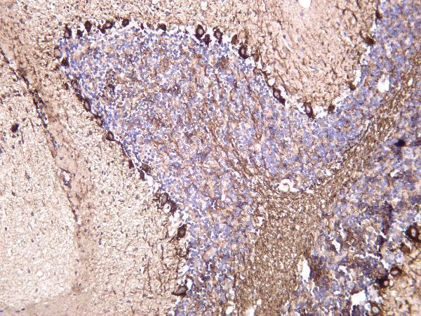
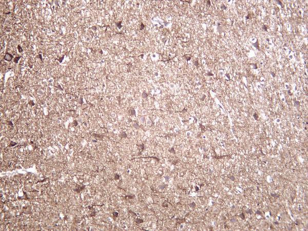
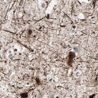
M05307-9 has IHC images of mouse, rat, and human brain (catalog IHC captions). IF is listed as an application, but no IF figure is supplied (catalog applications; image alts).

Real IHC data Mouse brain was stained with anti-Neurofilament heavy polypeptide rabbit antibody
Anti-Neurofilament heavy polypeptide Rabbit Monoclonal Antibody
Cat # M05307-9

M05307-9 is listed for IHC and IF with human, mouse, and rat reactivity (catalog applications; reactivity). Its own IHC captions show stained mouse, rat, and human brain; no IF image is supplied (catalog image alts).

Which to pick: For tissue IHC, choose M05307-9: its own captions show stained mouse, rat, and human brain, while section processing and fixative are unreported (catalog IHC captions). For IF, M05307-9 is listed at 1:200–1:1000; ICC validation and an IF figure are unreported (catalog applications; dilutions; image alts). For cross-species work, M05307-9 is listed as reactive with human, mouse, and rat (catalog reactivity).

Each figure is that product's own IHC / IF validation image from its datasheet.

References

  1. UniProt Consortium. UniProt entry P12036 (NFH_HUMAN, Neurofilament heavy polypeptide).
  2. Human Protein Atlas. NEFH tissue IHC expression (reliability: Enhanced).
  3. Human Protein Atlas. NEFH subcellular location (ICC-IF): Mainly localized to the nuclear bodies, centrosome and basal body. In addition localized to the nucleoplasm, cytosol, primary cilium and primary cilium transition zone..
  4. Human Protein Atlas. NEFH antibody validation summary (2 antibodies).
  5. Spatial patterns of FUS-immunoreactive neuronal cytoplasmic inclusions (NCI) in neuronal intermediate filament inclusion disease (NIFID). Journal of neural transmission (Vienna, Austria : 1996) 2011 — PMC3199334.
  6. Applicability of regenerative oligodendrocyte precursor cell optic nerve transplantation in rat model of demyelinating disease. Cell transplantation 2025 — PMC12536187.
  7. Neurofilament heavy polypeptide regulates the Akt-beta-catenin pathway in human esophageal squamous cell carcinoma. PloS one 2010 — PMC2815775.
  8. Bioinformatic and statistical analysis of the optic nerve head in a primate model of ocular hypertension. BMC neuroscience 2008 — PMC2567987.
  9. PubMed PMID:3138108 — UniProt-cited evidence.
  10. PubMed PMID:10048485 — UniProt-cited evidence.
  11. PubMed PMID:14702039 — UniProt-cited evidence.