This website uses cookies to ensure you get the best experience on our website.


- Table of Contents
Plan chromogenic NEURL1 IHC in paraffin sections using brain as a positive control and the catalog antibody at 1:100–1:300 (HPA tissue IHC; datasheet A30740). Assess cytoplasmic CNS staining, including high Bergmann glial signal, alongside a negative control (HPA tissue IHC).

Expected localisation, validated protocols, controls and antibodies — the at-a-glance facts below, then the full design guide.
| Expected localisation | Predominantly CNS cytoplasm; Bergmann glia also show membrane staining (HPA tissue IHC) | |
| Staining pattern | CNS glial cytoplasm; Bergmann glia also at the membrane (HPA tissue IHC) | |
| Antigen retrieval | Tris-EDTA pH 8.0 HIER, heat-mediated (datasheet A30740) | |
| Positive control | Cerebellum+3 more · see all | |
| Negative control | Adipose tissue+4 more · see all |
| Fixation | Keep fixation consistent across compared paraffin sections. (selected-SKU IHC image A30740) | |
| Caveat | Antibody staining and RNA show medium consistency (HPA tissue IHC) | |
| Regulation | Lower in medulloblastoma than cerebellum (UniProt) | |
| Isoform / epitope | 2 isoforms; map the epitope to both (UniProt) |
The catalog antibody has a datasheet IHC-P protocol (datasheet A30740); one published conjunctival melanoma protocol provides an additional starting point (PMC6849808).
| Sample | Paraffin-embedded Human brain tissue; fixative not specified (datasheet A30740) |
| Fixation | Image fixative and duration unreported (datasheet A30740); verify before use. |
| Sectioning | 4–5 µm sections on charged slides (standard) |
| Deparaffinisation | Xylene, graded ethanol series to water (standard) |
| Antigen retrieval | Heat retrieval: Tris-EDTA pH 8.0 (datasheet A30740); 20 min, 95–100 °C (standard) |
| Peroxidase block | 3% H2O2, 10 min, room temperature (standard) |
| Blocking | 10% normal serum of the secondary host, 30 min, room temperature (standard) |
| Primary antibody | Rabbit anti-NEURL1, 1:100 - 1:300 (datasheet A30740) |
| Primary incubation | Overnight at 4 °C (standard) |
| Detection | HRP-polymer secondary, DAB chromogen 5–10 min (standard) |
| Counterstain | Hematoxylin, blue, dehydrate and mount (standard) |
| Expected result | NEURL1-positive staining in bergmann glia - cytoplasm/membrane of cerebellum (HPA tissue IHC: High). HPA tissue profile: Cytoplasmic expression mainly in CNS. No signal in the no-primary control. |
NEURL1 staining is expected mainly in the cytoplasm of CNS cells, with high cytoplasmic or membrane staining in cerebellar Bergmann glia and medium staining in cortical and hippocampal glia (HPA tissue IHC). Perinuclear, dendritic and postsynaptic localization is also described, chiefly from neuronal evidence by similarity (UniProt O76050). NEURL1 has no transmembrane segment (UniProt O76050 topology). HPA reports medium consistency between tissue staining and RNA data (HPA tissue IHC).
| Strong cytoplasmic or membrane signal in cerebellar Bergmann glia. | This matches the clearest supplied tissue observation: Bergmann glia stain at a high level (HPA tissue IHC). Judge the pattern in the identified cells, alongside slide controls (general IHC practice). |
| Moderate signal in cortical or hippocampal glia, or pancreatic exocrine cells. | These are reported at medium levels (HPA tissue IHC). Signal outside the CNS can therefore be plausible; cell identity matters more than a whole-tissue positive call (HPA tissue IHC). |
| Predominantly nuclear staining with little cytoplasmic signal. | This conflicts with the reported cytoplasmic CNS profile and UniProt localization (HPA tissue IHC; UniProt O76050). Treat it as a possible artefact and review controls before scoring it as NEURL1. |
| Strong staining in a cell type reported as undetected, such as adipocytes. | Adipocytes are reported as undetected in adipose tissue (HPA tissue IHC). Check cell identification and controls; cross-reactivity or endogenous detection activity are possible explanations (general IHC practice), not established causes. |
| Widespread weak colour across cells and tissue spaces. | This is difficult to assign to the cell-specific HPA pattern (HPA tissue IHC). Compare the negative detection control and inspect tissue edges and deposits before interpreting it as expression (general IHC practice). |
| Tissue and cell selection | Cerebellar Bergmann glia provide the strongest reported tissue pattern; cortical and hippocampal glia and pancreatic exocrine cells are medium, while caudate glia are low (HPA tissue IHC). |
| Reported subcellular distribution | HPA describes mainly cytoplasmic CNS expression, including cytoplasmic or membrane staining in Bergmann glia (HPA tissue IHC). UniProt also lists perinuclear, dendritic and postsynaptic sites (UniProt O76050). |
| Antibody evidence and limits | The listed antibody, HPA044204, has IHC Enhanced validation; the tissue profile has medium staining-to-RNA consistency (HPA antibodies; HPA tissue IHC). These ratings support interpretation but do not establish every stained cell's identity. |
| Protein forms | UniProt lists 2 isoforms, no signal peptide or propeptide, and a chain spanning residues 2–574 (UniProt O76050). Without an antibody epitope, the supplied record cannot predict isoform-specific staining. |
| RNA and protein discordance | UniProt reports expression in bone marrow, while HPA reports hematopoietic cells there as undetected by tissue IHC (UniProt O76050; HPA tissue IHC). Do not treat the expression report as proof of visible IHC signal. |
| Situation | Likely cause | Next action |
|---|---|---|
| No signal in cerebellar Bergmann glia. | The result misses HPA's high-staining reference population (HPA tissue IHC); the supplied sources do not identify a target-specific fixation cause. | Confirm the cell population, then check positive-control performance, antibody dilution, retrieval and detection using the assay instructions (general IHC practice). |
| Only faint staining in caudate glia. | Low staining is reported for this population (HPA tissue IHC). | Use the reported high-staining Bergmann glia to judge whether the run worked; avoid calling a low-staining caudate section an assay failure on its own (HPA tissue IHC). |
| Strong, predominantly nuclear colour. | Nuclear dominance does not match the supplied NEURL1 localization (HPA tissue IHC; UniProt O76050). | Review morphology and the negative detection control; inspect counterstain and chromogen deposits before assigning target signal (general IHC practice). |
| Adipocytes or other reported negative cells stain strongly. | Adipocytes are undetected in the HPA tissue profile (HPA tissue IHC); cross-reactivity or endogenous activity remains possible (general IHC practice). | Verify cell identity and compare appropriate negative controls. Reassess blocking or detection conditions if controls show the same colour (general IHC practice). |
| Diffuse colour obscures cell boundaries. | Background can prevent assessment of the cell-specific HPA pattern (HPA tissue IHC); its source cannot be determined from appearance alone. | Compare the negative detection control and review blocking, washes and chromogen development under the assay instructions (general IHC practice). |
| Can IF/ICC confirm the same compartment? | HPA provides no main ICC-IF location or cell-line images for NEURL1 (HPA subcellular record). | Treat IF/ICC localization as unconfirmed by this HPA record; use the separate IF/ICC guide for its assay design, and do not transfer an IHC staining call directly to IF. |


Comprehensive Human Protein Atlas IHC scoring per tissue (reliability: Enhanced — Medium consistency between antibody staining and RNA expression data.). Rows are taken directly from the HPA tissue chart — click any row's HPA link to view the source.
| Tissue | Cell type | Level | Evidence | Source |
|---|---|---|---|---|
| Cerebellum | Bergmann glia - cytoplasm/membrane | High | Protein (IHC) | HPA → |
| Cerebral cortex | Glial cells | Medium | Protein (IHC) | HPA → |
| Hippocampus | Glial cells | Medium | Protein (IHC) | HPA → |
| Pancreas | Exocrine glandular cells | Medium | Protein (IHC) | HPA → |
| Tissue | Cell type | Level | Evidence | Source |
|---|---|---|---|---|
| Adipose tissue | Adipocytes | Not detected | Protein (IHC) | HPA → |
| Adrenal gland | Glandular cells | Not detected | Protein (IHC) | HPA → |
| Appendix | Endocrine cells | Not detected | Protein (IHC) | HPA → |
| Bone marrow | Hematopoietic cells | Not detected | Protein (IHC) | HPA → |
| Breast | Adipocytes | Not detected | Protein (IHC) | HPA → |
Troubleshoot NEURL1 staining in paraffin section chromogenic IHC; the IF entry addresses planning a separate fluorescence assay.
Two anti-NEURL1 antibodies have paraffin-section IHC images from human tissue (catalog image captions). Both list human, mouse, and rat reactivity; A30740 also lists IF/ICC applications (catalog applications/reactivity).
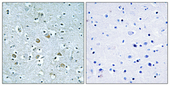
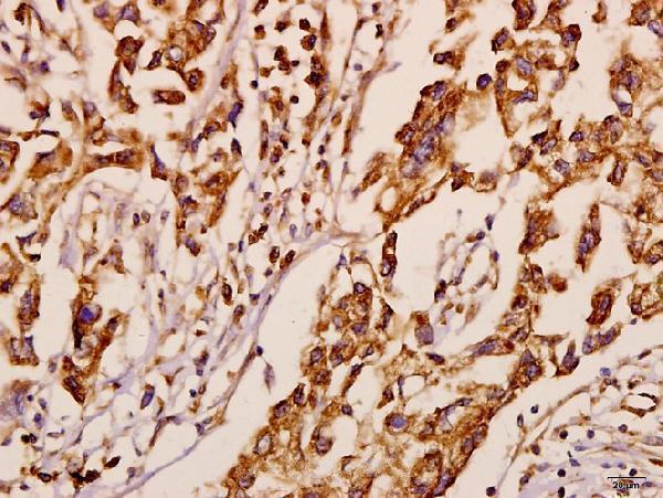
A30740 shows IHC in paraffin-embedded human brain at 1:100, with Tris-EDTA pH 8.0 retrieval and a peptide-preabsorption control (A30740 image caption). A11177 shows IHC in paraffin-embedded human breast carcinoma at 1:50 (A11177 image caption).
Which to pick: Choose A30740 for tissue IHC when its documented retrieval and peptide-preabsorption control are useful; its image shows paraffin-embedded human brain, but the fixative is unreported (A30740 image caption). Choose A30740 for IF/ICC because those applications are listed, although the payload provides no IF image (A30740 catalog applications/images). Both antibodies are rabbit polyclonals listing human, mouse, and rat reactivity; their IHC images document human tissue only, and A11177 offers a paraffin-embedded breast carcinoma example with fixative unreported (catalog host/type/reactivity; A11177 image caption).